Bifurcate Ligament Avulsion | Lateral Foot Pain | Usually Conservative
- Anterior process calcaneus fracture = bifurcate ligament avulsion injury (calcaneonavicular and calcaneocuboid ligaments)
- Conservative management effective
- “Bifurcate ligament avulsion = calcaneonavicular and calcaneocuboid ligaments
- “Usually conservative treatment - most heal with immobilization
- “Often misdiagnosed as ankle sprain - look carefully on lateral X-ray
- “Excision for small symptomatic fragments if conservative fails
Overview and Epidemiology
A fracture of the anterior process of the calcaneus is a rare injury at the attachment of the bifurcate ligament. It is usually treated conservatively, and it is often misdiagnosed as an ankle sprain.
Incidence. Classically quoted as under 1% of calcaneal fractures, but the radiology literature reports up to ~15% of calcaneal injuries (Hodge 1999). The true figure is uncertain (see Controversies).
Who. The peak age is 20-40 years, from sport and trauma. There is no clear sex predominance, although the largest functional-treatment cohort was 74% female (Massen 2019). The associated injuries are ankle sprains and other foot injuries.
Anatomy and Pathophysiology
The anterior process. The anterior aspect of the calcaneus, variable in size but typically 1-2 cm. It is the attachment site for the bifurcate ligament and forms part of the calcaneocuboid joint, the articulation between calcaneus and cuboid that provides lateral column stability.
The bifurcate ligament. Two limbs arise from the anterior process: the calcaneonavicular ligament runs to the navicular and the calcaneocuboid ligament to the cuboid. Together they stabilise the midfoot and prevent excessive motion. Forced inversion and plantarflexion put the ligament under excessive tension, and it avulses from the anterior process with a fragment of bone.
Why it is missed. The injury shares its mechanism and its symptoms with an ankle sprain, and it is often diagnosed as one. On the radiograph it is subtle and may be obscured or overlooked.
Why it is usually treated conservatively. The fragment is usually small and non-displaced, most heal with immobilisation, and the anterior process has low functional demand.
Two Mechanisms: Avulsion vs Compression (Impaction)
Recognising which mechanism produced the injury predicts the fracture morphology and the treatment lane.
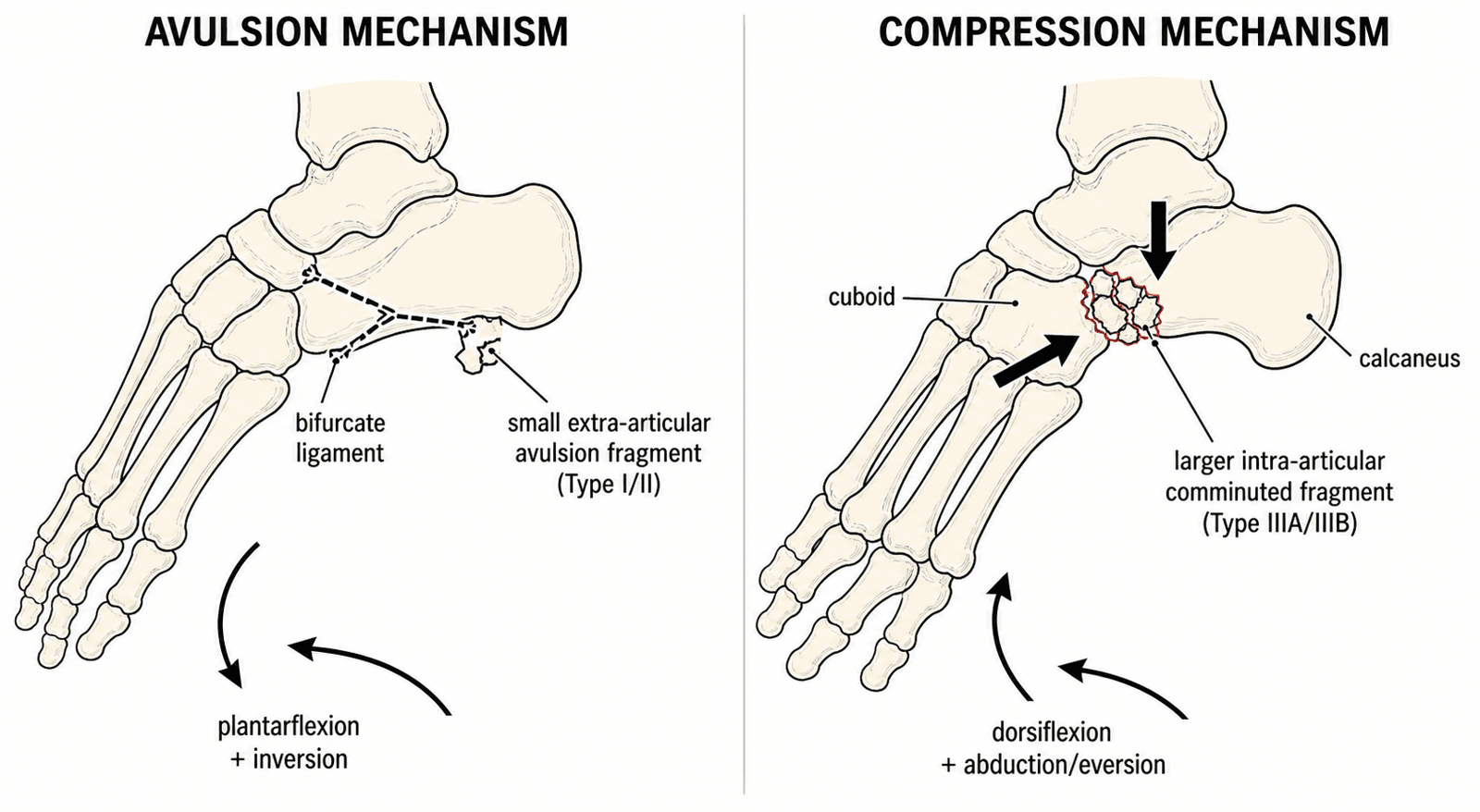
Avulsion, the common one. Forced plantarflexion combined with inversion (supination/adduction) tightens the bifurcate ligament, and it avulses its bony origin from the dorsal anterior process. The result is a small, extra-articular flake at the apex of the process, Degan type I/II, that typically heals with functional or conservative care.
Compression, the other pattern. Forced dorsiflexion with forefoot abduction/eversion, a pronation-abduction pattern, drives the anterior process against the cuboid and talar head. The process is crushed rather than pulled off, a shear/impaction injury of the process itself. The result is a larger, often comminuted, intra-articular fragment (Degan type IIIA/IIIB) that involves the calcaneocuboid articular surface, the group most likely to need CT and, when displaced, ORIF.
The cohort evidence. This mechanism is consistent with Massen's cohort, in which 82% of fractures were comminuted and 78% had concomitant Chopart-line injury. Those findings fit a higher-energy compression/midtarsal loading pattern rather than a pure ligamentous avulsion.
In the clinic. An avulsion history points to a small extra-articular fragment treated functionally. A dorsiflexion/abduction crush should raise suspicion of an intra-articular fragment and prompt CT to assess the calcaneocuboid joint.
Do not confuse compression of the anterior process with the cuboid "nutcracker" fracture, in which the cuboid is crushed between the calcaneus and the metatarsal bases. That is a separate lateral-column injury.
Classification Systems
Modified Degan. The original Degan classification (Degan, JBJS Am 1982) was modified by Cibura et al. (BMC Musculoskelet Disord 2022), who subdivided the intra-articular type III into displaced and non-displaced subgroups because the displaced subgroup is the one that most often needs surgery.
- Type I - small extra-articular fracture of the apex of the anterior process. Conservative / functional treatment.
- Type II - larger extra-articular fracture without calcaneocuboid joint involvement. Conservative / functional treatment.
- Type IIIA - large intra-articular fragment, non-displaced (no joint step-off). Conservative, but CT is advised: Cibura's series specifically flags missed type IIIA fractures as at risk of complications, and six missed IIIA fractures later required surgery for persistent symptoms.
- Type IIIB - large intra-articular fragment, displaced with a calcaneocuboid step-off. Most likely to require ORIF, or excision for an unreconstructable fragment.
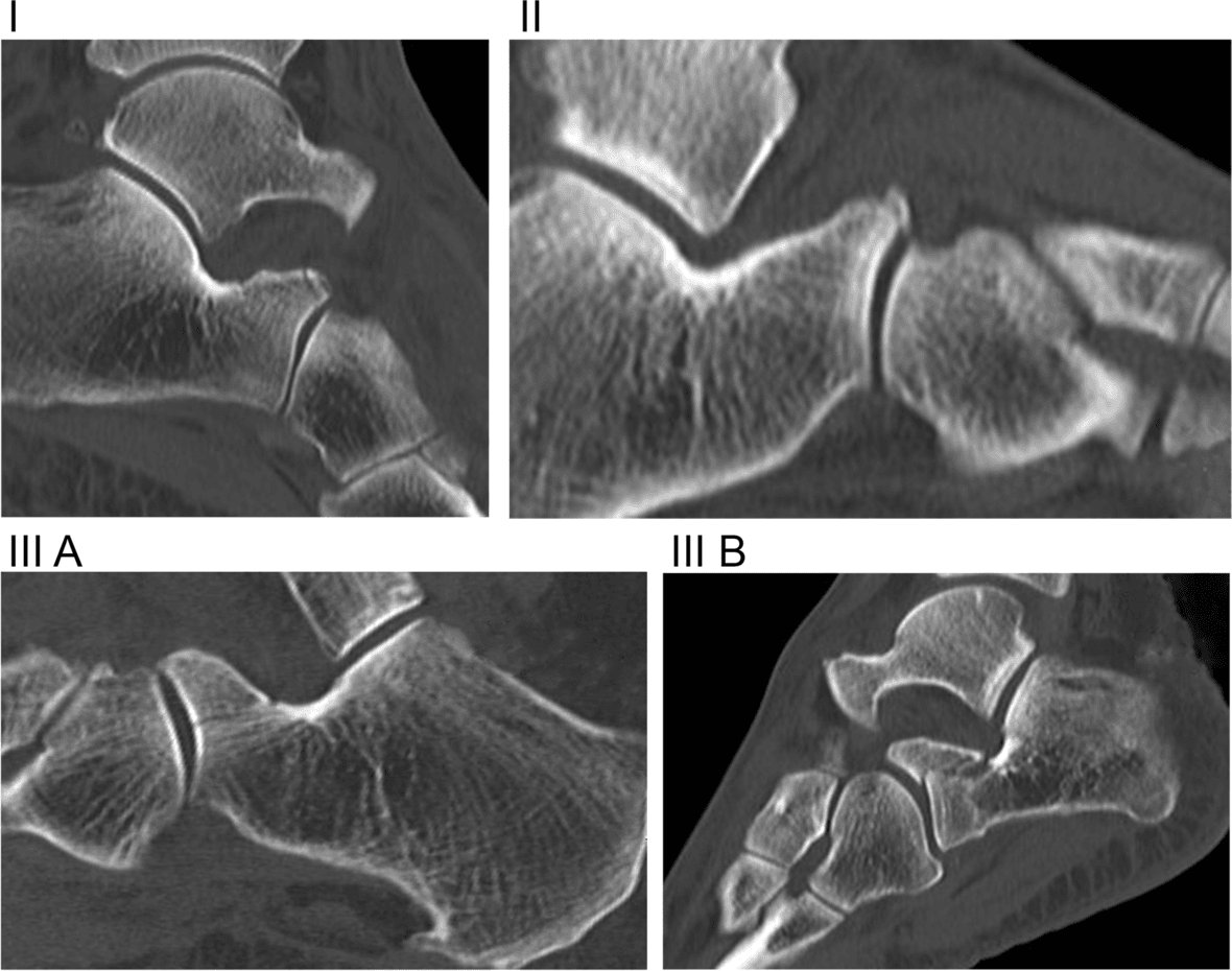
What the type predicts. Massen et al. (Injury 2019) found that fracture type did not predict functional outcome, so the classification informs the surgical decision more than the prognosis.
Displacement and fragment size. A 2 mm articular step is a commonly used operative threshold, extrapolated from articular-fracture principles and not validated specifically for this fracture. Fragment size, divided at 25% of the process, determines the treatment choice.
- Treatment
- Conservative / functional
- Outcome
- Good-to-excellent in the majority
- Treatment
- ORIF, or excision if unreconstructable
- Outcome
- Aim to restore the calcaneocuboid joint surface
- Treatment
- Conservative or excision
- Outcome
- Good if asymptomatic
- Treatment
- Conservative or ORIF
- Outcome
- Better with ORIF if displaced
Clinical Assessment
History. Lateral foot pain after forced inversion and plantarflexion, the ankle-sprain mechanism, with swelling localised to the lateral foot and pain on weight bearing. The risk factors are sports with inversion injuries, high-energy trauma and the ankle-sprain mechanism itself.
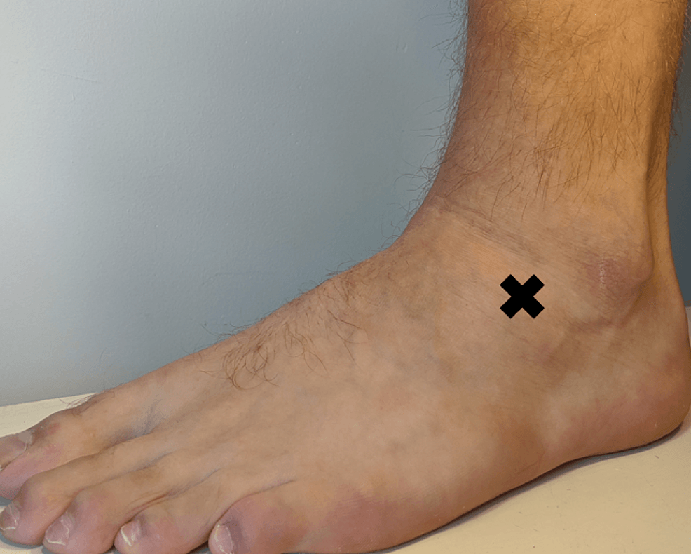
Examination. Swelling sits on the lateral foot; ecchymosis may be delayed and deformity is rare. The key finding is tenderness over the anterior process, anterior to the lateral malleolus, with calcaneocuboid joint tenderness and pain on calcaneocuboid stress, while the ankle joint is usually not tender. Ankle and midfoot range of motion may be limited, and inversion/eversion is painful.
Differential diagnosis. The classic trap is the "lateral ankle sprain that will not settle." Focal tenderness about 2 cm distal and anterior to the tip of the lateral malleolus, over the anterior process and calcaneocuboid joint, should redirect the differential.
- Mechanism / Clue
- Forced inversion + plantarflexion
- Key Tenderness
- Anterior process / calcaneocuboid joint
- Distinguishing Feature
- Irregular non-corticated fragment; oblique X-ray or CT positive
- Mechanism / Clue
- Inversion in plantarflexion
- Key Tenderness
- Anterior to lateral malleolus (ATFL)
- Distinguishing Feature
- Anterior drawer positive; normal foot films
- Mechanism / Clue
- None (incidental ossicle)
- Key Tenderness
- Usually asymptomatic
- Distinguishing Feature
- Smooth corticated margins; often bilateral; up to 5% prevalence
- Mechanism / Clue
- Inversion; peroneus brevis pull
- Key Tenderness
- Base of 5th metatarsal
- Distinguishing Feature
- Tenderness at styloid; proximal transverse line
- Mechanism / Clue
- Adolescent, recurrent sprains
- Key Tenderness
- Sinus tarsi / dorsolateral midfoot
- Distinguishing Feature
- Anteater-nose sign on lateral X-ray; CT confirms bar
- Mechanism / Clue
- Axial load / snowboarder mechanism
- Key Tenderness
- Cuboid or lateral talar process
- Distinguishing Feature
- Different tender point; CT localises
Investigations
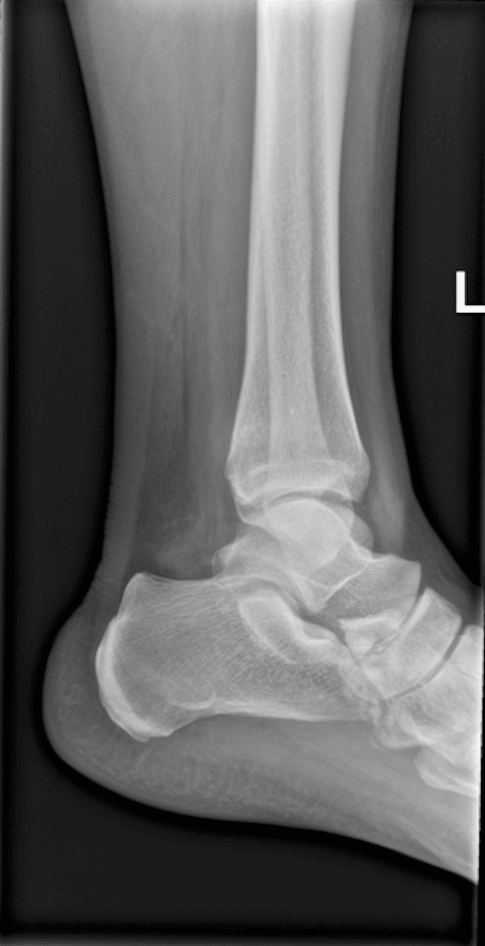
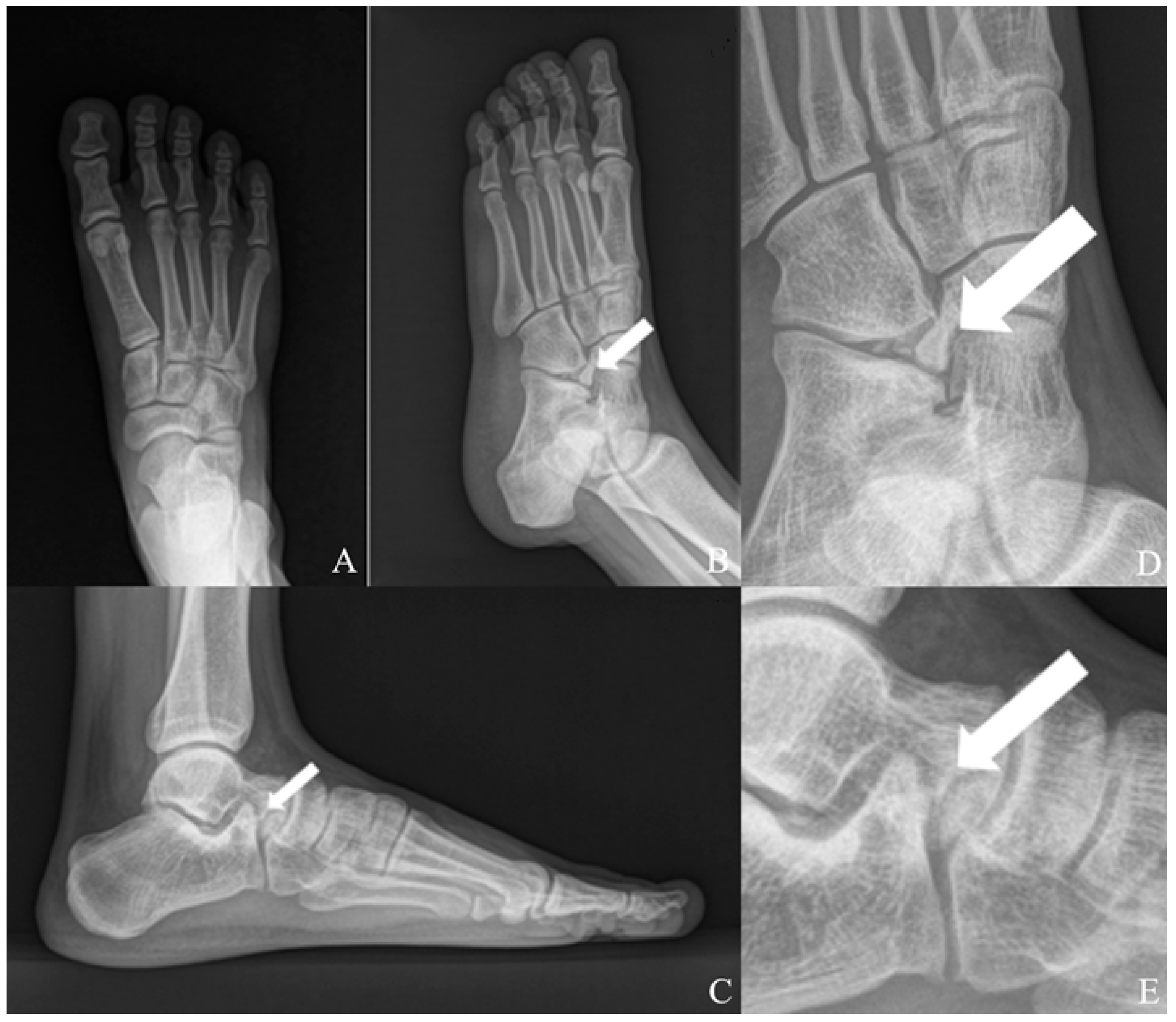
Radiographs. The fracture is often missed on the initial films, so a high index of suspicion is needed.
- Lateral - shows the anterior process, but the fracture is often subtle and may be obscured by overlapping structures
- Oblique - may show the fracture better; the calcaneocuboid joint view
- AP - may show the fracture, but less reliably than the lateral
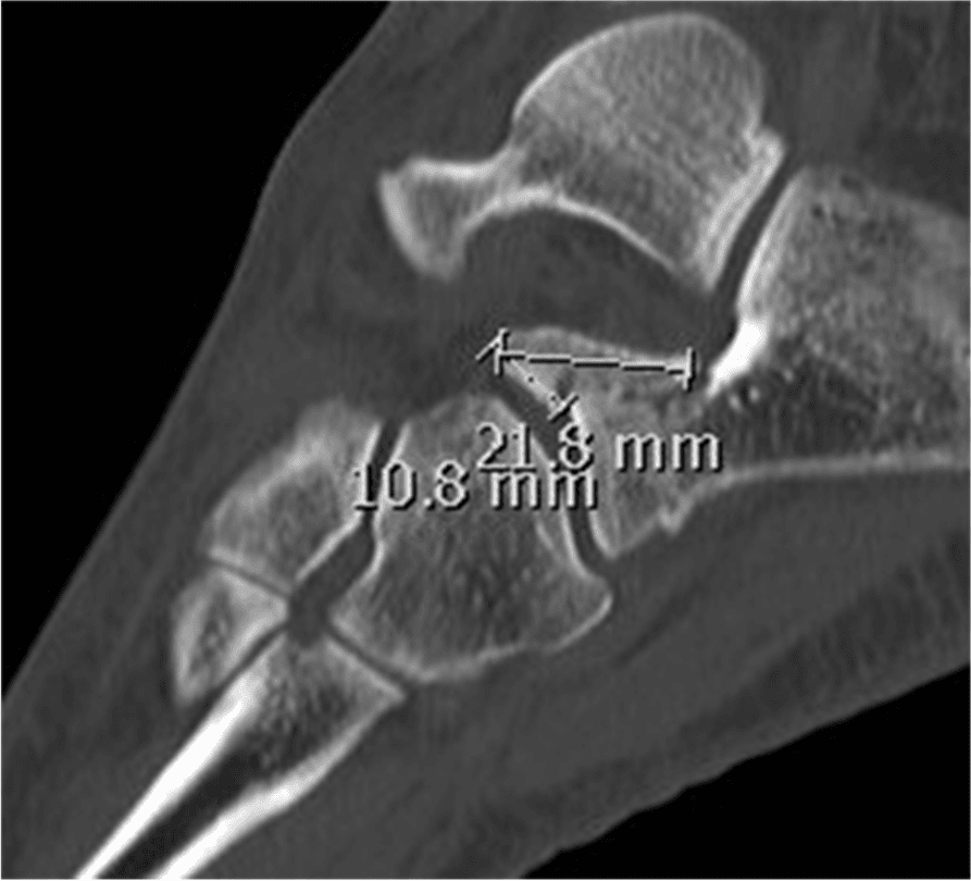
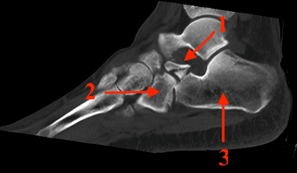
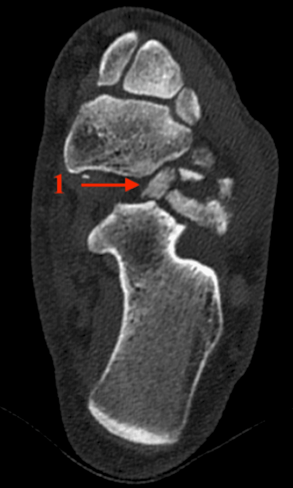
CT. Recommended when the fracture is suspected clinically but the radiographs are negative, when displacement is unclear, and when planning surgery; Cibura advises CT for any clinical suspicion. It shows the fracture pattern, measures the step-off, sizes the fragment and defines calcaneocuboid joint involvement, and it is often needed for diagnosis and planning.
MRI. Two problems are answered better by MRI than by plain films and CT.
- Occult fracture with normal radiographs. Where clinical suspicion is high but films are normal and CT is unavailable or negative for a cortical break, MRI shows bone-marrow oedema and a hypointense fracture line, confirming an acute injury.
- Associated soft-tissue and osteochondral injury. The same inversion/midtarsal mechanism injures the bifurcate ligament and the wider Chopart line, concomitant in a large proportion of cohorts. MRI can show bifurcate/calcaneocuboid ligament tears, osteochondral lesions and other midtarsal injuries that plain films miss and that flag a higher-risk, slower-recovering injury.
Bone scintigraphy. A technetium bone scan is sensitive but non-specific. Focal anterior-process uptake supports an occult acute fracture when cross-sectional imaging is unavailable, but it cannot characterise fragment size, displacement or joint involvement. In modern practice CT (for bony detail and surgical planning) and MRI (for acuity and soft tissue) have largely superseded it.
Fracture or calcaneus secundarius? A corticated density beside the anterior process may be an accessory ossicle, the calcaneus secundarius, rather than a fracture. On plain films and CT the distinction rests on the margins: an irregular non-corticated fragment favours fracture, a smooth corticated ossicle favours the variant, and CT shows the cortication best. MRI adds the physiological answer: an acute fracture has surrounding marrow oedema whereas an asymptomatic calcaneus secundarius is a quiescent, corticated, oedema-free ossicle. When trauma and focal symptoms create doubt, CT or MRI is needed.
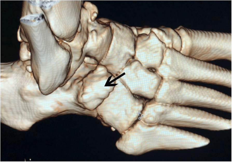
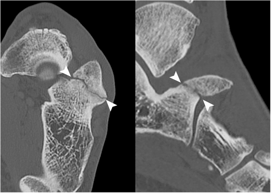
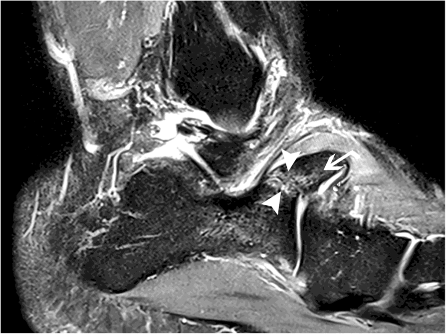
A systematic foot-and-ankle imaging approach is developed in the imaging-ankle-foot topic.
Management Algorithm
The decision. Most fractures do well with conservative or functional treatment, with good-to-excellent results in the majority. Surgery is reserved for the displaced intra-articular (type IIIB) fragment and the persistent symptomatic nonunion. Every pathway starts with the diagnosis: tenderness over the anterior process is the key finding, and CT is recommended when the fracture is suspected clinically but the radiographs are negative.
Non-operative treatment. First-line for most anterior process fractures: the non-displaced or minimally displaced fracture, the small fragment, and the patient who prefers it.
- Short leg cast or boot
- Non-weight bearing for 4-6 weeks
- Progressive weight bearing after healing
- Serial radiographs to monitor healing
Functional treatment, with immediate full weight-bearing, is a validated alternative for non-displaced fractures (Massen, Injury 2019).
Surgical indications. The absolute indications are a large displaced fragment (over 25% of the process), a persistent symptomatic nonunion and calcaneocuboid joint instability. The relative indications are a small fragment with persistent pain and failed conservative treatment; surgery for failed conservative treatment comes after 6-12 weeks.
Fix or excise. ORIF of a displaced intra-articular fragment protects the calcaneocuboid joint. Excision is appropriate for a small symptomatic fragment with established nonunion: it relieves pain and is preferable to unreliable fixation of a tiny fragment (Degan, JBJS Am 1982).
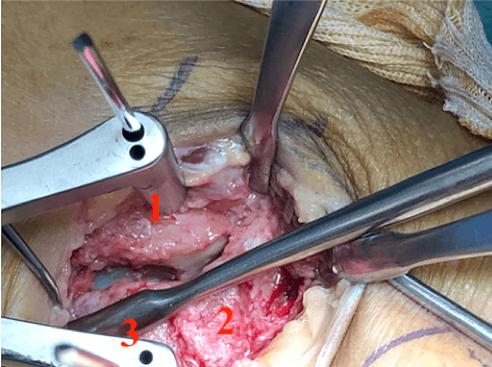
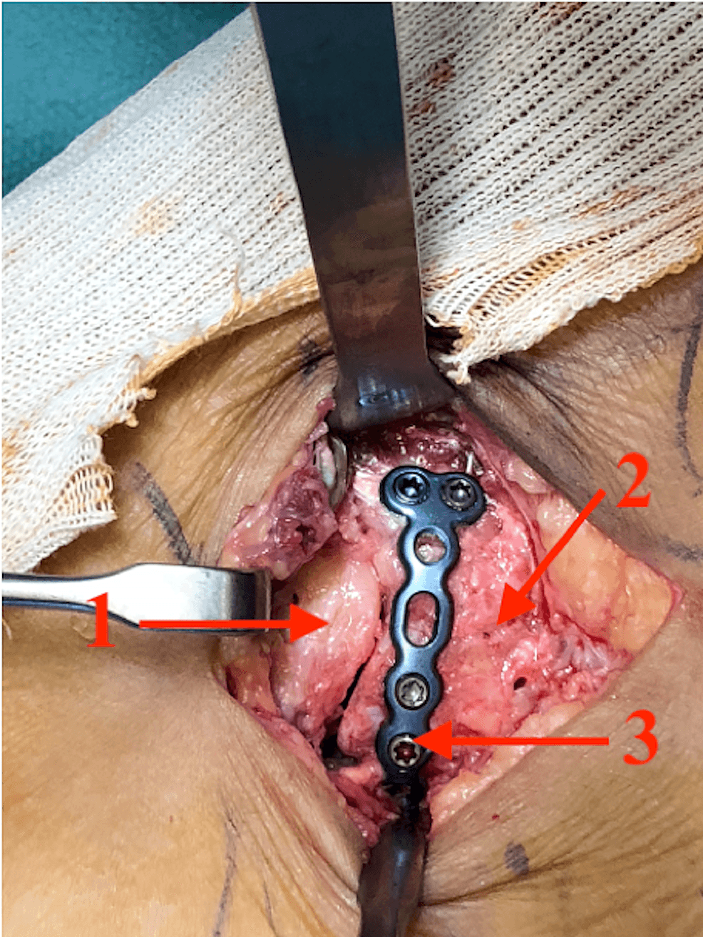
Surgical Technique
When to fix. ORIF is the operation for large fragments. It preserves the calcaneocuboid joint, prevents nonunion and gives better outcomes than excision for large fragments. The indications:
- Large fragment (over 25% of process)
- Displaced (greater than 2 mm)
- Calcaneocuboid joint instability
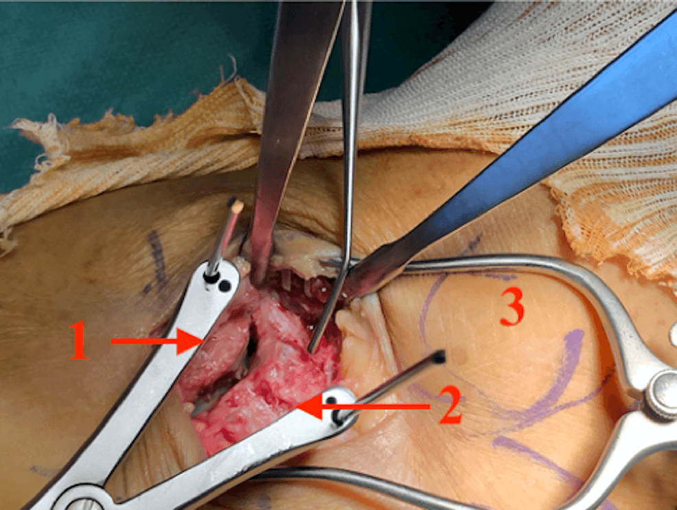
Technique.
- Exposure - lateral approach to the anterior process, protecting the peroneal tendons
- Reduction - anatomic reduction of the fragment
- Fixation - lag screws (2.0-2.7 mm) or mini-fragment screws
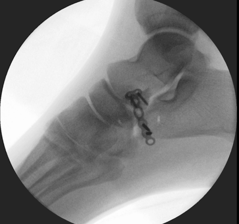
- Verification - confirm reduction and hardware position fluoroscopically
Complications
- Incidence
- 5-10%
- Risk Factors
- Displacement, inadequate immobilisation
- Prevention/Management
- Adequate immobilisation, ORIF if needed; excision if symptomatic, ORIF if large fragment
- Incidence
- 10-15%
- Risk Factors
- Nonunion, fragment size, joint involvement
- Prevention/Management
- Adequate treatment, anatomic reduction; excision if symptomatic, activity modification
- Incidence
- ~30% (Cibura 2022)
- Risk Factors
- Similar to ankle sprain
- Prevention/Management
- High index of suspicion, CT if needed
- Incidence
- 5-10%
- Risk Factors
- Malunion, joint involvement
- Prevention/Management
- Anatomic reduction
Incidence figures for nonunion, persistent pain and calcaneocuboid arthritis are conventional teaching ranges, not measured rates from a defined cohort. The measured figures in the literature are the ~30% missed-diagnosis rate (Cibura 2022) and the 31% unfavourable-outcome rate at 2 years with conservative care (Xiao 2024).
Postoperative Care
Immediately after surgery. A short leg cast or boot, non-weight bearing for 4-6 weeks. Ankle range of motion follows cast removal, and physiotherapy works on midfoot range of motion and strengthening.
- Weeks 0-4 - short leg cast, non-weight bearing, elevation to reduce swelling, ankle range-of-motion exercises if stable
- Weeks 4-6 - cast removal, transition to a walking boot, progressive weight bearing
- Weeks 6-12 - full weight bearing and progressive activity; return to sport at 3-4 months
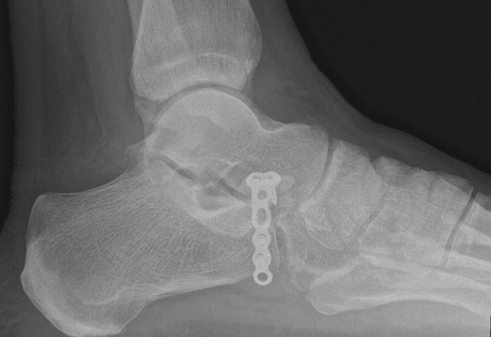
Outcomes and Prognosis
Conservative and functional treatment. The default for most fractures. Massen et al. (Injury 2019) reported good-to-excellent results with immediate full weight-bearing, a median VAS-FA of 95 and Karlsson of 90, with return to work at about 2 weeks and return to sport at about 3 months. The outcome is not uniformly perfect: of 84 conservatively treated patients in Xiao et al. (OTSR 2024), 31% had an unfavourable result (Karlsson ≤80) at 2 years.
Surgery. Reserved for displaced intra-articular fragments or symptomatic nonunion. Of 13 surgically treated type IIIB fractures in Cibura et al. (BMC 2022), only one nonunion occurred. In Degan's series (JBJS Am 1982) excision relieved symptoms in most but failed in patients with established missed nonunions, which argues for correct early diagnosis rather than primary excision.
Predictors of a poorer outcome. Concomitant talonavicular joint fracture (OR 3.6) and age ≥47.5 years (OR 5.0) independently predicted an unfavourable functional result (Xiao, OTSR 2024). Missed or delayed diagnosis is associated with higher complication rates and more frequent need for surgery (Cibura, BMC 2022).
Stratified single-figure "success rates" by treatment type are not supported by high-quality data; the numbers above are quoted directly from the published series.
Guidelines, Registries & Global Practice
Global Epidemiology
- Frequency: Classically described as rare; quoted figures range from under 1% of calcaneal fractures to as high as ~15% of calcaneal injuries in radiology series, the variation driven mainly by under-recognition (Hodge, J Emerg Med 1999).
- Demographics: Adults across a wide age range; one of the larger functional-treatment cohorts was 74% female with a median age of 38 years (Massen, Injury 2019). Mechanism is forced inversion and plantarflexion — the same as a lateral ankle sprain.
- Associated injuries: Concomitant Chopart-line / talonavicular injuries are common and predict slower recovery (Xiao, OTSR 2024).
Guidelines & Society Positions (side by side)
There is no dedicated AAOS / BOA-BOAST / NICE / AO / EFORT clinical practice guideline specifically for the anterior process calcaneal fracture — a point worth stating directly in a viva. Practice is therefore extrapolated from foot-and-ankle trauma principles and the small published series.
- Relevant contribution
- Anatomic restoration of articular surfaces; protect the lateral column
- Practical message
- Reduce and fix displaced intra-articular (type IIIB) fragments
- Relevant contribution
- Soft-tissue assessment, timely senior review, CT for intra-articular injury
- Practical message
- Low threshold for CT when an inversion injury stays focally tender
- Relevant contribution
- Classification literacy (Degan / modified Degan) and outcome data
- Practical message
- Match treatment to fragment size and displacement, not type alone
- Relevant contribution
- Best available Level IV evidence
- Practical message
- Conservative first; surgery for displaced intra-articular or symptomatic nonunion
Registry Evidence
National joint/trauma registries (NJR, AJRR, AOANJRR, SHAR/Swedish, Norwegian, NZJR) do not capture this non-arthroplasty fracture, so there is no registry-level survival or revision data. Evidence remains confined to single-centre retrospective series — a genuine limitation of the field.
High- vs Limited-Resource Practice
- High-resource settings: CT is readily available and is used liberally to confirm the diagnosis, size the fragment, and assess calcaneocuboid involvement (Cibura, BMC 2022). Displaced intra-articular fragments may be offered ORIF.
- Limited-resource settings: Diagnosis relies on lateral and oblique foot radiographs (Trnka, 1998); where CT is unavailable, clinical suspicion plus oblique films guides a default of functional/conservative care, which gives good outcomes in the majority. The main avoidable harm worldwide is the missed diagnosis mislabelled as an ankle sprain.
A common foot-and-ankle viva trap. Know the bifurcate-ligament avulsion mechanism, the modified Degan classification, and that most fractures do well with conservative/functional treatment. State clearly that the evidence is Level IV with no dedicated society guideline, reserve surgery for displaced intra-articular fragments or symptomatic established nonunion, and remember the calcaneus secundarius differential and the talonavicular-injury / older-age predictors of poorer outcome.
Controversies & Areas of Uncertainty
The evidence base is entirely Level IV-V (small retrospective series and case reports). There is no randomised trial and no validated, universally adopted classification, so the following questions remain genuinely open.
True incidence. The spread between the quoted figures, from under 1% of calcaneal fractures to up to 15% of calcaneal injuries, reflects how often the fracture is missed and whether Chopart-line avulsions are counted. Any quoted figure should be hedged.
Immobilise or mobilise? Traditional teaching is a non-weight-bearing cast for 4-6 weeks, set against Massen's good-to-excellent results with immediate full weight-bearing for non-displaced fractures. The optimal duration and the need for any immobilisation are unsettled.
The value of classification. Degan (and the modified Cibura version) is the most used scheme, yet Massen found that fracture type did not predict outcome. Whether classification should drive treatment, or only the size and displacement of an intra-articular fragment, is debated.
Excise or fix large fragments. For displaced intra-articular (type IIIB) fragments threatening the calcaneocuboid joint, some favour ORIF to preserve the joint while others excise. Numbers are too small for a definitive answer; surgeon experience and fragment quality drive the choice.
State explicitly that the evidence is low-level (no RCTs). Anchor your answer on principles: most fractures do well conservatively; image with CT when suspected; reserve surgery for displaced intra-articular fragments or symptomatic established nonunion; and counsel older patients and those with concomitant Chopart/talonavicular injury that recovery may be incomplete.
MCQ Practice Points
Q: What is the bifurcate ligament and how does it relate to anterior process calcaneus fractures? A: Bifurcate ligament consists of calcaneonavicular and calcaneocuboid ligaments - Attaches to anterior process of calcaneus. Forced inversion and plantarflexion causes avulsion, resulting in anterior process fracture. Usually treated conservatively.
Q: What is the treatment approach for anterior process calcaneus fractures? A: Usually conservative or functional treatment - Cast/boot with protected weight-bearing, or immediate full weight-bearing for non-displaced fractures, giving good-to-excellent results in the majority. Surgery is reserved for displaced intra-articular (type IIIB) fragments (ORIF) or persistent symptomatic nonunion (excision).
Q: Why are anterior process calcaneus fractures often missed initially? A: Similar mechanism and symptoms to ankle sprain - about 30% are missed at presentation (Cibura, BMC 2022). High index of suspicion needed, especially with lateral foot pain after inversion injury. CT recommended if suspected clinically but X-ray negative.
Q: When is surgery indicated for anterior process calcaneus fractures? A: Rarely needed - Only for displaced intra-articular fragments (modified Degan IIIB, ORIF) or persistent symptomatic nonunion (excision). Most fractures do well with conservative/functional treatment.
Q: When is excision appropriate for anterior process calcaneus fractures? A: Small symptomatic fragments with persistent pain or established nonunion - Excision relieves symptoms and is preferable to unreliable fixation of a tiny fragment (Degan, JBJS Am 1982). It is a salvage step, not a primary strategy.
Exam Viva Scenarios
Practise clinical reasoning and management decisions out loud
“A 28-year-old athlete presents with lateral foot pain after forced inversion and plantarflexion injury. Initial diagnosis was ankle sprain, but pain persists 3 weeks later. Clinical examination shows tenderness over anterior process of calcaneus (anterior to lateral malleolus). Lateral X-ray shows small fracture of anterior process. CT shows non-displaced fracture.”
“A 30-year-old patient presents with persistent lateral foot pain 3 months after anterior process calcaneus fracture treated conservatively. X-rays show nonunion of small fragment (under 25% of process). The fragment is causing persistent pain and calcaneocuboid joint irritation.”
“A 45-year-old recreational hiker presents with mild dorsolateral midfoot ache after twisting the foot a week ago. A lateral foot X-ray shows a small bony density anterior to the calcaneus, overlying the talar head. The emergency team has labelled this an anterior process fracture and applied a cast. The patient asks whether this is really a fracture.”
Key Concepts
- Bifurcate ligament avulsion = calcaneonavicular + calcaneocuboid ligaments
- Anterior process is attachment site for bifurcate ligament
- Forced inversion and plantarflexion mechanism
- Usually conservative/functional treatment with good-to-excellent results
Classification (modified Degan)
- Type I / II: Extra-articular (small / larger) - conservative or functional
- Type IIIA: Intra-articular, non-displaced - conservative; confirm with CT
- Type IIIB: Intra-articular, displaced (CC step-off) - ORIF or excision
- Type drives the SURGICAL decision more than prognosis (Massen 2019)
Treatment
- Most fractures: Conservative (cast or boot, NWB 4-6 weeks)
- Large displaced fragment: ORIF (preserves joint)
- Small symptomatic fragment: Excision (relieves pain)
- Surgery rarely needed - only for specific indications
Surgical Technique
- ORIF: Lateral approach, anatomic reduction, lag screws (2.0-2.7mm)
- Excision: Same approach, remove fragment, smooth edges
- Preserve calcaneocuboid joint stability
- Verify reduction fluoroscopically
Complications
- Nonunion: 5-10% conventional range (prevent with adequate immobilization)
- Persistent pain: 10-15% conventional range (excision if symptomatic)
- Missed diagnosis: ~30% missed at presentation (Cibura 2022)
- Calcaneocuboid arthritis: 5-10% conventional range (prevent with anatomic reduction)
Evidence Base
Degan: Surgical excision — classic series
- 18 of 25 healed with cast immobilisation
- Excision failures were both missed/untreated nonunions
- Early excision usually NOT necessary
- Correct initial diagnosis is the key determinant of outcome
Cibura: Modified Degan classification & treatment algorithm
- Modified Degan: type IIIA (undisplaced) vs IIIB (displaced) intra-articular
- 29.3% missed at presentation
- Missed fractures: higher complication rate, more surgery
- Low-threshold CT for any clinical suspicion
Massen: Functional (full weight-bearing) treatment outcomes
- Full weight-bearing gave good-to-excellent outcomes
- Fast return to work (~2 weeks), slower return to sport (~3 months)
- Fracture type did NOT predict outcome in this cohort
- Most fractures are comminuted and have Chopart-line associations
Xiao: Predictors of poor functional outcome
- 31% unfavourable outcome at 2 years despite conservative care
- Concomitant talonavicular fracture predicts poor outcome (OR 3.6)
- Age ≥47.5 years predicts poor outcome (OR 5.0)
- Outcome is not uniformly benign
Trnka: Often-misdiagnosed fracture, oblique views
- Oblique foot X-rays are the key plain-film view
- Diagnosis is commonly missed
- Large intra-articular fragments risk calcaneocuboid arthrosis
- Do not delay surgery for significant displaced fragments
Hodge: Fracture vs calcaneus secundarius
- Calcaneus secundarius is an accessory ossicle (up to 5% prevalence)
- Smooth corticated margins favour ossicle; irregular margins favour fracture
- Clinical tenderness and mechanism distinguish the two
- Bilateral symmetric ossicles support a normal variant
Sarrafian: Bifurcate ligament & calcaneocuboid anatomy
- Bifurcate ligament = calcaneonavicular + calcaneocuboid limbs
- Origin is the dorsal anterior process of the calcaneus
- Stabiliser of the Chopart (midtarsal) joint
- Anterior process contributes to the calcaneocuboid joint surface