FGFR2 Acrocephalosyndactyly
- FGFR2: Two recurrent mutations - Ser252Trp and Pro253Arg.
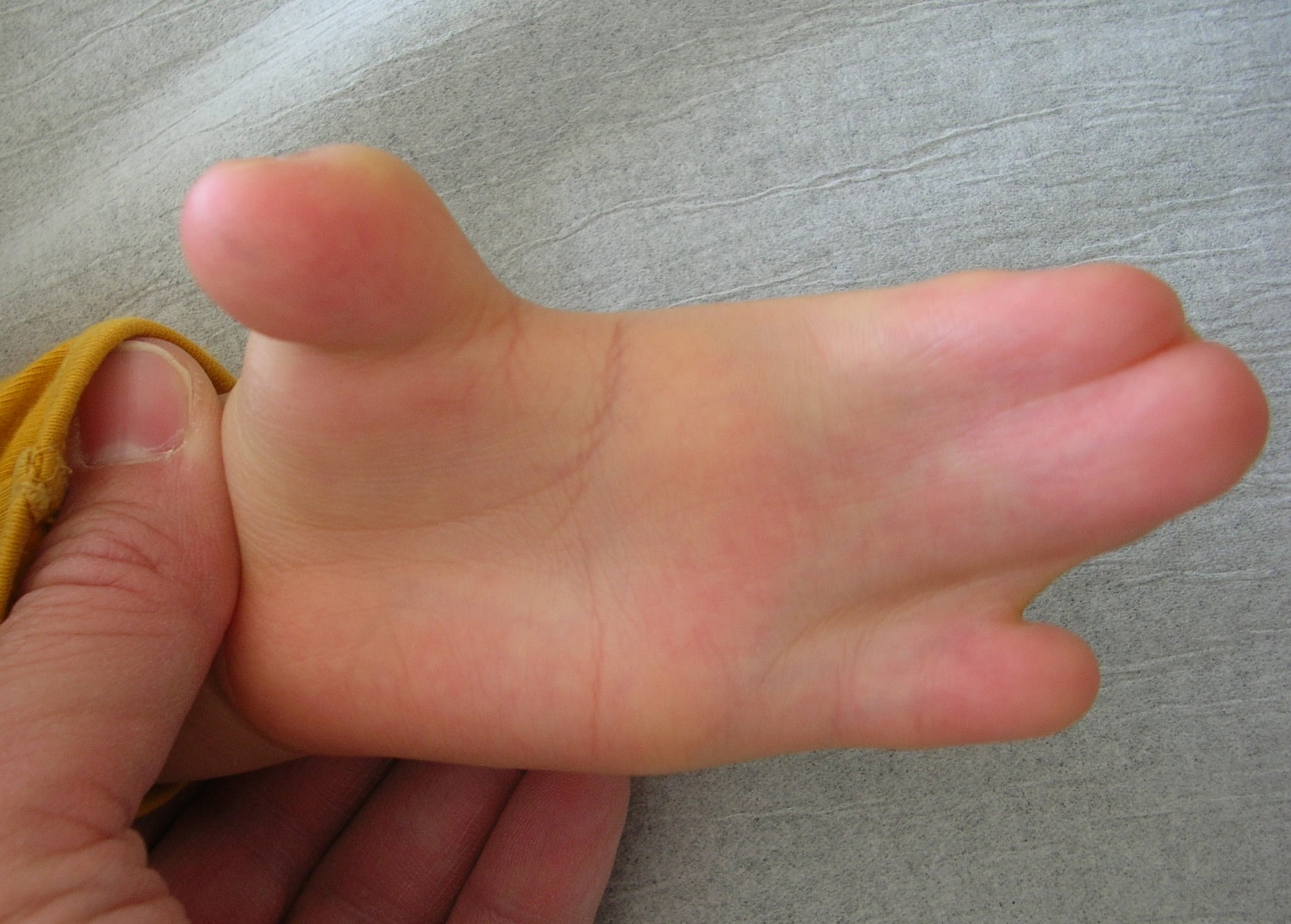
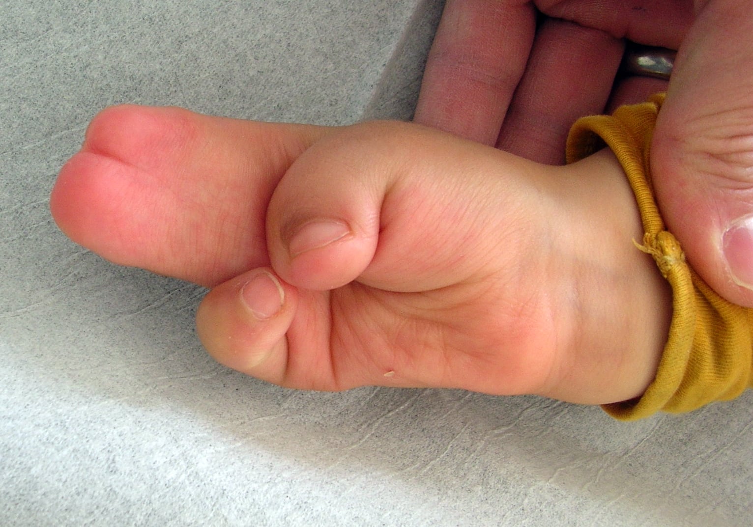
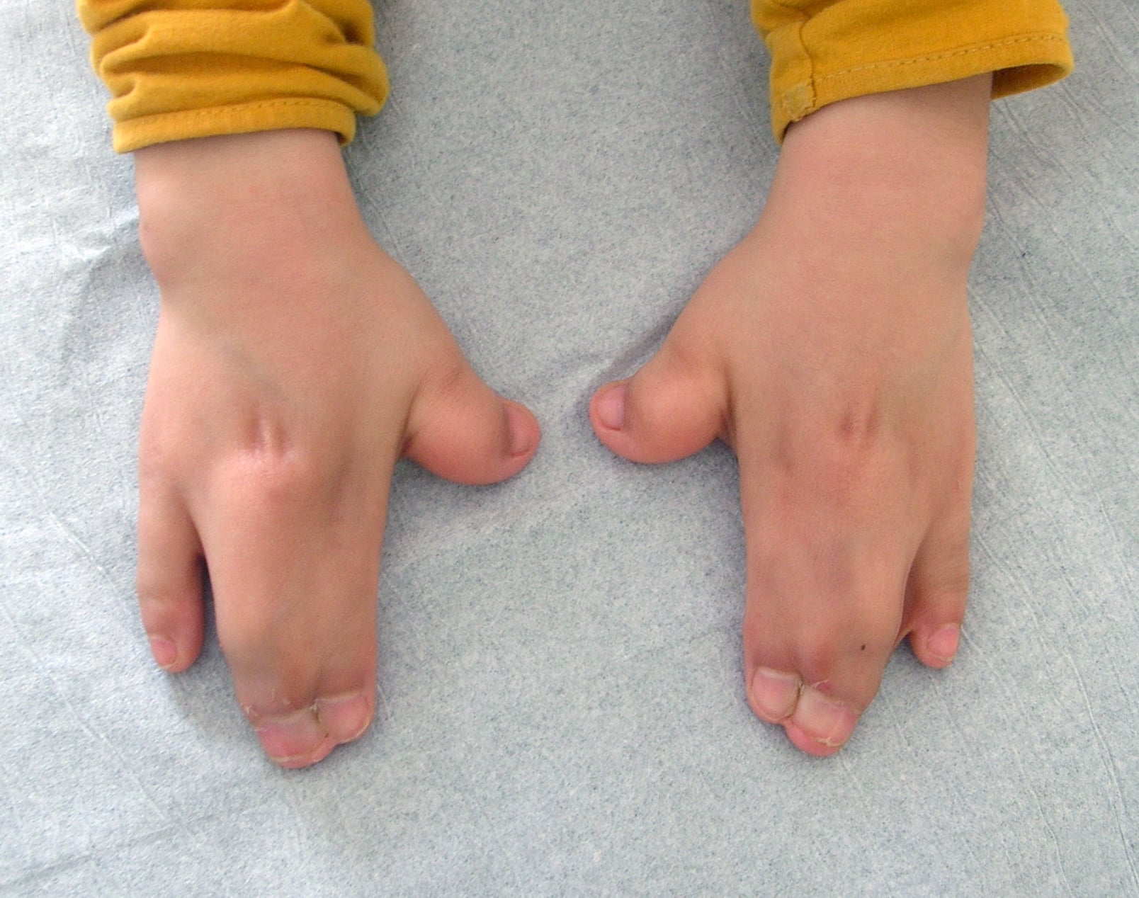
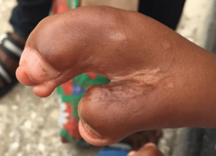
- Hand: Complex syndactyly with bony fusion - the mitten hand.
- Thumb: Short, broad, with radial clinodactyly.
- Symphalangism: Stiff fingers (except little finger) limit outcome.
- Staged surgery: Release border digits and thumb first.
- “FGFR2 Ser252Trp and Pro253Arg
- “Upton classification of the Apert hand (I-III)
- “Allelic with Crouzon syndrome
- “Symphalangism is the main limit on function
Overview/Epidemiology
Apert syndrome (acrocephalosyndactyly type I) is one of the most recognisable craniosynostosis syndromes. It is autosomal dominant, caused by mutations in FGFR2 (fibroblast growth factor receptor 2), and has a birth prevalence of roughly 1 in 65,000-80,000 live births.
The orthopaedic angle. Apert is the craniosynostosis syndrome with the most severe hand involvement: its complex (bony) syndactyly is more severe than in any other common craniosynostosis syndrome. It is the syndrome in which the hand surgeon plays a central role, and the classic viva case linking genetics, craniofacial surgery and hand surgery.
Pathophysiology and Genetics
FGFR2 gain of function. Wilkie and colleagues showed that essentially all Apert cases result from one of two adjacent missense mutations in FGFR2, Ser252Trp or Pro253Arg (Wilkie, Nat Genet 1995). Both sit in the linker between the second and third immunoglobulin-like domains of the receptor.
From receptor to phenotype. FGFR signalling normally coordinates suture and limb morphogenesis. The Apert mutations cause a gain of function, with enhanced ligand binding and prolonged signalling, and the result is premature fusion of the cranial sutures (craniosynostosis) and abnormal fusion of the developing digital rays (syndactyly).
Allelic with Crouzon. Crouzon syndrome is also caused by FGFR2 mutations, but these affect the third Ig domain and produce craniosynostosis with normal limbs. The Apert linker mutations produce craniosynostosis plus severe syndactyly (Wilkie, Nat Genet 1995).
Genotype and phenotype. The two mutations tend to weight the disease differently (Wilkie, Novartis Found Symp 2001):
- Ser252Trp tends to produce more severe craniofacial disease, with more cleft palate
- Pro253Arg tends to produce more severe syndactyly of the hands and feet
Why almost every case is new and paternal. Almost all cases arise as new (de novo) mutations, and Apert is the textbook example of a paternal age effect disorder driven by selfish spermatogonial selection. The two recurrent mutations arise on the paternally derived allele in essentially all cases, during spermatogenesis rather than oogenesis.
Selfish selection. These gain-of-function mutations give the mutant spermatogonial stem cell a selective growth, or clonal, advantage in the testis, so mutant clones expand and accumulate with the man's age, a process analogous to a benign tumour of spermatogonia. The risk therefore rises with advancing paternal age, and the small number of mutable nucleotides explains why the same two substitutions recur across unrelated families.
Counselling. For unaffected parents the recurrence risk is very low, because the child's mutation was a sporadic de novo event. An affected individual transmits it as autosomal dominant with a 50% risk, and that is the distinction families need.
Classification Systems
The Upton classification. The most widely used scheme grades the Apert hand from I to III (Upton, Clin Plast Surg 1991):
- Type I, the "spade" hand. Flattest and mildest. The central digits are fused; the thumb and little finger are relatively spared, and the first and fourth web spaces are more developed.
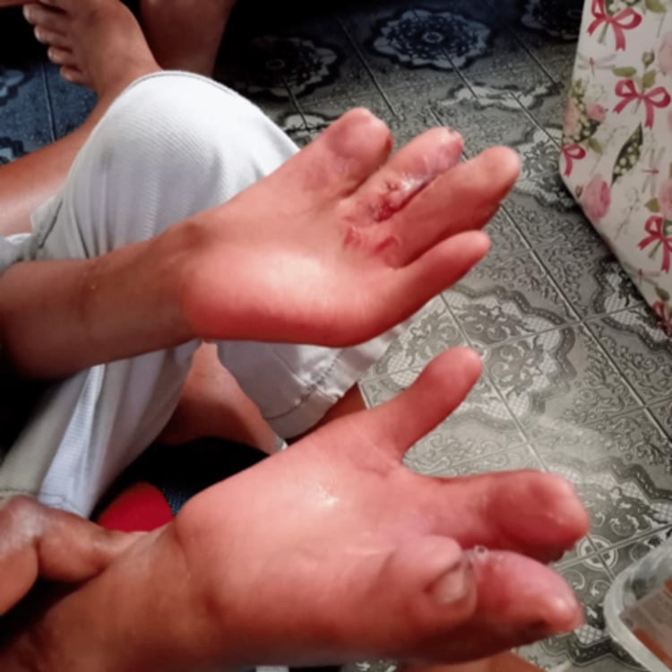
- Type II, the "spoon" or "mitten" hand. The palm becomes concave, and the thumb is joined to the index by simple syndactyly.
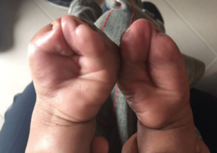
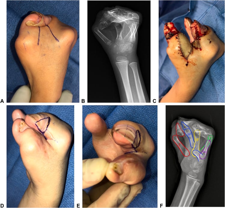
- Type III, the "rosebud" or "hoof" hand. Most severe and hardest to reconstruct: a tight central mass of fused digits with a single conjoined nail, with the thumb incorporated.
Spade to spoon to rosebud: the hand gets tighter from I to III.
Constant findings. Whatever the type, certain features are present in essentially every Apert hand (Roje, Acta Chir Plast 2012; Guero, Handchir Mikrochir Plast Chir 2004):
- Complex syndactyly of the index, long and ring fingers, with distal bony fusion
- Simple syndactyly of the fourth (little-finger) web space
- A short thumb with radial clinodactyly
- Symphalangism, stiff fused interphalangeal joints, sparing the little finger
Beyond the Hand: Progressive Proximal Upper-Limb Synostosis
The Apert upper limb is abnormal proximally as well as distally. Elbow and shoulder stiffness is easy to record as an incidental finding and miss the reason for it: there is progressive synostosis and dysplasia of the elbow and shoulder, and it compounds the hand disability rather than sitting alongside it.
Elbow. The hallmark is humeroradial (radiohumeral) synostosis. The radius and capitellum progressively fuse, so the elbow loses flexion-extension and, with it, the ability to bring the hand to the mouth and midline. This is distinct from proximal radioulnar synostosis, which limits forearm rotation and is covered in its own topic.
Shoulder. Glenohumeral dysplasia, with a small, abnormally shaped glenoid/proximal humerus, brings progressive loss of abduction and elevation, and so reduced overhead reach.
Progression. Like the cranial sutures and the cervical spine, these joints are often mobile in infancy and stiffen with growth, so range of motion documented early may not last.
Why it matters. The disability is a whole-limb problem. A technically excellent hand reconstruction can be undermined if the child cannot position the hand because the elbow and shoulder are stiff, so assess the proximal joints, set realistic goals and counsel the family accordingly.
Management. Surgery to the proximal joints is uncommon, and therapy, compensatory and adaptive strategies dominate. At the elbow, therapy aims to preserve function, and a derotation or positioning osteotomy is used only rarely.
Clinical Assessment
History. Craniosynostosis is often diagnosed at birth or on prenatal ultrasound, so start with the antenatal and birth history. Then cover:
- Airway, feeding and sleep: obstructive sleep apnoea is common with midface hypoplasia
- Developmental milestones and any features of raised intracranial pressure
- Functional use of the hands, and how the child grasps and pinches
Examination. Work from the head to the feet:
- Head and face. Acrocephaly (a tall, turret skull), midface hypoplasia, hypertelorism, proptosis with shallow orbits, and a flat facial profile.
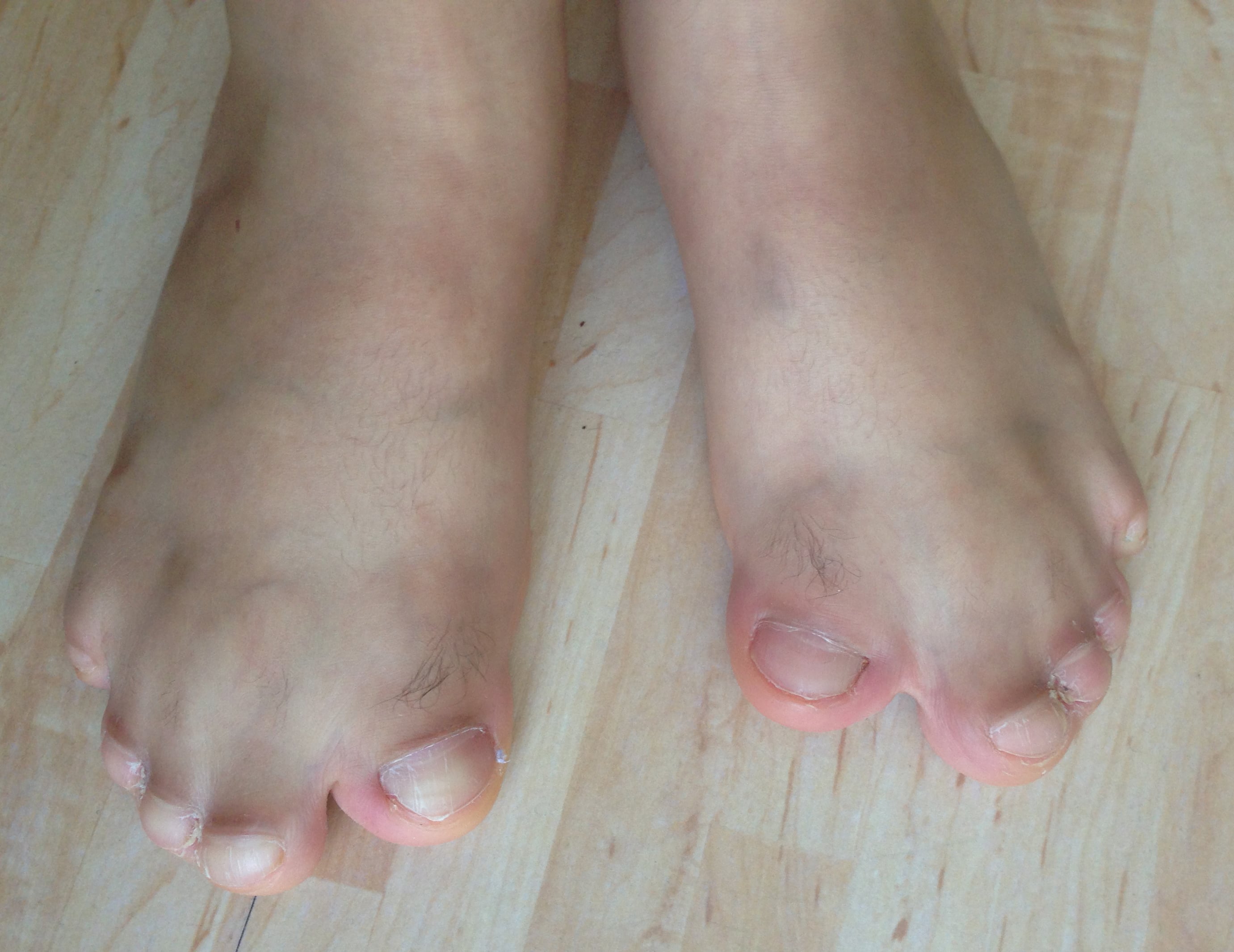
- Hands. Define the Upton type. Assess the thumb for length and clinodactyly, the first web, which may be narrow or absent, the border digits, which angle towards the centre, and the conjoined central mass, whose fused digits have minimal independent motion.
- Movement. Test interphalangeal motion, which symphalangism leaves stiff. Check elbow and shoulder range, which is often reduced.
- Feet. Toe syndactyly and broad great toes.
- Neurology. Screen for signs of cord compression if there is cervical fusion.
Investigations
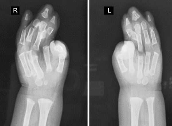
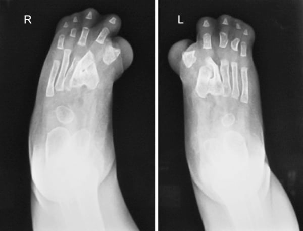
Imaging and genetics. The hand and foot radiographs are the ones that drive the surgical plan:
- Hand and foot radiographs map the bony fusions, the conjoined nail, symphalangism and the thumb anatomy
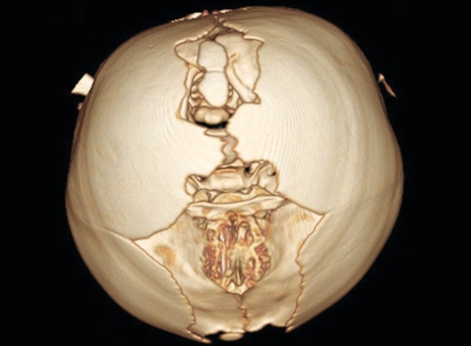
- CT head shows suture fusion, ventricular size (hydrocephalus) and midface anatomy for craniofacial planning
- Cervical spine imaging looks for progressive cervical vertebral fusion, commonly at C5-C6
- MRI brain, when indicated, for structural anomalies and ventriculomegaly
- FGFR2 mutation testing confirms the diagnosis (Ser252Trp or Pro253Arg) and guides genetic counselling
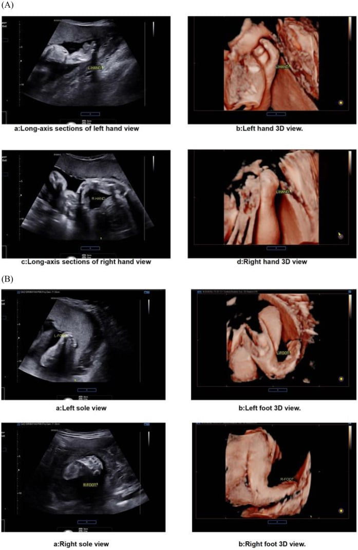
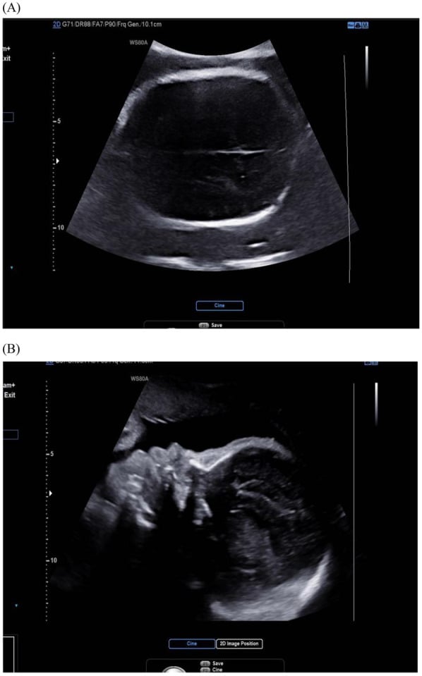
Prenatal diagnosis. Fixed glove-like hands and feet on multiple ultrasound views are a powerful prenatal clue, and loss of the expected coronal-suture gap accompanies frontal bossing and an altered skull contour. Once Apert is suspected, targeted cranial-suture assessment and FGFR2 testing distinguish syndromic craniosynostosis from isolated syndactyly, and limb and cranial findings together justify multidisciplinary counselling and molecular confirmation.
Differential Diagnosis
Other FGFR-related craniosynostosis syndromes:
- Gene
- FGFR2
- Key Differentiator
- Severe complex (bony) syndactyly - mitten hand
- Gene
- FGFR2
- Key Differentiator
- Craniosynostosis with normal limbs
- Gene
- FGFR1/FGFR2
- Key Differentiator
- Broad, deviated thumbs and big toes; partial syndactyly
- Gene
- TWIST1
- Key Differentiator
- Ptosis, low frontal hairline, mild cutaneous syndactyly
- Gene
- RAB23
- Key Differentiator
- Autosomal recessive; polydactyly with syndactyly
Key Distinguishing Points:
- Apert: The most severe hand syndactyly (bony fusion, conjoined nail).
- Crouzon: FGFR2 like Apert, but limbs are normal - this is the classic contrast.
- Pfeiffer: Broad thumbs and big toes are the clue.
- Saethre-Chotzen: Milder, with ptosis and a low frontal hairline.
Management Algorithm
Apert is a craniosynostosis syndrome. Raised intracranial pressure, hydrocephalus, obstructive sleep apnoea and a difficult airway take priority over the hands in infancy, and the craniofacial and neurosurgical teams lead early care.
The skull and face. Posterior vault expansion, often by distraction, and later midface advancement address the skull and face (Raposo-Amaral, J Craniofac Surg 2022).
When to start on the hand. Syndactyly release usually begins around 9-12 months and aims to be largely complete by 2-4 years, before fine-motor skills mature. In Roje's series the border digits were released bilaterally at 1-2 years and the central syndactyly was divided later, completed by about 4 years (Roje, Acta Chir Plast 2012).
Building a functional hand. The sequence follows from what the child needs to grasp and pinch:
- Release the border digits first, the thumb-index and ring-little webs, to create a thumb and an ulnar post for grasp
- Open the first web and correct the thumb's radial clinodactyly to give a usable pinch (Salazard, Chir Main 2008)
- Stage the central mass separately, and never release both sides of a single finger at one sitting, because of the risk of vascular compromise
Feet. Toe syndactyly is usually left alone because it rarely limits walking or shoe wear; surgery is reserved for specific functional problems.
Surgical Techniques
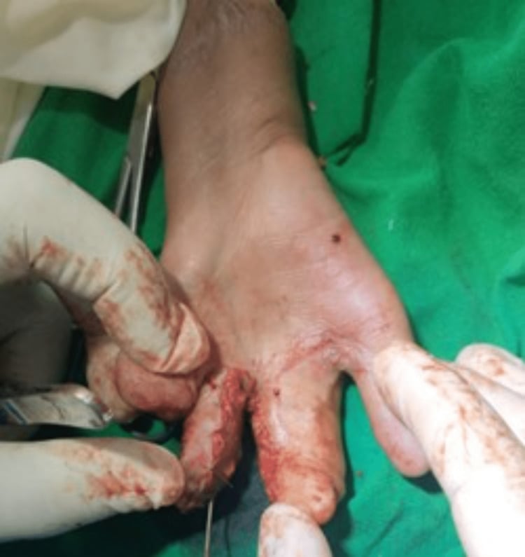
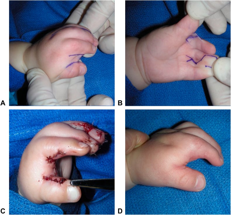
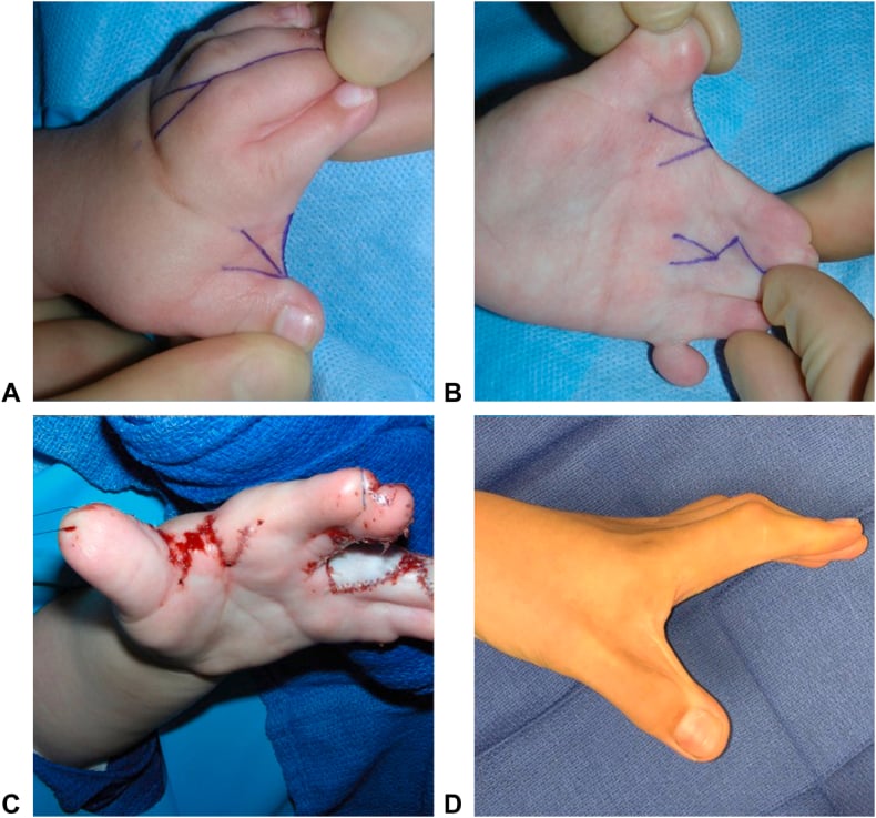
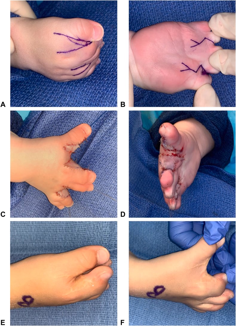
Staged, border-first release. Indicated for the complex syndactyly of the Apert hand. The digits are separated in stages, the border digits (thumb and little finger) at the first operations, and the central conjoined mass is then divided with osteotomy and bone grafting as required (Roje, Acta Chir Plast 2012).
Commissures and cover. Each web commissure is reconstructed with a local flap, and the sides of the digits are resurfaced with full-thickness skin grafts. Multiple operations, often 4-6 per hand, are the norm; Salazard's series averaged six per child (Salazard, Chir Main 2008).
First web and thumb. Indicated for a tight first web and a short thumb with radial clinodactyly. The first web is released and deepened, with a dorsal hand flap for the more severe Upton grades, and the clinodactyly is corrected by osteotomy of the abnormal (delta) phalanx with skin rearrangement (Salazard, Chir Main 2008). A mobile, abducted thumb opposite a stable ulnar digit is the single most important functional goal.
Central coalition osteotomy for the type III hand. Theman and Upton (J Hand Surg Am 2018) described limited retrograde axial osteotomies between the conjoined phalangeal segments during a three-stage, border-first release, producing a thumb and four fingers in nearly every type III hand in their series. The three-stage approach increases the number of operations but improves both appearance and function in the most severe hands.
Complications
Surgical Complications of Hand Reconstruction
- Frequency
- Uncommon if staged
- Risk Factors
- Releasing both sides of one digit at once
- Management
- Strict border-first staging
- Frequency
- Variable
- Risk Factors
- Tension, infection, mobile child
- Management
- Re-graft, splinting, hand therapy
- Frequency
- Common over growth
- Risk Factors
- Growth of the child, scar
- Management
- Revision web release (about 16% in some series)
- Frequency
- High
- Risk Factors
- Severe (Upton III) hands, symphalangism
- Management
- Planned multiple stages
Functional and Long-Term Issues
- Why it Happens
- Symphalangism (fused IP joints)
- Implication
- Limits final motion despite good release
- Why it Happens
- Short thumb, stiff fingers
- Implication
- Pinch is rudimentary but usually functional
- Why it Happens
- Proximal joint involvement
- Implication
- Compensatory strategies, therapy
Before any anaesthetic. Four problems must be planned for in any surgery in Apert syndrome:
- Difficult airway. Midface hypoplasia and a high-arched or cleft palate make intubation challenging, so plan ahead.
- Cervical fusion. Progressive vertebral fusion limits safe neck extension: monitor it, flag it to the anaesthetic team and avoid forced positioning.
- Obstructive sleep apnoea. Optimise and monitor it in the perioperative period.
- Raised intracranial pressure. It must be excluded or treated by the craniofacial team before elective limb surgery.
Clinical Relevance and Outcomes
What reconstruction achieves. Even with expert staged reconstruction the goal is a functional, opposable hand with a rudimentary but useful pinch, not normal fingers. Early, specialist, staged surgery gives the best functional and aesthetic results with acceptable revision rates (Roje, Acta Chir Plast 2012).
What limits it. Even after perfect syndactyly release the fingers stay stiff because of symphalangism, which is repeatedly cited as the factor that "darkens" the functional prognosis (Salazard, Chir Main 2008).
Counselling families. Families should hear three things:
- The hand will not look or move normally, but most children achieve independent grasp and pinch
- Multiple operations are expected; this is a planned, staged journey, not a single fix
- Care is lifelong and multidisciplinary: craniofacial, neurosurgery, hand, ENT, genetics and development
Guidelines, Registries & Global Practice
Global Epidemiology
- Birth prevalence around 1 in 65,000-80,000; one of the more common craniosynostosis syndromes worldwide.
- Almost all cases are de novo FGFR2 mutations with a strong paternal age effect.
- Two recurrent mutations (Ser252Trp, Pro253Arg) account for the overwhelming majority of cases globally.
Side-by-Side Guidance
- Emphasis
- Brain and airway first; staged vault expansion (often posterior distraction) and midface advancement
- Emphasis
- Early, staged, border-first syndactyly release completed by 2-4 years; Upton classification to plan
- Emphasis
- FGFR2 confirmatory testing; counselling for de novo recurrence risk and 50% transmission risk
- Emphasis
- Anticipate difficult airway, cervical fusion and OSA in all procedures
Practice Variation
- High-resource settings: Specialist craniofacial units, distraction osteogenesis, multi-stage hand reconstruction and FGFR2 testing are available.
- Limited-resource settings: Diagnosis is clinical; care prioritises airway/feeding safety and the most functionally important hand release (thumb and first web) where full multi-stage programmes are not feasible.
- Family support organisations (craniofacial and Apert-specific groups) are valuable everywhere for psychosocial support and shared decision-making.
Controversies and Areas of Uncertainty
Timing and number of hand operations There is no single agreed protocol. Most centres favour an early, border-first, staged release completed by 2-4 years, but the exact age to start, the number of stages, and how aggressively to pursue a five-fingered hand in type III cases vary between units (Roje 2012; Theman/Upton 2018).
How far to go in the type III hand Central coalition osteotomy can create five digits in severe hands, but it requires more operations. Whether the extra aesthetic and functional gain justifies the additional surgery is a judgement made with each family (Theman/Upton 2018).
Feet Toe syndactyly is usually left alone because it rarely impairs function; routine separation is not recommended, and indications for foot surgery remain selective.
Genotype-guided counselling The Ser252Trp/Pro253Arg phenotype gradient (worse face vs worse hands) is real but not absolute, so genotype informs but does not dictate individual prognosis (Wilkie 2001).
MCQ Practice Points
Q: Which gene is mutated in Apert syndrome? A: FGFR2 - specifically the Ser252Trp or Pro253Arg mutations.
Q: Apert is allelic with which other craniosynostosis syndrome? A: Crouzon syndrome (also FGFR2, but with normal limbs).
Q: What is the hand hallmark of Apert syndrome? A: Complex (bony) syndactyly of the central digits - the spade or mitten hand.
Q: Which classification is used for the Apert hand? A: The Upton classification (types I spade, II spoon/mitten, III rosebud/hoof).
Q: What limits the functional outcome after syndactyly release in Apert? A: Symphalangism - the fused, stiff interphalangeal joints (the little finger is usually spared).
Q: What is the key safety rule when releasing Apert syndactyly? A: Never release both sides of a single digit at one operation - stage the release to protect the digital blood supply.
Self-Assessment Quiz
Viva Scenarios
Practise clinical reasoning and management decisions out loud
“A newborn has a turret-shaped skull, midface hypoplasia and both hands fused into a single mass. What is the diagnosis and what are your immediate priorities?”
“A 1-year-old with Apert syndrome has a tight central mass of fused fingers with a single conjoined nail. How do you classify and plan the hand?”
“The anaesthetist asks what they should know before a hand operation in a 4-year-old with Apert syndrome. What do you tell them?”
GENETICS
- FGFR2 mutation
- Ser252Trp / Pro253Arg
- Autosomal dominant
- Allelic with Crouzon
CRANIOFACIAL
- Acrocephaly (turret skull)
- Midface hypoplasia
- Hypertelorism, proptosis
- Craniosynostosis
HAND
- Complex bony syndactyly
- Mitten/spade hand
- Short radial thumb
- Symphalangism (spares little finger)
CLASSIFICATION
- Upton I - spade
- Upton II - spoon/mitten
- Upton III - rosebud/hoof
- Drives surgical plan
SURGERY
- Staged, border-first release
- Open first web, fix thumb
- Never both sides of one digit
- Complete by 2-4 years
PERIOP
- Difficult airway
- Cervical fusion (C5-C6)
- OSA
- Exclude raised ICP first
Evidence Base
Wilkie et al
- Identified two adjacent FGFR2 missense mutations - Ser252Trp and Pro253Arg - in all 40 unrelated Apert cases studied
- Mutations sit in the linker between the second and third Ig domains of FGFR2
- Apert is allelic with Crouzon syndrome (third Ig-domain mutations, normal limbs)
Wilkie et al
- Reviewed the contrasting craniofacial vs limb severity of the two Apert mutations
- Ser252Trp tends to give more severe craniofacial disease; Pro253Arg more severe syndactyly
- Proposed alternative FGFR2 splice forms mediate the distinct cranial and limb effects