Arthrogryposis Multiplex Congenita | Multiple Contractures
- Definition: Non-progressive condition with multiple congenital joint contractures.
- Pathogenesis: Fetal akinesia (lack of movement) from any cause leads to contractures.
- Amyoplasia (30%): Classic type. Symmetric. Normal intelligence. Sporadic.
- Foot: Clubfoot is common and rigid; begin with Ponseti-style casting, expect recurrence and individualise limited versus salvage surgery
- Upper Limb Goal: Elbow flexion + Hand to mouth. Elbow is key.
- “Arthrogryposis is a DESCRIPTION, not a diagnosis. Find the underlying cause.
- “Amyoplasia children are intelligent - treat like normal children cognitively.
- “AMC clubfoot is rigid but still deserves Ponseti-style casting first; avoid treating posteromedial release or talectomy as inevitable.
- “Prioritize function over appearance. Elbow flexion is key for UL function.
Overview and Epidemiology
Definition. Arthrogryposis multiplex congenita (AMC) is a descriptive term for conditions with multiple congenital joint contractures affecting two or more body areas. It is a clinical finding, not a specific diagnosis, so the task is to find the cause behind it. The contractures are present from birth, often in all four limbs, and the condition is non-progressive.
Frequency. AMC affects about 1 in 3000 live births. Amyoplasia is the most common recognisable type, at about 30% of cases.
The common pathway. Any condition that decreases fetal movement leads to contractures. Whatever the cause, every form of arthrogryposis shares this final pathway: fetal akinesia during the critical period of joint development.
Anatomy and Joint-Specific Considerations
Soft tissue, not bone. The pathology is periarticular. Fibro-fatty replacement of muscle, capsular fibrosis and ligamentous shortening fix the joint, while the articular surfaces and ossification centres are usually structurally normal at birth. Bony deformity is secondary and develops with growth against unbalanced soft tissues.
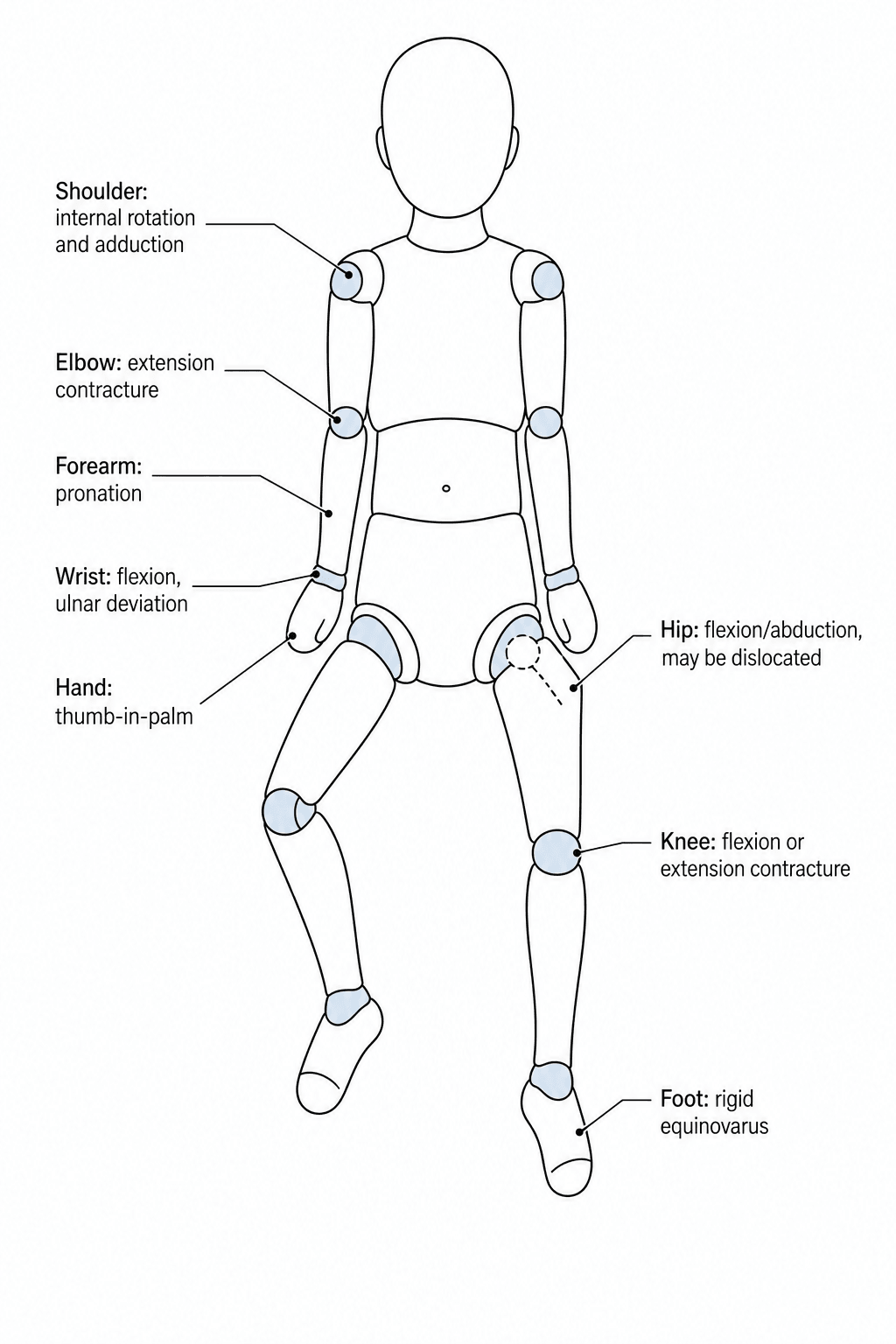
The pattern in classic amyoplasia. Each joint has a typical position, a structure that holds it there, and a place in the order of priorities:
- Typical position
- Internal rotation, adduction
- Limiting structure
- Subscapularis, capsule
- Functional priority
- Low – usually accommodated
- Typical position
- Extension (most common)
- Limiting structure
- Triceps, posterior capsule
- Functional priority
- High – flexion for hand-to-mouth
- Typical position
- Flexion, ulnar deviation
- Limiting structure
- Flexor tendons, volar capsule
- Functional priority
- Moderate – position for grasp
- Typical position
- Thumb-in-palm, camptodactyly
- Limiting structure
- Intrinsics, skin, A1 region
- Functional priority
- Moderate – pinch and release
- Typical position
- Flexion, abduction, ext rotation; dislocation
- Limiting structure
- Capsule, iliopsoas, adductors
- Functional priority
- High – stable base for sitting/standing
- Typical position
- Flexion or extension contracture
- Limiting structure
- Hamstrings or quadriceps/capsule
- Functional priority
- High – determines bracing and gait
- Typical position
- Rigid equinovarus (or vertical talus)
- Limiting structure
- Posteromedial structures, talus
- Functional priority
- High – plantigrade, braceable foot
- Typical position
- Neuromuscular scoliosis
- Limiting structure
- –
- Functional priority
- Monitor; affects sitting balance
Pathomechanics. Muscle is deficient rather than merely tight, so the deforming forces are weak but a corrected position is also poorly maintained by active power. This dual problem, a rigid contracture with weak motors, is why recurrence is high and why surgery aims for a balanced, braceable position rather than a normal arc of motion.
Pathophysiology of Fetal Akinesia
Normal joint development. Joints form through cavitation of mesenchyme in weeks 8-12, and movement is essential for the joint cavity to form. Fetal movement shapes the articular surfaces and prevents contractures, and muscles develop in response to neural input and use.
Timing and duration. What the akinesia produces depends on how long it lasts and when it begins:
- Gestational Age
- Early
- Consequence
- Mild contractures (correctable)
- Gestational Age
- Early (8-12 weeks)
- Consequence
- Severe fixed contractures
- Gestational Age
- Third trimester
- Consequence
- Milder deformities
Why the contractures are fixed. Lack of fetal movement leads in turn to failure of joint cavity formation (ankylosis), capsular and ligamentous contracture, muscle fibrosis and shortening, and secondary bony deformity. That is why the contractures are rigid and resistant to simple stretching.
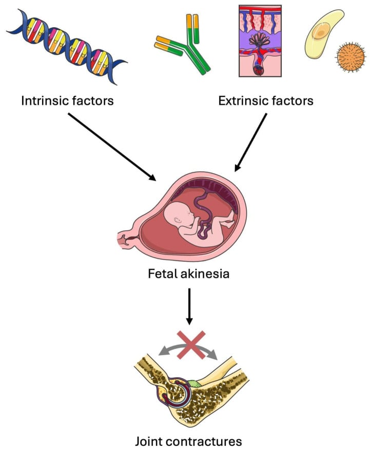
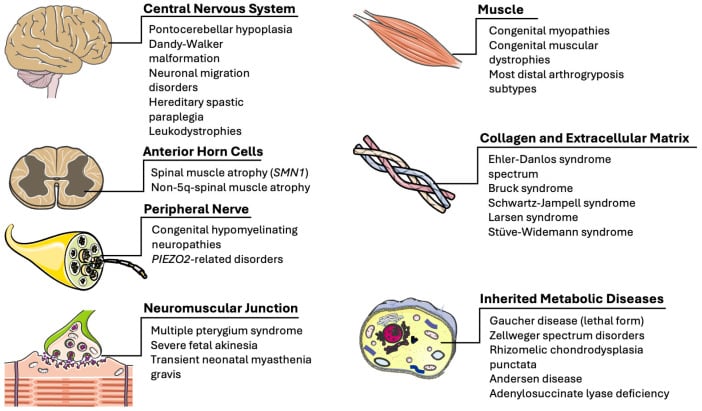
The causes. Intrinsic genetic or neuromuscular causes and extrinsic maternal or mechanical factors converge on fetal akinesia. Neurogenic causes are the most common.
- Neurogenic - anterior horn cell dysfunction (amyoplasia, a vascular insult), spinal muscular atrophy, myelomeningocele, CNS malformations
- Myopathic - congenital myopathies (nemaline, central core), congenital muscular dystrophy, maternal myotonic dystrophy
- Mechanical restriction - oligohydramnios (renal agenesis, PROM), multiple pregnancy, uterine anomalies, amniotic bands
- Connective tissue disorders - diastrophic dysplasia, Larsen syndrome
Amyoplasia. Amyoplasia is caused by anterior horn cell ischaemia in early gestation. Its symmetric involvement suggests a vascular aetiology; anterior horn cells are particularly vulnerable to hypoxia. The result is muscle hypoplasia or aplasia with fatty replacement, while sensory function is preserved, the posterior horn being spared.
Amyoplasia Is More Than the Limbs: Associated Anomalies and Birth Fractures
A vascular disruption. Amyoplasia is thought to follow an early vascular insult, the same compromise that depletes anterior horn cells and replaces muscle with fibro-fat. That insult produces a recognised cluster of associated anomalies elsewhere, and they change both the newborn work-up and the way the child is handled. The newborn with amyoplasia needs a whole-child screen, not just a limb assessment. Hall's defining series of 560 individuals sets the anomalies out:
- Approximate frequency
- About 9%
- Why it matters
- Needs abdominal examination/imaging in the neonate
- Approximate frequency
- Roughly 12% and 4%
- Why it matters
- Count and document digits; part of the disruption picture
- Approximate frequency
- About 3%
- Why it matters
- Affects core strength and surgical planning
- Approximate frequency
- About 10%
- Why it matters
- The fragile osteopenic limb can FRACTURE at birth or on handling/casting
- Approximate frequency
- About 7% (odds ratio ~11)
- Why it matters
- Supports a vascular/twin-related pathogenesis; reassure on recurrence
The midline frontal capillary haemangioma belongs to the same vascular picture.
The orthopaedic point. The amyoplasia limb is osteopenic and fragile, so it must be handled and cast gently, and a fresh "deformity" at birth may actually be a fracture. Screen the abdomen for an atresia or wall defect and count the digits. Despite the cluster of anomalies, amyoplasia is sporadic, with negligible recurrence risk.
Classification
AMC is sorted into amyoplasia, distal arthrogryposis and the syndromic forms.
- Features
- Symmetric limb contractures, 'Waiter's Tip'
- Inheritance
- Sporadic
- Intelligence
- Normal
- Features
- Hands and Feet primarily
- Inheritance
- AD
- Intelligence
- Normal
- Features
- CNS involvement
- Inheritance
- Variable
- Intelligence
- Often impaired
- Features
- Muscle disease
- Inheritance
- Variable
- Intelligence
- Variable
Amyoplasia (classic AMC). The most common recognisable type. It is sporadic, with no inheritance, and the limb involvement is symmetric. Intelligence is normal: do not assume cognitive impairment; these children are typically intelligent and motivated. Its features:
- Upper limb - internal rotation, elbow extension and wrist flexion, the "waiter's tip"
- Lower limb - hip dislocation (30%), knee flexion or extension, clubfoot
- Face - micrognathia, depressed nasal bridge
- Skin - dimpling, lack of creases
Distal arthrogryposis. Primarily the hands and feet, with the proximal joints relatively spared. Inheritance is autosomal dominant across many types (DA1, DA2A Freeman-Sheldon and others), and intelligence is normal. The hands show ulnar deviation, camptodactyly and thumb-in-palm, the feet clubfoot or vertical talus, and treatment focuses on hand and foot function.
Syndromic AMC. Here the contractures are part of a broader syndrome, drawn from the neurogenic, myopathic and connective-tissue causes above. In the neurogenic group (SMA, myelomeningocele, CNS malformations) intelligence is often impaired. Prognosis depends on the underlying syndrome, and multidisciplinary care is essential.
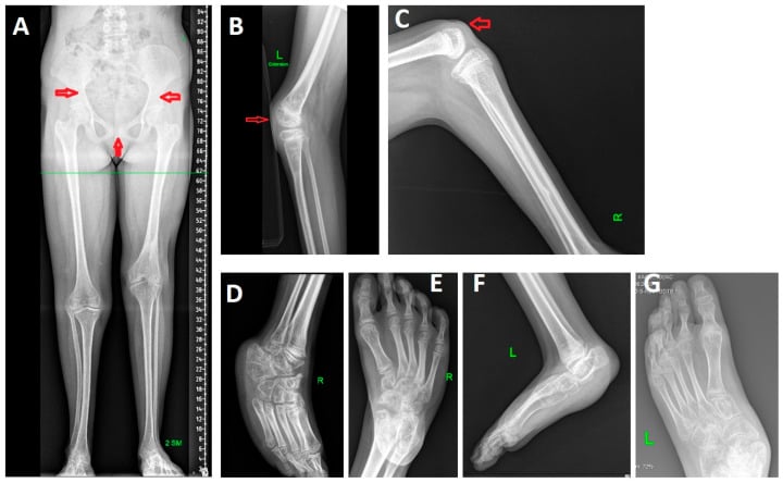
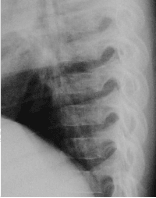
Abnormal bone points to a syndrome. Structural skeletal abnormalities signal syndromic arthrogryposis rather than classic amyoplasia. Primary bone changes are not features of classic amyoplasia and should trigger syndromic investigation. PITX1-related mandibular-pelvic-patellar syndrome, for example, produces limb-length discrepancy, abnormal pelvis and patellae, knee contracture, carpal/tarsal abnormalities and equinovarus.
Clinical Assessment
History. The questions that place the child in a category:
- Pregnancy - decreased fetal movements, oligohydramnios
- Birth - breech presentation, complications
- Family history - any inheritance pattern
- Development - milestones, intelligence
Examination. Examine every joint against the pattern in the anatomy table. The elbow may be held in flexion rather than extension, the hip may be dislocated or have a flexion contracture, the foot may show a rigid clubfoot or a vertical talus, and there may be a congenital knee dislocation. Then look beyond the limbs: the facies and skin, the spine for scoliosis, which is common, and a neurological assessment of muscle bulk, tone and reflexes.
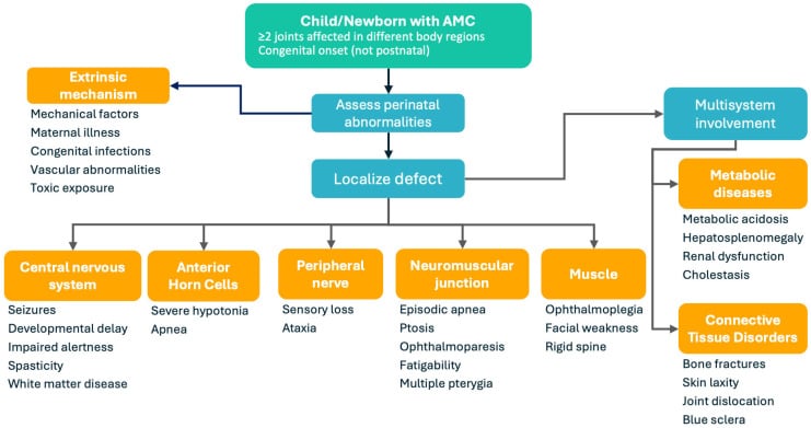
Differential diagnosis. Arthrogryposis is a sign, not a diagnosis. The task is to place the child into a prognostic and genetic category.
- Distinguishing Features
- Symmetric, 4-limb, extended elbows, midline facial haemangioma, fatty muscle replacement
- Intelligence
- Normal
- Inheritance
- Sporadic
- Distinguishing Features
- Hands/feet predominant, proximal sparing, camptodactyly + clubfoot
- Intelligence
- Normal
- Inheritance
- Autosomal dominant
- Distinguishing Features
- Whistling face, microstomia, ulnar deviation; MYH3 mutation
- Intelligence
- Usually normal
- Inheritance
- Autosomal dominant
- Distinguishing Features
- Multiple large-joint dislocations, flat facies, cervical kyphosis (cord risk)
- Intelligence
- Normal
- Inheritance
- AD (FLNB)
- Distinguishing Features
- Short limbs, hitchhiker thumb, cauliflower ear, cleft palate
- Intelligence
- Normal
- Inheritance
- AR (SLC26A2)
- Distinguishing Features
- CNS signs, progressive weakness, abnormal imaging/EMG
- Intelligence
- Often impaired
- Inheritance
- Variable
Symmetric four-limb contractures + midline frontal capillary haemangioma + normal cognition strongly suggests amyoplasia. Hall's 2014 series of 560 patients confirmed this is the single most common recognisable form and is entirely sporadic — reassure parents regarding recurrence risk.
The Waiter's Tip Trap: Amyoplasia versus Obstetric Brachial Plexus (Erb's) Palsy
The trap. The amyoplasia upper limb is classically described as the "waiter's tip": an internally rotated, adducted shoulder, extended elbow, pronated forearm and flexed wrist. That is exactly the posture of an obstetric brachial plexus (Erb's, C5-6) palsy, and getting it wrong sends the child down the wrong pathway entirely.
The discriminator is the joints. In amyoplasia they are stiff, a fixed contracture limiting passive as well as active motion. In Erb's palsy the limb is floppy with full passive motion, a paralysis rather than a contracture.
- Amyoplasia
- Fixed joint CONTRACTURE
- Erb's palsy (obstetric brachial plexus)
- Flaccid NERVE palsy (C5-6)
- Amyoplasia
- Limited / stiff
- Erb's palsy (obstetric brachial plexus)
- Full and free (the joints are not contracted)
- Amyoplasia
- Bilateral and symmetric
- Erb's palsy (obstetric brachial plexus)
- Usually unilateral
- Amyoplasia
- Multiple — feet, hips, knees also involved
- Erb's palsy (obstetric brachial plexus)
- Isolated to the affected arm
- Amyoplasia
- No birth trauma; reduced fatty muscle bulk, sensation preserved
- Erb's palsy (obstetric brachial plexus)
- Difficult/instrumented delivery, shoulder dystocia; hypotonic, absent Moro/biceps reflex
Why it matters. The management diverges completely. Erb's palsy is a nerve problem, treated with physiotherapy; it often partially recovers, and microsurgical nerve reconstruction is for those that do not. Amyoplasia needs contracture management: stretching, serial casting and later tendon transfers such as triceps-to-biceps. A waiter's tip arm that is bilateral, stiff and accompanied by clubfeet is amyoplasia until proven otherwise, and should never be labelled a birth palsy.
Investigations
The diagnosis is clinical, resting on the multiple congenital contractures. Investigations aim to find the underlying cause:
- Genetic testing - gene panels for distal arthrogryposis, SMA and others
- MRI brain and spine - if a neurogenic cause is suspected
- EMG/NCS - to differentiate myopathic from neurogenic
- Muscle biopsy - if a myopathy is suspected
- Ophthalmology review - for associated eye anomalies
- Cardiac echo - for associated cardiac anomalies
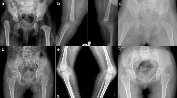
Orthopaedic imaging. A hip radiograph or ultrasound assesses dislocation, a spine radiograph looks for scoliosis, and foot radiographs assess the clubfoot.
Management Algorithm
Principles. AMC involves many joints, so prioritise them for function: elbows, knees, hips and feet. Function comes before appearance.
Stretching still gains range. The contractures are often rigid, yet early stretching and serial casting can still gain useful range. Escalate selectively, when residual deformity prevents function, bracing, hygiene or comfortable positioning.
Goals. Comfortable positioning, transfers, standing or walking potential and plantigrade braceable feet are individualised goals.
Hip. Decide from pain, stability, range, pelvic obliquity, sitting and standing goals, and the effect of reduction on adjacent contractures. Laterality alone does not mandate reduction or observation: bilateral painless dislocations are often accepted, while symptomatic or function-limiting hips may need reconstruction.
Knee. Start with gentle stretching or casting when safe, protecting the osteopenic limb. Choose quadriceps lengthening, posterior release, guided growth or osteotomy only for the specific direction, rigidity, age and functional barrier.
Foot. The AMC clubfoot is rigid, unlike the idiopathic foot, but Ponseti-style serial casting remains the starting point, with tenotomy when indicated. Relapse and repeat casting are common: treat recurrence with repeat casting where possible, and select limited release or osteotomy for the residual components. Talectomy and other salvage procedures are reserved for severe rigid deformity that cannot become plantigrade and braceable by less destructive means. The goal is a plantigrade, painless and braceable foot that can be shoe-fitted.
Casting first is a change in practice. Historically the AMC clubfoot went straight to extensive release, and Södergård and Ryöppy's series, in which primary treatment was operative, predates the Ponseti method entirely. Boehm and Dobbs achieved initial correction by casting in every distal-arthrogrypotic foot, with more casts than for idiopathic feet, though that subtype is the mildest and the result should not be transferred to amyoplasia.
Surgical Technique
Casting and tenotomy. Ponseti-style serial casting often achieves partial correction, and more casts may be needed than for an idiopathic foot. An Achilles tenotomy is almost always needed.
Residual deformity. It is common, and is dealt with by operation:
- Posteromedial release (PMR) - extensive release of the hindfoot soft tissues
- Talectomy - removal of the talus for severe rigid clubfoot, creating a plantigrade foot
Complications
- Risk Factor
- Rigid deformity
- Management
- Revision surgery
- Risk Factor
- Inherent in AMC
- Management
- Accept / Physio
- Risk Factor
- Forced reduction
- Management
- Avoid aggressive reduction
- Risk Factor
- Atypical creases
- Management
- Careful incision planning
- Risk Factor
- Natural history
- Management
- Monitor / Fusion
Postoperative Care
After clubfoot surgery. Cast for 6-8 weeks, then brace long term (AFO, UCBL) and monitor for recurrence.
After elbow surgery. Immobilise in flexion in a splint for 6 weeks, then regain range gradually, with long-term physiotherapy.
Outcomes
Amyoplasia. Outcomes are excellent with appropriate management and comprehensive multidisciplinary care. Most children walk, and intelligence and lifespan are normal.
Syndromic AMC. The outcome depends on the underlying condition.
Function. Children adapt remarkably and achieve independence.
Guidelines, Registries & Global Practice
Global epidemiology: Arthrogryposis affects roughly 1 in 3,000 to 1 in 5,000 live births worldwide; amyoplasia is the most common recognisable form (Hall 2014, N=560). Distribution is broadly similar across regions; apparent variation reflects ascertainment rather than true geography.
Side-by-side guidance:
- Position relevant to AMC
- Multidisciplinary, function-led care; Ponseti casting first-line for clubfoot of any aetiology
- Position relevant to AMC
- Casting-first with rigorous foot-abduction bracing; surgery reserved for relapse/failure
- Position relevant to AMC
- Soft-tissue balancing and staged correction; aim for a plantigrade, braceable foot, not a normal arc
- Position relevant to AMC
- Early genetic work-up to define subtype and recurrence risk before committing to a surgical plan
There is no dedicated AMC implant registry; arthroplasty registries (NJR, AJRR, AOANJRR) are not relevant in childhood. Outcome data come from specialist-centre cohorts and clubfoot relapse audits, which consistently identify brace non-compliance as the dominant predictor of recurrence.
In high-resource settings, early MDT clinics (orthopaedics, physiotherapy, occupational therapy, genetics) and serial casting from the neonatal period are standard. In limited-resource settings, late presentation is common, Ponseti programmes are the most scalable and cost-effective intervention, and talectomy remains a valuable single-stage salvage where staged reconstruction and long-term bracing are impractical.
Controversies and Areas of Uncertainty
No consensus. Bilateral teratologic dislocations are often left unreduced if symmetric and painless, as motion may matter more than location for sitting and gait. Others reduce via the medial approach citing satisfactory acetabular development (Szöke 1996). AVN risk is the deciding factor.
Historically AMC clubfoot went straight to extensive release. Boehm/Dobbs (2008) showed Ponseti casting works (more casts, strict bracing), shifting practice toward casting-first. Talectomy/release is now reserved for true failure.
Whether to correct knee flexion before or after hip and foot is debated. Many correct the knee first because residual knee flexion undermines foot bracing and gait, but sequencing is individualised.
For fixed knee extension (hyperextension/dislocation), VY quadricepsplasty versus distal femoral extension osteotomy or shortening is unsettled; choice depends on age, severity and reducibility.
Viva Scenarios
Clinical Decision Scenarios
Practise clinical reasoning and management decisions out loud
“What is your diagnosis and approach?”
“What are your options?”
“How would you help this child?”
MCQ Practice Points
Q: What is Arthrogryposis? A: A descriptive term for conditions with multiple congenital joint contractures affecting two or more body areas. It is NOT a specific diagnosis.
Q: What is the most common recognizable type of AMC? A: Amyoplasia (~30% of cases). Sporadic, Normal intelligence, Symmetric limb involvement.
Q: Why is clubfoot in AMC different from idiopathic clubfoot? A: AMC clubfoot is RIGID and resistant to conservative treatment. It often requires extensive surgery (Posteromedial Release or Talectomy).
Q: What is the priority for upper limb surgery in AMC? A: Elbow flexion - to allow hand-to-mouth function for feeding and self-care.
Q: What is the procedure to restore elbow flexion in AMC? A: Triceps to Biceps transfer - the triceps is detached and rerouted to act as an elbow flexor.
Key Features
- Multiple congenital contractures
- Fetal akinesia is cause
- Amyoplasia = Normal IQ
- Non-progressive
Lower Limb
- Clubfoot: Casting then PMR/Talectomy
- Hip: Bilateral may leave dislocated
- Knee: Serial casting, Release
- Goal: Independent Mobility
- Vertical Talus: Common, Surgical
Upper Limb
- Elbow Flexion = Priority
- Triceps to Biceps Transfer
- Wrist Fusion for function
- Thumb-in-Palm Release
Prognosis
- Amyoplasia: Excellent (Normal IQ)
- Most walk (conventional teaching figure ~85%)
- Children adapt well
- Distal Arthrogryposis: Best outcomes
- Ongoing PT/OT essential for function
Evidence Base
Amyoplasia Revisited — the defining series
- 560 individuals collated from over 600 reports — the largest amyoplasia cohort.
- Most common recognisable form; 55.9% had symmetric four-limb involvement; equinovarus almost always present and elbows characteristically extended.
- Completely sporadic; fatty-fibrous muscle replacement and vascular-compromise anomalies (bowel atresia, gastroschisis, digit loss) support a vascular pathogenesis.
Foot deformities in AMC — surgery is usually needed
- 43 of 52 AMC patients had foot deformities; talipes equinovarus was the commonest (72 feet), bilateral in all cases.
- Primary treatment was operative in 52 patients; recurrence was frequent (36 reoperations across 15 feet).
- Talectomy and bony decancellation were both effective for recurrent deformity; knee/hip deformity influenced foot outcomes.
Ponseti method for distal arthrogrypotic clubfoot
- 12 infants (24 feet) with distal-arthrogryposis clubfoot; initial correction achieved in all feet.
- Required a mean of 6.9 casts versus 4.5 for idiopathic clubfeet (p=0.002) — more casts but still effective.
- All six relapses were linked to brace non-compliance; most were salvaged by repeat casting/tenotomy.
Tendon transfer to restore elbow flexion
- 18 tendon transfers in 14 children; triceps-to-biceps gave the best results (7 of 9 arms good).
- Capsulotomy with triceps lengthening improved the motion arc from 17° to 67°.
- Best candidates: age over 4 years, full passive elbow motion in the dominant arm, and at least grade-4 donor strength.
Medial-approach open reduction of the arthrogrypotic hip
- 40 dislocations in 26 amyoplasia patients; medial-approach open reduction at a mean 8.9 months.
- 92% good/fair outcomes with satisfactory acetabular development; bilateral cases showed no stiffness or asymmetry.
- AVN occurred in 4 of 25 hips — the principal risk to weigh against reduction.
Hip dislocation management — approach matters
- 18 of 131 arthrogryposis children had hip dislocation; 14 underwent open reduction.
- Medial-approach reduction gave better range of motion and acetabular development than anterolateral or bilateral closed reduction.
- Only one AVN and no redislocations after the medial approach.
BSCOS / AO consensus on paediatric clubfoot and neuromuscular foot
- Ponseti casting is first-line for all clubfoot types, including syndromic/arthrogrypotic, before any soft-tissue surgery.
- Bracing adherence is the dominant predictor of relapse across registries and cohorts.
- Soft-tissue release and talectomy are reserved for true Ponseti failure, not used as primary treatment.