Osteonecrosis | Progressive Collapse | Joint-Preserving vs Arthroplasty
- Bilateral in 50-80% - always image contralateral hip, counsel about second hip
- MRI gold standard - detects Stage I (pre-radiographic) disease before collapse
- Core decompression best for Ficat I-II (pre-collapse), controversial for Stage III
- Crescent sign (subchondral fracture) = collapse imminent, poor outcomes with joint preservation
- THA outcomes worse than primary OA: younger age, higher dislocation, more osteolysis
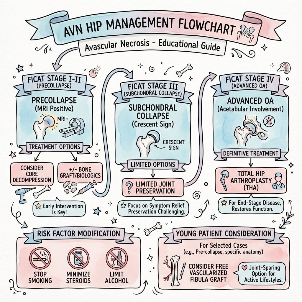
- “Stage I-II = joint preservation window (core decompression ± grafting)
- “Stage III-IV = arthroplasty preferred in most patients over 40 years
- “Steinberg adds lesion size (under 15%, 15-30%, over 30% of head) to staging
- “ARCO classification most comprehensive: adds location (medial/central/lateral columns)

Vascular insult disrupts femoral head blood supply. Three mechanisms: direct vessel injury (trauma, surgery), thrombosis/embolism (coagulopathy, sickle cell), extravascular compression (Gaucher, steroids cause fat hypertrophy). Final common pathway = ischemia, osteocyte death, trabecular collapse.
Pre-collapse (Ficat I-II) vs Post-collapse (III-IV). Joint preservation (core decompression, grafting, osteotomy) only works before crescent sign appears. Once collapse starts (Stage III), progression to arthritis is inevitable without arthroplasty.
50-80% bilateral within 2 years. Always MRI contralateral hip even if asymptomatic. Counsel about risk of second hip. Stagger surgeries if bilateral decompression needed (infection, anesthetic risk).
Age under 40 = arthroplasty complications. Higher activity, longer life expectancy = revision burden. Consider joint preservation even in Stage III if patient motivated. Inform about THA revision rates (15-20% at 10 years in young AVN).
- Ficat Stage
- I-II (pre-collapse)
- Lesion Size
- Under 30% head
- Treatment
- Core decompression ± NVBG
- Key Pearl
- 80% success in Stage I, 65% in Stage II
- Ficat Stage
- III (early collapse)
- Lesion Size
- Any size
- Treatment
- Valgus osteotomy or THA
- Key Pearl
- Osteotomy rotates necrotic segment - needs intact lateral column
- Ficat Stage
- III (collapse) or IV
- Lesion Size
- Over 30% head
- Treatment
- Total hip arthroplasty
- Key Pearl
- Cementless fixation preferred, beware osteolysis in young
- Ficat Stage
- Any stage
- Lesion Size
- Any size
- Treatment
- THA with special considerations
- Key Pearl
- Exchange transfusion pre-op, cemented femoral stem
ASEPTICRisk Factors for AVN
Hook:ASEPTIC necrosis = sterile bone death from vascular insult, not infection!
MEDALFemoral Head Blood Supply (Why AVN Happens)
Hook:MEDAL = Medial circumflex is the GOLD standard blood supply - lose it, lose the head!
DRILLCore Decompression Principles
Hook:DRILL holes in the femoral head to decompress and revascularize before collapse!
Overview and Epidemiology
AVN is the great imitator - can present like hip OA, but affects young patients (30-50 years) with devastating functional impact. Unlike OA, AVN is often bilateral (50-80%), progressive (2-5 years to collapse), and has modifiable risk factors (steroids, alcohol). Early diagnosis with MRI can identify pre-radiographic disease (Stage I) when joint-preserving surgery (core decompression) has 80% success. Once collapse occurs (crescent sign), arthroplasty is inevitable but outcomes are worse than primary OA due to young age and poor bone quality.
Age: Peak incidence 30-50 years (productive working age)
Gender: Males 4-8 times more common (alcohol, steroid use patterns)
Bilateral: 50-80% develop contralateral hip AVN within 2 years
Progression: 80-90% of untreated hips collapse within 2-5 years
Economic: Young patients = decades of disability, multiple revisions, high healthcare costs
Steroid-induced: 30-40% of AVN cases (most common non-traumatic cause)
Alcohol-related: 20-30% of AVN cases (over 400mL ethanol per week)
Idiopathic: 30-40% no clear cause despite workup
Traumatic: Femoral neck fracture (30% AVN), hip dislocation (10-25% AVN)
Sickle cell: 10-50% of sickle cell patients develop AVN by age 35
Pathophysiology and Vascular Anatomy
Tenuous perfusion + extracapsular vessels = AVN susceptibility. The femoral head relies on retinacular vessels ascending the posterior neck (from medial circumflex femoral artery) - these are extracapsular and easily disrupted by: (1) Intracapsular fractures (hemarthrosis, vessel kinking), (2) Hip dislocation (vessel stretch), (3) Intraosseous pressure over 30mmHg (venous outflow obstruction), (4) Thrombosis (hypercoagulable states, sickle cell), (5) Fat emboli (steroids, alcohol, Gaucher). Once vessels are damaged, ischemia leads to osteocyte death within 12-48 hours, followed by trabecular collapse (2-5 years).
Blood Supply Architecture
- Contribution
- 70-80%
- Pathway
- From profunda femoris → posterior neck → posterosuperior retinacular vessels
- Clinical Significance
- Primary blood supply - injury = AVN
- Contribution
- Under 20%
- Pathway
- From profunda femoris → anterior neck, metaphysis
- Clinical Significance
- Minor femoral head contribution
- Contribution
- Variable (0-30%)
- Pathway
- From obturator artery → fovea centralis
- Clinical Significance
- Minimal in adults, cannot sustain head alone
Pathophysiological Mechanisms
Direct Vessel Injury
Trauma:
- Femoral neck fracture: Displaces head, stretches/tears retinacular vessels (30% AVN rate)
- Hip dislocation: Posterior dislocations stretch MFCA branches (10-25% AVN, higher if delayed reduction)
- Surgical iatrogenic: Hip pinning, osteotomy, excessive retractor pressure
Radiation: Vessel fibrosis and obliteration in cancer treatment
Understanding these mechanisms helps explain why urgent reduction of hip dislocations and anatomic fracture reduction are critical.

Histopathological Progression
Natural History of Untreated AVN
Vascular insult → cessation of blood flow → osteocyte death within 12-48 hours. Bone matrix remains intact initially. MRI shows marrow edema (dark T1, bright T2) before any structural change.
Dead osteocytes → empty lacunae on histology. No acute inflammation (sterile process). Surrounding viable bone attempts revascularization → interface zone forms. Sclerotic rim appears on XR as new bone is laid down at interface (Stage II).
Granulation tissue invades from periphery → osteoclastic resorption of dead bone → weakens trabeculae. "Creeping substitution" - new bone forms on dead trabeculae scaffolds. Head still spherical but mechanically weakened.
Mechanical failure of weakened trabeculae → subchondral fracture → crescent sign on XR/MRI. Fracture propagates → segmental collapse → head loses sphericity. Cartilage remains viable initially but shear forces develop.
Incongruent joint → cartilage degeneration → joint space narrowing → osteophytes → acetabular changes. Secondary OA develops. Both femoral and acetabular surfaces involved.
Classification Systems
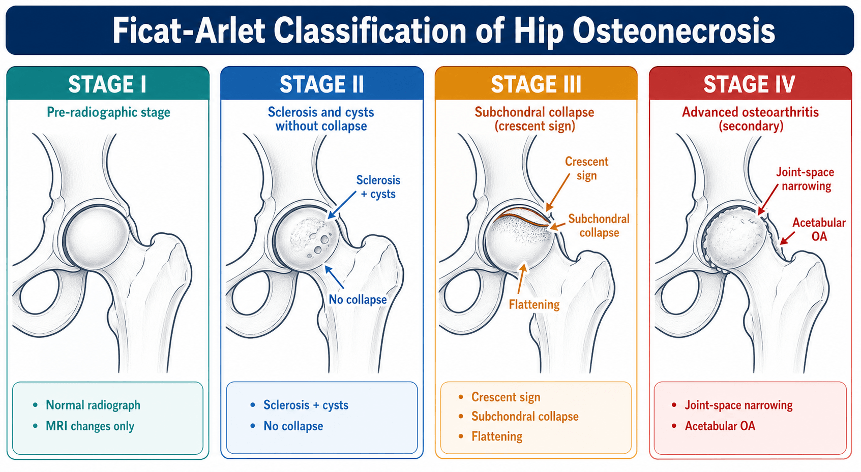
Ficat-Arlet Classification
Original 1985, Modified by Steinberg 1995
- Symptoms
- Pain or asymptomatic
- XR Findings
- Normal
- MRI Findings
- Positive (band sign)
- Treatment Options
- Core decompression ± NVBG
- Prognosis
- 80% success with surgery
- Symptoms
- Pain with activity
- XR Findings
- Sclerosis, cysts, no collapse
- MRI Findings
- Positive, demarcated lesion
- Treatment Options
- Core decompression ± grafting/osteotomy
- Prognosis
- 60-70% success, depends on lesion size
- Symptoms
- Pain at rest and activity
- XR Findings
- Crescent sign, collapse
- MRI Findings
- Subchondral fracture, fluid
- Treatment Options
- Osteotomy (young) or THA
- Prognosis
- Poor with preservation, THA preferred
- Symptoms
- Severe pain, limp
- XR Findings
- Collapse + acetabular changes
- MRI Findings
- Arthritis, joint effusion
- Treatment Options
- Total hip arthroplasty
- Prognosis
- THA definitive, revision risk 15-20% at 10 years

Crescent sign (subchondral fracture) = Stage III = poor prognosis for joint preservation. This is the critical inflection point. Before crescent sign (Stages I-II), trabecular architecture is intact and core decompression can relieve pressure and promote revascularization (80% success in Stage I). Once crescent sign appears, subchondral bone has fractured, collapse is underway, and joint preservation fails in 70-80% of cases. In patients over 40 years, THA is preferred at Stage III.
Quantifying Collapse Risk: The Kerboul Combined Necrotic Angle
Lesion size and location (Steinberg extent, ARCO zones) predict collapse, but the most widely quoted single number is the Kerboul combined necrotic angle - a simple way to express how much of the weight-bearing head is dead.
How it is measured: the arc of the necrotic segment is measured on the mid-coronal image and on the mid-sagittal image, and the two angles are added together. The original description used plain radiographs (AP and lateral); the modified Kerboul angle measures the same two arcs on MRI, which is now the standard.
- Risk category
- Low risk
- Implication
- Head likely to survive; joint preservation favourable
- Risk category
- Moderate risk
- Implication
- Intermediate; weigh lesion size, location and patient factors
- Risk category
- High risk
- Implication
- Collapse likely despite isolated core decompression; consider grafting or arthroplasty
Add the arc of necrosis on the mid-coronal and mid-sagittal views: a combined angle over 240 degrees is high-risk for collapse and predicts failure of isolated core decompression, whereas under 190 degrees is low-risk. It complements - rather than replaces - Steinberg lesion size and ARCO location, and is the quantitative threshold examiners look for when you justify offering or withholding joint preservation.
Clinical Assessment
Onset: Insidious groin pain over weeks to months (vs acute trauma)
Character: Dull ache progressing to sharp pain with weight-bearing
Radiation: Groin to anterior thigh, knee (L3 referred pain)
Aggravating: Weight-bearing, stairs, rising from chair, internal rotation
Relieving: Rest, non-weight-bearing, analgesia
Functional: Limp, reduced walking distance, difficulty with shoes/socks
Risk Factors: ASEPTIC mnemonic - systematically ask about steroids (dose, duration), alcohol (quantity per week), trauma, clotting disorders, sickle cell, Gaucher, pancreatitis
Gait: Antalgic (shortened stance phase on affected side), Trendelenburg if chronic
Look: Usually normal (no swelling unless Stage IV OA), leg length discrepancy if chronic collapse
Feel: Groin tenderness, no effusion palpable
Move:
- Active ROM: Painful limitation (especially internal rotation and abduction)
- Passive ROM: Loss of internal rotation earliest sign, flexion-adduction-internal rotation (FADIR) painful
- Specific tests: Log roll test painful, FABER positive
Neurovascular: Intact (unless chronic with nerve compression)
Other joints: Examine shoulders, knees (multifocal AVN common in sickle cell, steroids)
Always examine both hips and image both sides. AVN is bilateral in 50-80% of cases, often asymptomatic initially. Patients presenting with unilateral symptoms may have MRI-positive AVN in the contralateral hip (Stage I) requiring surveillance or prophylactic treatment. Failure to image the contralateral side is medicolegal risk - patient develops second hip AVN 2 years later asking why it was not detected earlier.
Differential Diagnosis
- Key Distinguishing Features
- Older age (over 60), gradual onset, no risk factors, unilateral
- Imaging Findings
- Joint space narrowing, osteophytes, subchondral sclerosis
- Key Distinguishing Features
- Third trimester pregnancy or middle-aged men, self-limiting (6-12 months)
- Imaging Findings
- Diffuse marrow edema on MRI, NO band sign, XR normal or osteopenic
- Key Distinguishing Features
- Athletes, military recruits, acute pain, no risk factors
- Imaging Findings
- MRI shows fracture line (not band), XR may show cortical break
- Key Distinguishing Features
- Older age, night pain, weight loss, no trauma
- Imaging Findings
- MRI shows mass lesion, XR shows lytic/blastic lesion
- Key Distinguishing Features
- Mechanical symptoms (clicking, catching), younger athletic patients
- Imaging Findings
- MRI arthrogram shows labral tear, no bone marrow edema
Investigations
Imaging Protocol for Suspected AVN
Views: AP pelvis (both hips for comparison), lateral affected hip, frog-leg lateral
Stage I: Normal (pre-radiographic - MRI needed)
Stage II: Sclerosis (increased density at interface), cystic changes, preservation of sphericity
Stage III: Crescent sign (subchondral lucency = fracture), flattening of head
Stage IV: Collapse, secondary OA changes (joint space narrowing, osteophytes, acetabular sclerosis)
Utility: Staging for treatment decisions, monitoring progression, excluding other pathology.
Sensitivity: 99% for early AVN (can detect Stage I before XR changes)
Specificity: 95% when band sign present
Findings:
- Band sign: Low signal line on T1 and T2 (interface between necrotic and viable bone)
- Double line sign: Low signal outer rim + high signal inner rim on T2 (granulation tissue)
- Geographic pattern: Wedge-shaped or band-like lesion in anterosuperior head
Protocol: Coronal and axial T1, T2, STIR sequences of both hips
Value: Detects pre-radiographic disease, determines lesion size (Steinberg substaging), identifies bilateral disease, monitors treatment response.
CT: Detects sclerosis, cysts, crescent sign, but less sensitive than MRI for Stage I. Useful for surgical planning (valgus osteotomy, measuring lesion location).
Bone Scan: Cold in center (no uptake in necrotic bone), hot rim peripherally (reparative response). Less specific than MRI, radiation exposure, no anatomic detail.
Role: Second-line if MRI unavailable or contraindicated (pacemaker, severe claustrophobia).
Baseline: FBC (sickle cell, Gaucher), ESR/CRP (exclude infection), LFTs (alcohol-related liver disease), lipid profile
Coagulation Screen: Protein C, Protein S, Antithrombin III, Factor V Leiden, Lupus anticoagulant, Anticardiolipin antibodies (if young, idiopathic, bilateral)
Specific Tests: Hemoglobin electrophoresis (sickle cell), Gaucher enzyme assay, HIV serology
Rationale: Identify modifiable risk factors (cease steroids if possible, alcohol cessation), screen for systemic disease requiring treatment, counsel about contralateral hip risk.

Management Algorithm
Treatment Decision Framework
Joint Preservation Strategy
Goal: Prevent collapse, promote revascularization, preserve native joint
Non-Operative Management (Observation)
- Small lesions (under 15% of head - Steinberg A)
- Medial location (non-weight-bearing zone)
- Asymptomatic or minimal symptoms
- Patient refuses surgery
Observation alone has 80% progression rate in Stage I-II, so reserved for very small medial lesions.
- Activity modification: Reduce impact loading, crutches if symptomatic
- Medications: NSAIDs for pain, bisphosphonates (alendronate) may slow progression
- Risk factor modification: Cease alcohol, reduce/taper steroids if possible
- Surveillance: MRI every 6 months for 2 years, then annually if stable
Inform patient that 80% progress without surgery - observation is not benign neglect.
Core Decompression (Gold Standard Stage I-II)
- Reduces intraosseous pressure (from over 30mmHg to under 10mmHg)
- Creates drill track for revascularization (granulation tissue)
- Removes necrotic marrow (decompresses venous congestion)
- Stimulates bone remodeling
Success rate: 80% in Stage I (prevents collapse), 60-70% in Stage II (depends on lesion size).
- Positioning: Supine on radiolucent table, hip in neutral or slight flexion
- Entry point: Lateral cortex 2-3cm distal to greater trochanter, just above lesser trochanter
- Trajectory: Fluoroscopy guidance, aim for center-center position on AP and lateral
- Drill: 8-10mm cannulated drill, advance to subchondral bone (leave 5mm intact)
- Grafting: Consider non-vascularized bone graft (from iliac crest) to fill defect
- Closure: Deep fascia, subcutaneous, skin, drain usually not needed
Complications: Femoral neck fracture (under 1% - avoid multiple tracts, lateral cortex violation), infection, neurovascular injury.
- Weight-bearing: Touch weight-bearing (10-20kg) on crutches for 6-8 weeks
- Progression: Gradual increase to full weight-bearing at 8-12 weeks based on pain
- Surveillance: XR at 6 weeks, 3 months, 6 months, then annually for 2 years
- Success criteria: No progression of collapse, pain resolution, MRI shows revascularization
Failure indicators: Crescent sign develops, increasing pain, progression to Stage III = consider arthroplasty.
Non-vascularized bone graft (NVBG) + core decompression improves outcomes in Stage II large lesions. Cortical strut graft from fibula or iliac crest provides structural support to weakened subchondral bone (80-90% success in Stage II B-C vs 60% with core decompression alone). Technique: Insert strut graft through drill tract to contact subchondral plate (tantalum rod is modern alternative - inert, porous, promotes bone ingrowth).
Biologic Augmentation: Bone Marrow Aspirate Concentrate (Cell Therapy)
A major refinement of core decompression is to add bone marrow aspirate concentrate (BMAC) to the drill tract. The rationale is that osteonecrosis - particularly steroid- and alcohol-related disease - may involve a depleted or impaired pool of marrow osteoprogenitor cells; concentrating autologous iliac-crest marrow re-delivers mesenchymal stem cells, osteoprogenitors and growth factors to the necrotic segment to support repair and revascularisation. (The underlying cell biology is covered in the dedicated mesenchymal stem cell topic.)
- Technique: aspirate marrow from the iliac crest, concentrate it by centrifugation, and deliver the concentrate down the core-decompression channel into the necrotic zone (often combined with small or multiple percutaneous drill channels rather than a single large tract).
- Evidence: in this topic's own evidence base, core decompression plus bone marrow concentrate reduced femoral-head collapse compared with core decompression alone (relative risk roughly 0.55 over short-term follow-up in the ARCO systematic review), and core decompression plus cell therapy ranked best for limiting radiographic progression in a network meta-analysis (SUCRA 96.4%) - although the certainty of the evidence remains low.
Core decompression relieves intraosseous pressure and opens a revascularisation channel; bone marrow aspirate concentrate adds the biology - osteoprogenitor cells and growth factors to a segment that may be deficient in them. The best current evidence (ARCO review; network meta-analysis) suggests biologically augmented decompression slows collapse and radiographic progression in pre-collapse disease more than decompression alone, but the certainty is low - counsel honestly rather than overpromise.
Surgical Technique
Pre-operative Planning
Risks:
- Success rate: 80% Stage I, 60-70% Stage II, under 30% Stage III
- Femoral neck fracture: Under 1% (requires protected weight-bearing 6-8 weeks)
- Infection: Under 1% (prophylactic antibiotics)
- Neurovascular injury: Under 0.5% (sciatic nerve, femoral vessels)
- Progression despite surgery: 20-40% require THA in future
- DVT/PE: Standard surgical risk (LMWH prophylaxis)
Emphasize that this is joint preservation attempt - not guaranteed to prevent collapse.
Required:
- Radiolucent table (fracture table or standard OR table)
- C-arm fluoroscopy (AP and lateral views)
- 8-10mm cannulated drill system with guidewire
- Power drill with variable speed
- Bone graft harvesting set (if adding NVBG)
- Standard hip surgical tray
Optional Adjuncts:
- Tantalum rod (10mm diameter, 100-120mm length)
- Iliac crest bone graft instruments
- Fibular strut graft instruments (if adding structural support)
Patient Positioning
Setup
Supine on radiolucent table (fracture table not essential but helpful for traction control).
- Hip position: Neutral rotation or slight internal rotation (opens lateral cortex access)
- Knee: Flexed 10-20 degrees (relaxes iliopsoas, improves fluoroscopy)
- Contralateral leg: Abducted and externally rotated (allows C-arm access)
- Arms: Across chest or on arm boards (away from operative field)
Ensure full range of C-arm movement for AP and lateral views before draping.
- Skin prep: Chlorhexidine or iodine from umbilicus to knee, lateral to midline
- Draping: Standard hip draping exposing proximal femur to mid-thigh
- C-arm draping: Sterile cover over C-arm intensifier
- Landmarks: Palpate greater trochanter, mark entry point 2-3cm distal to GT on lateral thigh
Confirm adequate fluoroscopy images (AP pelvis showing both hips, perfect lateral of affected hip) before incision.
Surgical Approach and Technique
Step-by-Step Core Decompression
Location: Lateral thigh, 2-3cm distal to tip of greater trochanter, just above lesser trochanter level
Size: 3-4cm longitudinal incision parallel to femoral shaft
Depth: Through skin and subcutaneous tissue to fascia lata
Fluoroscopy confirms position - entry point should be proximal to lesser trochanter to avoid stress riser at narrowest femoral neck.
- Incise fascia lata in line with skin incision
- Split vastus lateralis fibers bluntly (no intermuscular plane - direct muscle split)
- Expose lateral femoral cortex with retractors (Hohmann on anterior and posterior edges)
- Periosteum: Minimal stripping (reduces devascularization)
Critical: Entry point must be distal to greater trochanter but proximal to lesser trochanter. Too distal = stress riser at narrow femoral neck = fracture risk. Confirm position on AP and lateral fluoroscopy before drilling.
Trajectory Planning:
- AP view: Aim for center of femoral head (bisects head diameter)
- Lateral view: Aim for center of femoral head (anterior-posterior midpoint)
- Target: Subchondral bone 5mm deep to articular cartilage (DO NOT breach cartilage)
Technique:
- Use 2.4-2.8mm guidewire through cannulated system
- Hand-drill guidewire through lateral cortex (careful control)
- Advance under fluoroscopy guidance to center-center position
- Stop 5mm short of subchondral plate on lateral view
- Confirm position on AP and lateral before proceeding
Multiple passes to "find" the correct trajectory waste bone and weaken neck - plan trajectory carefully before drilling.
- Drill size: 8-10mm cannulated drill over guidewire
- Speed: Slow (100-200 RPM) to avoid thermal necrosis
- Irrigation: Copious saline irrigation during drilling (cool bit, clear debris)
- Depth: Advance to 5mm from subchondral plate (measure on fluoroscopy)
- Confirmation: Final AP and lateral images confirm center-center position, 5mm margin
DO NOT:
- Drill multiple tracts (weakens femoral neck = fracture risk)
- Breach lateral cortex widely (stress riser)
- Penetrate subchondral plate (articular cartilage damage, intra-articular communication)
Suction through drill to remove necrotic marrow and blood (decompress intraosseous pressure).
Indications for Adding Bone Graft:
- Large lesion (over 30% of head - Steinberg C)
- Stage II with significant cystic changes
- Structural support needed (tantalum rod or fibular strut)
Non-Vascularized Bone Graft (NVBG):
- Harvest corticocancellous graft from ipsilateral iliac crest (2-3cm incision)
- Morselized cancellous bone OR cortical strut (from fibula or iliac crest)
- Pack through drill tract to contact subchondral plate
- Provides structural support and osteogenic cells
Tantalum Rod:
- 10mm diameter porous tantalum rod (100-120mm length)
- Insert through drill tract to contact subchondral plate
- Promotes bone ingrowth (porous structure), inert, radiopaque (easy follow-up)
- More expensive than NVBG but easier to insert, less donor site morbidity
Confirm graft or rod position on final fluoroscopy images.
- Fascia lata: Absorbable suture (Vicryl 1 or 0) in interrupted or continuous fashion
- Subcutaneous: 2-0 Vicryl to close dead space
- Skin: Subcuticular 3-0 Monocryl or staples
- Dressing: Standard sterile dressing, no drain needed (minimal dead space)
Final check: Ensure no retained swabs, instrument count correct, fluoroscopy images saved.
Intraoperative Pearls and Pitfalls
- Single drill tract only (multiple tracts weaken neck)
- Center-center position on AP and lateral (maximizes decompression)
- 5mm subchondral margin (prevents articular cartilage damage)
- Slow drilling speed with irrigation (prevents thermal necrosis)
- Measure guidewire depth before drilling (prevents overpenetration)
- Save fluoroscopy images for medicolegal documentation
- Do NOT drill multiple tracts (femoral neck fracture risk)
- Do NOT violate lateral cortex widely (stress riser)
- Do NOT breach subchondral plate (articular damage, joint communication)
- Do NOT drill without fluoroscopy confirmation (malposition common)
- Do NOT attempt in Stage III-IV with collapse (failure rate over 70%)
- Do NOT skip postoperative protected weight-bearing (fracture risk)
Understanding these technical details helps anticipate complications and optimize outcomes.
Complications
- Incidence
- 80-90% at 5 years
- Risk Factors
- Large lesion (over 30%), lateral location, Stage II, no treatment
- Prevention/Management
- Early diagnosis (MRI), core decompression in Stage I-II, risk factor modification
- Incidence
- 50-80% within 2 years
- Risk Factors
- Systemic cause (steroids, alcohol, sickle cell, coagulopathy)
- Prevention/Management
- MRI both hips at diagnosis, surveillance imaging, prophylactic treatment if Stage I contralateral
- Incidence
- Under 1%
- Risk Factors
- Multiple drill tracts, lateral cortex violation, early weight-bearing
- Prevention/Management
- Single central tract, avoid lateral cortex, protected weight-bearing 6-8 weeks
- Incidence
- 20-40% (Stage I-II)
- Risk Factors
- Large lesion (over 30%), Stage III, lateral location, delayed diagnosis
- Prevention/Management
- Patient selection (Stage I-II only), add bone grafting if large lesion, realistic expectations
- Incidence
- 15-20% at 10 years
- Risk Factors
- Age under 40, high activity, poor bone quality, polyethylene wear
- Prevention/Management
- Counsel about revision risk, cross-linked poly, large head size, activity modification
- Incidence
- 2-5%
- Risk Factors
- Young active patients, posterior approach, component malposition
- Prevention/Management
- Optimize component position, repair posterior capsule, large head (36mm+), patient education
Core decompression does not guarantee success - 20-40% of Stage I-II patients still progress to collapse. Monitor with clinical review and imaging (XR at 6, 12, 24 months) for signs of failure: (1) Increasing pain despite initial improvement, (2) Crescent sign develops on XR, (3) MRI shows progression of necrosis. If failure detected early (before major collapse), consider salvage with vascularized fibular graft or proceed to arthroplasty. Delaying arthroplasty until severe collapse makes surgery more difficult (bone loss, acetabular involvement).
REVISIONComplications of THA in AVN
Hook:AVN THA outcomes need REVISION more than primary OA - counsel young patients!
Postoperative Care and Rehabilitation
Rehabilitation After Core Decompression
- Analgesia: PCA or oral opioids, transition to paracetamol + NSAIDs
- DVT prophylaxis: LMWH (enoxaparin 40mg daily) for 14 days, TED stockings
- Mobilization: Physiotherapy Day 1, non-weight-bearing on crutches
- Wound: Check drain (if used), remove at 24 hours, dry dressing
- Discharge: Usually Day 1-2 once mobile on crutches, safe at home
- Weight-bearing: Touch weight-bearing (10-20kg) on two crutches
- Exercises: Quadriceps isometric, ankle pumps, hip abduction (prevent stiffness)
- Precautions: Avoid pivoting, twisting, impact activities
- Pain: Expect groin discomfort for 2-4 weeks (surgical pain)
- Follow-up: Clinic at 6 weeks with XR (check for fracture, progression)
- Weight-bearing: Increase to 50% at 6 weeks, progress to full weight-bearing by 12 weeks
- Crutches: Wean from two crutches → one crutch → stick → unaided
- Exercises: Progressive resistance (hip abduction, flexion), stationary bike, pool
- Return to work: Sedentary work at 6 weeks, manual work at 12 weeks
- Imaging: XR at 3 months (assess for healing, progression)
- Activity: Gradual return to impact activities (running, sport) at 6 months if pain-free
- Surveillance: XR at 6, 12, 24 months, then annually for 5 years
- Success criteria: No pain, no progression on XR, return to activities
- Failure signs: Increasing pain, crescent sign develops, MRI shows progression → consider arthroplasty
Understanding this timeline helps set patient expectations - core decompression is a slow recovery (3-6 months to full activity) but preserves the native joint.
Outcomes and Prognosis
- Success Definition
- No collapse, pain relief
- 5-Year Outcome
- 80%
- 10-Year Outcome
- 70%
- Failure Predictors
- Large lesion (over 30%), lateral location, delayed diagnosis
- Success Definition
- No progression to THA
- 5-Year Outcome
- 65-70%
- 10-Year Outcome
- 60%
- Failure Predictors
- Lesion over 30%, crescent sign present, lateral location
- Success Definition
- Hip preservation, no THA
- 5-Year Outcome
- 60-70%
- 10-Year Outcome
- 40-50%
- Failure Predictors
- Age over 40, collapse over 2mm, inadequate intact arc (under 90 degrees)
- Success Definition
- Implant survival, no revision
- 5-Year Outcome
- 95%
- 10-Year Outcome
- 85-90%
- Failure Predictors
- Age under 40, cementless stem in poor bone, high activity level
Large lateral lesions in young patients = highest risk of progression. The combination of (1) lesion over 30% of head (Steinberg C), (2) lateral location (maximal weight-bearing forces), (3) age under 40 (high activity), (4) Stage II or worse at diagnosis predicts 80-90% progression to collapse despite core decompression. Consider free vascularized fibular graft in this group if patient refuses arthroplasty. Conversely, small (under 15%) medial lesions detected early (Stage I) have 80-90% success with core decompression - emphasize importance of early diagnosis.
Quality of Life Considerations
Advantages:
- Preserves native anatomy and proprioception
- No implant-related complications (dislocation, wear, loosening)
- No activity restrictions lifelong
- Delays or avoids THA (and future revisions)
Disadvantages:
- 20-40% failure rate requires future THA
- Prolonged recovery (6-12 months to full activity)
- Risk of collapse during healing period
- Not suitable for all patients (age, lesion size, stage)
Advantages:
- Predictable pain relief (95% at 2 years)
- Rapid functional recovery (6 weeks to normal activities)
- Durable (85-90% survival at 10 years)
- Definitive solution for failed joint preservation
Disadvantages:
- Activity restrictions lifelong (no high impact sports)
- Revision risk in young patients (15-20% at 10 years)
- Dislocation risk (2-5% in AVN patients)
- Psychological impact of "artificial joint" in young patients
Guidelines, Registries & Global Practice
Worldwide Burden and Demographics
United States: ONFH underlies roughly 3-12% of all total hip arthroplasties; an estimated greater than 20,000 patients are treated surgically each year [PMID 31483399, 37001624]
Germany: estimated 5,000-7,000 new cases of non-traumatic AVN per annum (national S3 guideline) [PMID 26667621]
East Asia: disproportionately high non-traumatic burden, partly reflecting alcohol-related and idiopathic disease and large nationwide cohorts [PMID 38830431]
Age: predominantly the third to fifth decades - far younger than primary osteoarthritis
Corticosteroids: a Korean nationwide nested case-control study found ONFH risk rose sharply once cumulative prednisolone reached 1,800mg or more [PMID 38830431]
Alcohol: heavy intake (more than 3-7 standard drinks per week) independently associated with ONFH in the same cohort [PMID 38830431]
Other: sickle cell disease and haemoglobinopathies (a major cause in sub-Saharan Africa, the Caribbean and India), Gaucher disease, dysbaric exposure (caisson disease), thrombophilias and post-traumatic disruption
MCQ Practice Points
Q: What is the main blood supply to the adult femoral head? A: Medial circumflex femoral artery (MFCA) provides 70-80% of blood to the femoral head via posterosuperior retinacular vessels. The MFCA arises from the profunda femoris artery, forms an extracapsular ring with the lateral circumflex femoral artery at the femoral neck base, then sends deep branches (retinacular vessels) that ascend the posterior femoral neck under the capsule to reach the femoral head. The artery of ligamentum teres (from obturator artery) contributes minimally in adults (under 20%). This anatomy explains why intracapsular femoral neck fractures and posterior hip dislocations cause AVN (retinacular vessels are disrupted).
Q: What is the significance of the crescent sign in AVN? A: Crescent sign = subchondral fracture = Ficat Stage III = poor prognosis for joint preservation. The crescent sign appears as a radiolucent line beneath the subchondral plate on XR or MRI, representing a fracture through weakened necrotic bone. Once present, the femoral head has begun to collapse, trabecular architecture is irreversibly damaged, and joint preservation surgery (core decompression) fails in 70-80% of cases. The crescent sign is the critical inflection point between pre-collapse disease (Stages I-II where core decompression has 60-80% success) and post-collapse disease (Stages III-IV where arthroplasty is preferred). In patients over 40 years with crescent sign, THA is recommended over attempted joint preservation.
Q: What cumulative steroid dose increases AVN risk? A: Over 2000mg cumulative prednisone equivalent or over 20mg/day for over 3 months. Steroid-induced AVN is the most common non-traumatic cause (30-40% of all AVN cases). The mechanism involves adipocyte hypertrophy causing increased intraosseous pressure (over 30mmHg normal under 10mmHg) and venous outflow obstruction, leading to ischemia. Risk is dose-dependent and time-dependent. Other high-risk factors include chronic alcohol use (over 400mL ethanol per week), sickle cell disease (10-50% develop AVN by age 35), and coagulopathies (Factor V Leiden, Protein C/S deficiency). Idiopathic AVN accounts for 30-40% despite extensive workup.
Q: What is the success rate of core decompression for Stage I AVN? A: 80% success in preventing collapse at 5-10 years. Core decompression reduces intraosseous pressure (from over 30mmHg to under 10mmHg), creates a drill tract for revascularization, and removes necrotic marrow. Success rates decline with advancing stage: Stage I (pre-radiographic) 80%, Stage II (sclerosis/cysts, no collapse) 60-70%, Stage III (collapse/crescent sign) under 30%. Adding non-vascularized bone graft or tantalum rod improves outcomes in Stage II large lesions (over 30% of head) to 75-80%. The procedure involves a lateral approach, fluoroscopy-guided drilling from above the lesser trochanter to center-center position, leaving 5mm of subchondral bone intact. Postoperative protected weight-bearing (touch weight-bearing for 6-8 weeks) is essential to prevent iatrogenic femoral neck fracture.
Q: How do THA outcomes in AVN patients compare to primary OA? A: Worse outcomes - 15-20% revision rate at 10 years (vs 5-10% in OA). AVN patients are younger (mean age 50 vs 70 in OA), more active, have poorer bone quality (necrotic sclerotic bone), and higher complication rates. Specific risks: (1) Dislocation 2-5% (vs under 1% in OA) due to younger age and higher activity, (2) Aseptic loosening and osteolysis from polyethylene wear (high activity, long life expectancy), (3) Periprosthetic fracture (osteoporotic or sclerotic bone), (4) Infection (immunosuppression from steroids, SLE, or underlying disease). Registry data shows implant survival at 10 years is 85-90% for AVN vs 94-95% for OA. Counsel extensively about revision burden, activity restrictions, and realistic expectations. Use highly cross-linked polyethylene, large femoral heads (36mm or larger), and optimize component position to minimize complications.
Q: What percentage of AVN cases are bilateral? A: 50-80% develop contralateral hip involvement within 2 years. Bilateral disease is the rule, not the exception, in AVN (especially with systemic causes like steroids, alcohol, sickle cell, coagulopathy). This has important management implications: (1) Always MRI both hips at diagnosis even if contralateral hip is asymptomatic (may detect Stage I disease amenable to prophylactic core decompression), (2) Counsel patients about risk of second hip requiring treatment, (3) Consider prophylactic core decompression or close surveillance (MRI every 6 months) for asymptomatic contralateral Stage I AVN, (4) Stage bilateral surgeries if both hips need intervention (infection risk, anesthetic burden, mobilization challenges). Idiopathic AVN has lower bilateral rate (30-40%) compared to steroid-induced (70-80%) or sickle cell disease (over 80%).
Exam Viva Scenarios
Practise clinical reasoning and management decisions out loud
“A 38-year-old man presents with a 4-month history of right groin pain. He is on long-term steroids for Crohn's disease (prednisolone 20mg daily for 18 months). Examination reveals painful internal rotation. Plain XR of the hip is normal. What is your assessment and management?”
“A 35-year-old woman with bilateral hip AVN (Ficat Stage IIIA on right, Stage II on left) secondary to SLE and long-term steroids presents with severe right hip pain and collapse on XR. She is desperate to avoid hip replacement. Walk me through your management options and preferred approach.”
“A 42-year-old man underwent core decompression for Stage II AVN 18 months ago. He presents with worsening groin pain over the last 3 months. How do you assess and manage this?”
Key Anatomy
- Medial circumflex femoral artery = 70-80% of femoral head blood supply
- Posterosuperior retinacular vessels ascend posterior neck (at risk in displaced #NOF)
- Artery of ligamentum teres = minimal contribution in adults (under 20%)
- Extracapsular vessels vulnerable to trauma, thrombosis, and compression
Classification (Ficat-Arlet)
- Stage I = Normal XR, MRI positive (band sign), 80% core decompression success
- Stage II = Sclerosis/cysts, no collapse, 60-70% core decompression success
- Stage III = Crescent sign (subchondral fracture), collapse started, under 30% core decompression success
- Stage IV = Secondary OA, acetabular involvement, THA definitive treatment
Treatment Algorithm
- Stage I-II small lesion (under 30%) = Core decompression ± grafting
- Stage II large lesion (over 30%) = Core decompression + NVBG or tantalum rod
- Stage III under 40 years = Valgus osteotomy or vascularized fibular graft (consider THA)
- Stage III over 40 years or Stage IV any age = Total hip arthroplasty
Surgical Pearls
- Core decompression: Lateral entry 2-3cm distal to GT, center-center position, leave 5mm subchondral bone
- Protected weight-bearing (touch WB) for 6-8 weeks post-decompression (prevent femoral neck fracture)
- THA in AVN: Cementless fixation, large head (36mm+), cross-linked poly, counsel about 15-20% revision at 10 years
- Valgus osteotomy: Requires intact medial column, rotates lateral necrotic segment out of weight-bearing
Complications
- Bilateral disease in 50-80% within 2 years (always MRI both hips)
- Core decompression failure in 20-40% of Stage II (progression to collapse despite surgery)
- Femoral neck fracture under 1% (avoid multiple tracts, lateral cortex, early weight-bearing)
- THA revision in 15-20% at 10 years (young age, high activity, poor bone quality)
Evidence Base and Key Trials
ARCO Clinical Practice Guideline: Diagnosis and Treatment of ONFH
- 36 studies synthesised to inform the ARCO guideline for ARCO stage I-III nontraumatic ONFH
- MRI had the highest pooled sensitivity (0.91, 95% CI 0.87-0.94) and specificity (0.96, 95% CI 0.87-0.99) for diagnosis
- CT and MRI detected subchondral fractures better than plain radiography
- Lower collapse with core decompression plus bone marrow concentrate vs core decompression alone (RR 0.55, 95% CI 0.36-0.83) and with vascularised vs non-vascularised bone grafting (RR 0.35, 95% CI 0.14-0.84) at 5 years or less
Core Decompression for ONFH: Systematic Review
- Systematic review of core decompression for hip osteonecrosis (4 studies meeting criteria, 139 hips)
- Overall 25.8% of hips required conversion to total hip arthroplasty
- Best outcomes in hips with a necrotic lesion under 50% of the head
- No rigorous studies providing long-term outcome measures were identified
Joint-Preserving Surgery vs Non-Operative Treatment: Network Meta-Analysis
- Network meta-analysis of 17 RCTs (784 patients, 918 hips) across 7 interventions for ONFH
- No statistically significant difference between joint-preserving methods and non-surgical treatment for conversion to THA
- Core decompression plus cell therapy ranked best for limiting radiographic progression (SUCRA 96.4%)
- No significant between-group difference in Harris Hip Score overall
Free Vascularised Fibular Graft in Precollapse ONFH: Long-Term Survivorship
- 61 patients (65 hips) with precollapse ONFH, minimum 10.5-year follow-up (mean 14.4 years)
- 49 of 65 hips (75%) had a surviving graft for at least 10 years
- 39 of 65 hips (60%) had a surviving graft at last follow-up; 26 of 65 (40%) converted to THA at a mean of 8 years
- Patients with a surviving graft were more likely to return to impact sport than those converted to THA
THA with Highly Cross-Linked Polyethylene: Osteonecrosis vs Osteoarthritis
- 461 osteonecrosis hips matched 1:1 to 461 osteoarthritis hips, median 10-year follow-up
- 15-year cumulative revision 6.6% (osteonecrosis) vs 4.5% (osteoarthritis), HR 1.8 (p=0.09)
- 15-year cumulative reoperation 10.5% vs 6.4%, HR 2.2 (p=0.008)
- Reoperation varied by aetiology: 0% radiation-induced, 6.3% alcohol, 12.1% steroid, 25% idiopathic
Alendronate to Prevent Femoral Head Collapse: RCT and Its Refutation
- Single-centre RCT (Lai 2005, PMID 16203877): in Steinberg II-IIIC necrosis over 30% of the head, only 2 of 29 alendronate hips collapsed vs 19 of 25 controls (p under 0.001)
- THA was needed in 1 alendronate hip vs 16 control hips in that trial (p under 0.001)
- Multicentre double-blind RCT (Chen 2012, PMID 22127729): alendronate did NOT reduce collapse, THA conversion, or improve function vs placebo
- The early single-centre benefit was not reproduced in the later multicentre placebo-controlled trial