Wear Debris → Osteolysis → Implant Migration | Rule Out Infection | Paprosky Classification
- Aseptic loosening = implant failure due to particle-induced osteolysis, NOT infection
- Wear debris (polyethylene, metal, cement) triggers macrophage activation
- Gruen zones (1-7) for femur, DeLee/Charnley (I-III) for acetabulum
- Rule out infection with ESR, CRP, aspiration before labelling as aseptic
- Paprosky classification guides bone loss management in revision surgery
- “Aseptic loosening is a biologic process - particle disease
- “Progressive radiolucent lines, migration, subsidence on serial X-rays
- “Must exclude infection - aspirate if any doubt
- “Revision complexity depends on bone loss (Paprosky)
Overview and Epidemiology
Aseptic loosening is loss of mechanical fixation between implant and bone, driven by a biologic reaction to wear debris (particle disease) rather than by infection. Progressive bone resorption around the implant, osteolysis, leads to pain, migration and eventual mechanical failure.
How common. It is the most common indication for revision total hip arthroplasty in the late postoperative period, beyond 5 years, and with its associated osteolysis the single most frequent indication for revision in joint registry data. [1] It progresses over years and most often becomes symptomatic beyond 5-10 years from the primary procedure.
What polyethylene cost, and what cross-linking saved. A 10-year double-blinded RCT put numbers on the conventional bearing. [2] The conventional UHMWPE arm wore at a mean 3D rate of 0.27 mm/year, with 38% osteolysis and a 14.6% revision rate; highly cross-linked polyethylene (HXLPE) reduced these to 0.03 mm/year, 8% and 1.9%. HXLPE dramatically reduces particle generation, and that lower particle load is the principal modern defence against aseptic loosening.
Risk factors. Patient, implant and technique all contribute:
- Young, active patients - higher wear rates
- Obesity - increased joint forces
- Poor implant positioning - edge loading, impingement
- Conventional polyethylene - higher wear
- Thin cement mantles - cement fracture
- Undersized components - inadequate fixation
- Osteolysis-prone patients - genetic factors (cytokine polymorphisms, below)
Pathophysiology - Particle Disease
Aseptic loosening is fundamentally a biologic process driven by wear particles, not a purely mechanical phenomenon. The cascade runs from the bearing to the bone.
Particle generation. Polyethylene wear is the most common source. The others:
- Metal-on-metal wear (cobalt and chromium particles)
- Ceramic particles, if a liner fractures
- Cement particles (PMMA debris)
- Corrosion products at modular junctions
Particle distribution. Particles migrate along the effective joint space and enter the periprosthetic tissues through joint fluid pumping. Size matters, and the biologically critical window is narrower than the phagocytosable one: particles up to roughly 10 microns can be taken up by a macrophage, but the range that most potently drives osteolytic cytokine release from UHMWPE debris is about 0.1 to 1.0 microns. That is sub-micron, and the fraction that dominates polyethylene wear by particle number.
Do not quote "0.1 to 10 microns" as the active range. That welds the critical band to the whole phagocytosable one, and it obscures the design paradox.
The design paradox. Reducing bulk wear volume does not automatically reduce biological activity, because making particles smaller can make each one more provocative; this is the examinable subtlety. Cobalt-chrome debris from metal-on-metal bearings is nanometre-scale (roughly 10 to 120 nm, mean about 40 nm), an order of magnitude below the critical UHMWPE size. That is one reason those bearings failed by a different mechanism, metal ion release, hypersensitivity and adverse reaction to metal debris, rather than by classical particle osteolysis.
Macrophage activation. Macrophages phagocytose the particles but cannot digest polyethylene, a frustrated phagocytosis that activates them to release cytokines, chiefly TNF-α, IL-1β, IL-6 and RANKL. T cells are involved too, an adaptive immune response.
Osteoclast recruitment. RANKL binds RANK on osteoclast precursors. Osteoprotegerin (OPG) normally inhibits RANKL; here the RANKL/OPG ratio rises, and activated osteoclasts resorb bone.
Osteolysis and the membrane. Chronic inflammation forms a periprosthetic membrane of granulation tissue, containing macrophages, giant cells and particles, which extends along the implant-bone interface. Progressive resorption creates cystic lesions.
Mechanical loosening. With bone support lost, micromotion increases and the implant migrates or subsides.
Loosening creates more micromotion → generates more particles → drives more osteolysis → creates more loosening. This is why early detection and intervention are important - the process accelerates once established.
Genetic factors. Polymorphisms in cytokine genes (TNF-α, IL-1) make some patients high responders to particles. This explains the variability in osteolysis rates with similar wear.
When periprosthetic osteolysis was first described around cemented implants it was attributed to the cement itself and christened "cement disease", on the reasoning that PMMA was the foreign material and therefore the culprit. The term is obsolete and knowing why is a fair viva question. Cementless implants turned out to develop identical osteolysis, with the same macrophage-rich membrane and the same lytic lesions - and the common factor across both was not cement but particulate debris, overwhelmingly polyethylene. Hence the modern name, particle disease.
The correction is not merely historical housekeeping. It relocated the therapeutic target from the fixation method to the bearing surface, and everything that has actually worked since - cross-linking the polyethylene, hard-on-hard bearings, reducing third-body wear - follows from getting the causal agent right. It also explains why the strategies aimed at the biological cascade instead (anti-TNF agents, bisphosphonates, anti-RANKL) remain experimental: you can suppress the response to particles, but the only intervention proven in patients is generating fewer particles in the first place.
Classification Systems and Radiographic Assessment
Loosening is described by where the radiolucent lines, osteolysis or component migration occur, using two standard zone systems on the AP radiograph.
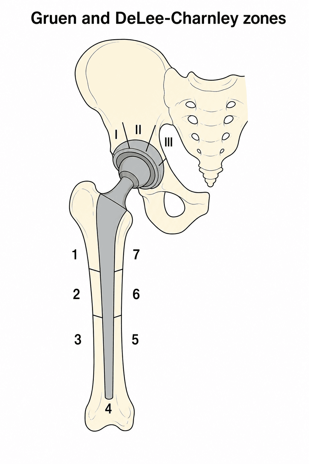
Gruen zones (femoral). The femur around the stem is divided into 7 zones: 1 to 3 down the lateral side from the greater trochanter, 4 at the stem tip, and 5 to 7 up the medial side, ending at the calcar in zone 7. Where the lucency lies tells you what it means:
- Zone 7 (calcar) lucency - calcar resorption, with a risk of subsidence
- Progressive lucency in zones 1 and 7 - suggests proximal loosening
- Progressive lucency, increasing on serial films - loosening
- Lucency more than 2mm - concerning for loosening
- Circumferential lucency, around all seven zones - definitely loose
Stress shielding in zone 7 is common with stiff stems and may be stable if non-progressive and the stem is otherwise well fixed distally. True loosening differs: it is progressive and often circumferential.
DeLee and Charnley zones (acetabular). The acetabulum is divided into 3 zones. Zone I is superolateral, the dome over the weight-bearing region and the most critical for load transfer, so lucency there is of high concern. Zone II is central, over the medial wall, and lucency there may indicate medial migration. Zone III is inferomedial, towards the ischium, and is a less common site for initial loosening.
Lucency in all three zones means the cup is definitely loose, and progressive lucency means loosening is in process. Look also for:
- Migration - a superior or medial shift of the cup, superior migration measured from Köhler's line
- Cup tilt - a change in anteversion or retroversion
- Rotation around screws, if present
Migration and subsidence. Migration is a change in position: lateral shift, rotation or tilt. Subsidence is distal, vertical settling. The criteria grade the certainty of loosening.
A single finding from this list means definite loosening:
- Migration, a change in position of more than 5mm in 2 years
- Subsidence of more than 5mm
- Component fracture
- Cement fracture
- A complete, circumferential radiolucent line more than 2mm
These make loosening probable:
- Progressive radiolucent lines, expanding over time
- Expanding osteolysis
- Lucency over more than 50% of the interface, short of circumferential
- Lucency more than 2mm thick anywhere
These mean the component is possibly loose, and it is monitored:
- Stable thin lucent lines, under 2mm and not progressing
- Focal osteolysis that is not expanding
- Early subsidence under 2mm in the first year, then stable; in an uncemented stem this early settling may be stable
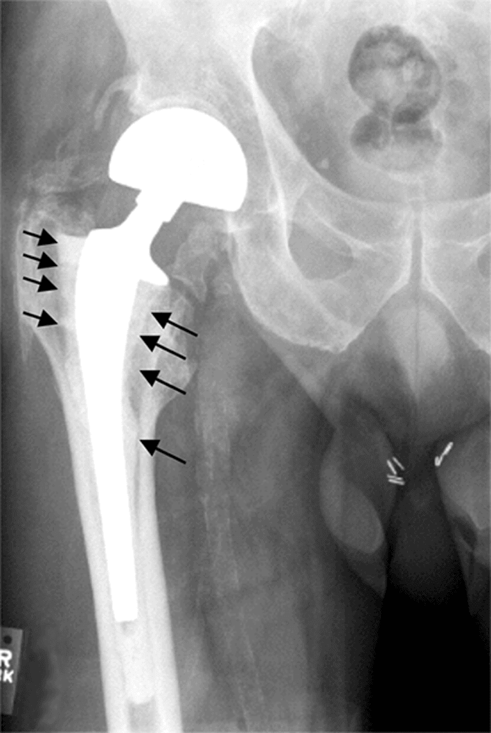
Serial films. Compare every film with the immediate postoperative radiograph, repeat annually to detect progression, measure migration and subsidence quantitatively, and document lucency zone by zone.
Single X-ray cannot diagnose loosening unless gross migration or fracture present. Serial films showing progression of lucent lines or increasing migration are the hallmark of aseptic loosening. Always request comparison films.
Paprosky classification. Used in revision planning to characterise the severity of bone loss, and each grade points to a reconstruction.
- Bone loss
- Intact rim, minimal bone loss (under 25%)
- Reconstruction
- Hemispheric cup with screws
- Bone loss
- Intact rim, superior/medial bone loss (25-50%)
- Reconstruction
- Hemispheric cup with screws
- Bone loss
- Intact rim, superior-lateral migration (under 3cm)
- Reconstruction
- Jumbo cup, cluster screws
- Bone loss
- Medial wall deficient
- Reconstruction
- Jumbo cup, cluster screws
- Bone loss
- Superior migration (more than 3cm), intact Köhler's line
- Reconstruction
- High hip centre, augments, or trabecular metal
- Bone loss
- Superior migration, ischial/teardrop destruction
- Reconstruction
- Reconstruction cage, cup-cage construct, custom triflange
- Bone loss
- Minimal metaphyseal bone loss, intact diaphysis
- Reconstruction
- Cemented or proximally coated stem
- Bone loss
- Extensive metaphyseal loss, intact diaphysis (more than 4cm)
- Reconstruction
- Cemented or proximally coated stem
- Bone loss
- Metaphyseal loss, diaphysis supportive (more than 4cm)
- Reconstruction
- Extensively coated stem (diaphyseal fit)
- Bone loss
- Extensive metaphyseal and diaphyseal damage
- Reconstruction
- Long extensively coated stem, impaction grafting
- Bone loss
- Extensive diaphyseal loss, non-supportive isthmus
- Reconstruction
- Extensively coated stems, impaction grafting, allograft-prosthesis composite, modular tumour prosthesis
Clinical Presentation and Assessment
History. The onset is gradual and progressive, over years not weeks, typically more than 5-10 years after surgery in a THA that often functioned well at first. Pain is in the groin (acetabular) or the thigh (femoral), and activities become progressively harder. Start-up pain, worse after rest and improving with walking, is typical.
Aseptic or septic. The history is the first discriminator. Aseptic loosening comes on gradually over months to years, the pain is mechanical and improves with activity, and there are no systemic or constitutional symptoms such as fever, malaise or night sweats. Acute or subacute onset, persistent pain despite rest, night or rest pain, systemic symptoms and a history of wound problems all raise the suspicion of infection.
Examination. The findings and what each suggests:
- Significance
- Pain with weight-bearing
- Interpretation
- Mechanical pain from loosening
- Significance
- Acetabular component loosening
- Interpretation
- Positive impingement test
- Significance
- Femoral component loosening
- Interpretation
- Start-up pain typical
- Significance
- Component migration
- Interpretation
- Measure and compare to prior
- Significance
- Suggests aseptic
- Interpretation
- Infection would show inflammation
- Significance
- Suggests septic
- Interpretation
- Requires infection workup
Range of motion. Usually preserved unless migration is severe. Pain at end range suggests impingement, and stiffness suggests other pathology such as infection or heterotopic ossification.
Neurovascular examination. Document distal pulses and sensation, and in particular the sciatic nerve after a previous posterior approach or the femoral nerve after an anterior one.
Investigations and Differential Diagnosis
Radiographs. AP pelvis, AP hip and lateral hip, always compared with prior films. Read them zone by zone, Gruen for the femur and DeLee/Charnley for the acetabulum: the width and extent of lucent lines, migration quantified, focal osteolysis, and the cement mantle if cemented.
CT, if planning revision. It assesses bone loss better for Paprosky classification, identifies osteolysis not visible on X-ray, evaluates pelvic discontinuity, and with 3D reconstruction supports templating and the reconstruction plan.
Differential diagnosis of the painful THA. Aseptic loosening is a diagnosis of exclusion. Systematically consider intrinsic and extrinsic causes before attributing pain to loosening.
- Discriminating features
- Gradual start-up/thigh or groin pain, progressive lucent lines, normal-to-mildly raised markers
- Key investigation
- Serial radiographs; aspiration to exclude PJI
- Discriminating features
- Rest/night pain, sometimes acute, raised ESR/CRP, possible sinus
- Key investigation
- Serum ESR/CRP then aspiration; 2018 ICM score
- Discriminating features
- Acute pain after a fall, deformity, inability to weight-bear
- Key investigation
- Radiographs (Vancouver classification)
- Discriminating features
- Pain with specific positions, episodes of giving way
- Key investigation
- Examination, radiographs, component-position CT
- Discriminating features
- MoM or modular-junction implant, groin mass, effusion
- Key investigation
- Serum cobalt/chromium, MARS MRI / ultrasound
- Discriminating features
- Groin pain on active hip flexion/stairs, oversized or prominent cup
- Key investigation
- Examination, CT for cup overhang, diagnostic injection
- Discriminating features
- Back-dominant pain, radicular features, normal hip on exam
- Key investigation
- Spine examination, lumbar imaging
- Discriminating features
- Lateral tenderness, pain lying on side, Trendelenburg
- Key investigation
- Examination, ultrasound/MRI of abductors
- Discriminating features
- Activity-related thigh pain with stiff stem, no progression
- Key investigation
- Serial radiographs showing stable fixation
Management Algorithm
The work-up runs in order, and the order matters:
- Confirm loosening - serial radiographs showing progression, definite radiographic criteria (migration, lucency), and other causes of pain, referred or spinal, excluded
- Rule out infection - ESR and CRP, aspiration if they are elevated or if there is any suspicion, and the MSIS criteria to classify the hip as aseptic or septic
- Assess symptoms and function - pain severity, functional limitation, impact on activities of daily living, and the patient's expectations and goals
The decision. Symptoms and radiographic behaviour together choose the path:
- Symptomatic, with radiographic progression - surgery
- Asymptomatic, with early changes - observation with serial X-rays
- Minimal symptoms, stable - conservative management
Surgical Technique - Revision for Aseptic Loosening
The sequence. Expose, identifying and protecting the neurovascular structures, with a complete capsulectomy, and identify and remove previous hardware. Remove the femoral head (disassociating it if modular), the acetabular liner and component and the femoral stem, then all cement, membrane and debris. Reconstruct the acetabulum and then the femur. A trial reduction checks leg length, offset and stability through a range of motion before the capsule and soft tissues are repaired and the wound closed in layers.
The previous approach is usually used, if possible. Adequate exposure is critical: plan an extensile approach if needed rather than fighting through a small exposure, and protect the neurovascular structures.
Posterior - the most common. Good exposure to the acetabulum and femur, extensile through a trochanteric slide or osteotomy, and familiar to most surgeons. The risk is dislocation, especially after a prior posterior approach.
Direct anterior - after an anterior primary. Good acetabular exposure, but femoral exposure for revision is difficult and the extensile options are limited.
Lateral - less common for revisions. It puts the abductors at risk; transtrochanteric extension is possible.
Extended trochanteric osteotomy (ETO). For difficult femoral extraction, well-fixed uncemented stems and removal of a cement column. It allows distal access to the femur, and extended approaches allow safer component removal and better access for reconstruction. The osteotomy must be repaired and protected postoperatively.
Complications of Revision THA
- Incidence
- 5-15% (higher than primary)
- Prevention/Management
- Restore offset, tension; constrained liner if needed
- Incidence
- 5-10%
- Prevention/Management
- Careful extraction; ETO for well-fixed stems
- Incidence
- 2-5%
- Prevention/Management
- Protected weight-bearing; adequate fixation
- Incidence
- 1-3%
- Prevention/Management
- Careful retraction; document preop exam
- Incidence
- 2-5%
- Prevention/Management
- Antibiotic prophylaxis; minimise operative time
- Incidence
- Common
- Prevention/Management
- Templating; intraoperative measurement
- Incidence
- 10-15% at 10 years
- Prevention/Management
- Address bone loss adequately; stable fixation
- Incidence
- 1-2%
- Prevention/Management
- Chemical and mechanical prophylaxis
Dislocation. Higher after revision than after primary THA, 5-15% against 1-3%, because of soft-tissue damage and laxity, abductor insufficiency, and poor bone stock affecting component position. Restore offset and leg length, repair the posterior soft tissues after a posterior approach, and consider a dual-mobility bearing or a constrained liner in high-risk patients.
Intraoperative fracture. Most often of the femur during stem removal, treated with cerclage cables and/or revision to a longer stem. On the acetabular side a medial wall blow-out may need a cage or column plating.
Nerve injury. The sciatic nerve is most at risk, from a posterior approach and retraction; the femoral nerve with an anterior approach. Document the preoperative examination, and avoid excessive retraction and leg lengthening.
Infection. The risk is higher in revision than in primary surgery. Debride meticulously and give antibiotic prophylaxis; local vancomycin powder is an option, though a controversial one.
Postoperative Care and Rehabilitation
- ICU/HDU if complex or prolonged case
- DVT prophylaxis (chemical and mechanical)
- Pain management (multimodal, avoid excessive opioids)
- Monitor haemoglobin (transfuse if needed)
- Foley catheter typically removed day 1
- Early mobilisation to chair
- Physiotherapy with walking frame, progressing to crutches or walker
- Weight-bearing as restricted below
- Hip precautions (if posterior approach)
- Wound inspection
- Progressive weight-bearing (if stable fixation)
- Outpatient physiotherapy
- Wound check at 2 weeks (remove sutures/staples)
- First follow-up X-rays at 6 weeks
- Advance to full weight-bearing (if healing well)
- Wean from walking aids
- Progressive strengthening exercises
- Return to normal activities gradually
- Avoid high-impact sports indefinitely
Weight-bearing. The reconstruction sets the restriction:
- Uncemented femur - partial weight-bearing; an extensively coated femoral stem may allow immediate weight-bearing
- Uncemented acetabulum - partial weight-bearing for 6 weeks
- Extended trochanteric osteotomy - toe-touch for 6 weeks, then partial from 6 to 12 weeks, protected until union (usually 12 weeks)
- Bone grafting - protected weight-bearing until the graft incorporates
Hip precautions after a posterior approach. No hip flexion beyond 90 degrees, no adduction past the midline and no internal rotation, for 6-12 weeks according to surgeon preference.
Watching for complications. Signs of wound infection (erythema, drainage), symptoms of DVT or PE (leg swelling, chest pain), neurovascular status, and pain out of proportion (compartment syndrome is rare).
Long-term follow-up. Radiographs at 6 weeks, 3 months and 1 year, then annually, monitoring for loosening, wear and osteolysis, documenting stable fixation (osseointegration) and checking for heterotopic ossification.
Outcomes and Prognosis
Survivorship. Modern techniques and implants have improved revision THA outcomes substantially. Freedom from re-revision is 80-90% at 10 years in modern series and 70-80% at 15 years, and worse with severe bone loss (Paprosky IIIB/IV).
What predicts it. Patient, surgeon and implant all contribute:
- Patient - age (younger patients have a higher revision risk), activity level, BMI (obesity increases failure risk) and bone quality
- Surgery - severity of bone loss (Paprosky grade), surgeon experience with revisions, achieving stable fixation, and managing soft-tissue and abductor integrity
- Implant - extensively coated stems (better outcomes than cemented in Paprosky III), tantalum augments and cups (good outcomes in the deficient acetabulum), and constrained liners (higher dislocation rate but necessary in some)
Implant-specific series. Extensively porous-coated diaphyseal femoral stems have re-revision for aseptic femoral loosening of 3% at 20 years, and any-cause femoral re-revision of 6.4% at 20 years. [4] Trabecular-metal and titanium revision cups for severe acetabular bone loss report survivorship of 100% at 5 years and 88% at 10 years. [5]
Function. Most patients achieve pain relief, functional scores improve (Harris Hip Score, WOMAC), and patients return to low-impact activities.
Across the major registries, aseptic loosening revision rates have fallen with modern bearings (HXLPE) and porous fixation. [1][2] However, once loosening occurs revision is complex and outcomes remain inferior to primary THA — prevention through optimal bearing choice and primary technique is the key message.
Guidelines, Registries & Global Practice
This section frames aseptic loosening as a global problem, drawing on the major national joint registries as cited evidence rather than the practice of any single country.
Global epidemiology
- THA volume — and therefore the at-risk population for late loosening — is rising worldwide. Registry-based modelling has projected a 208% increase in primary THA for osteoarthritis between 2013 and 2030 in high-income settings. [3] Ageing populations and rising obesity drive similar trends across high-income registries.
- Aseptic loosening / osteolysis is the leading recorded indication for revision THA in registry data; in the National Joint Registry of England and Wales it ranks ahead of dislocation, pain and periprosthetic fracture as a cause of single-stage revision. [1]
- The dominant modifiable driver historically was conventional polyethylene wear; the shift to HXLPE has reduced osteolysis and revision substantially. [2]
Major guidelines and consensus, side by side
- Position
- Exclude PJI in EVERY painful/loose THA using the 2018 weighted scoring system before calling it aseptic
- Evidence basis
- Multicentre diagnostic validation, sensitivity 97.7%
- Position
- Routine serum ESR and CRP first-line in suspected PJI; aspirate if raised or clinical suspicion
- Evidence basis
- Evidence-based CPG (moderate-strong)
- Position
- Surveillance imaging of THA and prompt specialist referral for a painful prosthesis; revision in units with appropriate expertise
- Evidence basis
- Guideline / consensus
- Position
- Bearing-surface optimisation (HXLPE, ceramic) to reduce wear-driven osteolysis; classify bone loss before revision
- Evidence basis
- Consensus, registry-supported
Registry evidence (implant survival and revision)
- Region
- England, Wales, NI
- Relevance to aseptic loosening
- Aseptic loosening/osteolysis the leading cause of revision; basis for ODEP implant benchmarking
- Region
- Australia
- Relevance to aseptic loosening
- Mandatory registry; rising THA burden and reduced wear-driven failure with HXLPE
- Region
- USA
- Relevance to aseptic loosening
- Largest by annual volume; tracks bearing-surface and fixation trends
- Region
- Sweden
- Relevance to aseptic loosening
- Longest follow-up; pioneered registry feedback driving lower long-term loosening
- Region
- New Zealand
- Relevance to aseptic loosening
- Uses revision rate per 100 component-years; bearing comparisons
Independent series corroborate registry signals: extensively porous-coated diaphyseal stems show only 3% re-revision for aseptic femoral loosening at 20 years [4], and trabecular-metal/titanium revision cups achieve ~88% survivorship at 10 years in deficient acetabula. [5]
Global practice variation
- Fixation philosophy differs by region: Scandinavian registries retain a strong cemented-stem tradition with excellent long-term results, while practice in North America and Australasia favours uncemented fixation — both achieve low aseptic-loosening rates in modern data.
- Resource setting changes the toolkit: porous-metal augments, cup-cage constructs and custom triflanges [4][5] are standard in high-resource centres, whereas impaction grafting and structural allograft remain important where modular porous metal is less available.
- Diagnostic access to alpha-defensin, leukocyte esterase and extended cultures varies, but the principle — exclude infection before revising a loose THA — is universal. [6]
Whatever board you sit, know the world standard: (1) aseptic loosening is the leading registry-recorded cause of THA revision [1]; (2) HXLPE markedly lowered wear-driven osteolysis and revision [2]; (3) both cemented (Scandinavian) and uncemented (North American/Australasian) fixation achieve low loosening in modern registries; (4) always apply the 2018 PJI scoring system before labelling loosening aseptic. [6]
MCQ Practice Points
Q: What is the primary mechanism by which polyethylene wear debris causes osteolysis in aseptic loosening?
A: Macrophage activation leading to cytokine release (TNF-α, IL-1, IL-6, RANKL). These cytokines recruit and activate osteoclasts, causing bone resorption. This is a biologic process (particle disease), not purely mechanical loosening.
Q: A patient has a 3mm lucent line in Gruen zone 7. What does this indicate?
A: Gruen zone 7 is the proximal medial (calcar) region. A 3mm lucent line (threshold is 2mm) indicates calcar resorption and suggests femoral component loosening or impending subsidence. This is concerning and warrants close follow-up or consideration of revision.
Q: What is the threshold synovial fluid white blood cell count that suggests periprosthetic joint infection rather than aseptic loosening?
A: More than 3000 cells/μL OR more than 80% polymorphonuclear cells (PMNs). Either of these thresholds is a minor criterion in the MSIS criteria for PJI. Aseptic loosening typically has synovial WBC less than 3000 with less than 80% PMNs.
Q: A revision THA CT shows acetabular superior migration of 4cm with destruction of the ischium and teardrop. What Paprosky classification is this?
A: Paprosky Type IIIB. Type III is defined by superior migration more than 3cm. Type IIIB specifically has ischial and teardrop destruction (loss of Köhler's line), indicating severe bone loss. This requires complex reconstruction (cup-cage or triflange).
Q: What is the primary advantage of highly cross-linked polyethylene (HXLPE) over conventional polyethylene in THA?
A: A roughly tenfold reduction in wear, and - the part that actually matters - a fall in osteolysis and revision to match. Quote the double-blinded randomised trial with 10-year follow-up rather than a generic figure: 3D wear fell from 0.27 mm/year with conventional UHMWPE to 0.03 mm/year with cross-linked, osteolysis from 38% to 8%, and revision from 14.6% to 1.9%. The reason to give all three numbers is that a wear-rate reduction on its own is a surrogate; this trial is valuable precisely because the surrogate and the clinical endpoints moved together, which is what licenses HXLPE as the standard bearing rather than merely a promising one.
Q: What radiographic finding is definitive for component loosening?
A: Migration of the component (change in position more than 5mm in 2 years) or component/cement fracture. Progressive lucent lines suggest loosening but are not definitive until migration occurs or lucency becomes circumferential (more than 2mm).
Clinical Decision Scenarios
Practise clinical reasoning and management decisions out loud
“A 68-year-old man presents with progressive groin pain 12 years after cemented THA. He had good function until 18 months ago when pain gradually worsened. X-rays show progressive lucent lines in DeLee/Charnley zones I and II around the acetabular component, measuring 3mm. Gruen zone 1 and 7 show 2mm lucent lines on the femoral side. ESR is 18, CRP is 6. How do you assess and manage this patient?”
“A 72-year-old woman presents with 6 months of progressive hip pain, 8 years after uncemented THA. X-rays show lucent lines around the femoral stem in all Gruen zones. ESR is 45, CRP is 18. She has no fever or systemic symptoms. How do you proceed?”
“You are planning revision THA for aseptic loosening. The CT scan shows Paprosky Type IIIB acetabular bone loss with superior migration more than 4cm and destruction of the ischium and teardrop. There is also complete loss of the anterior and posterior columns. How do you manage this acetabular defect?”
DEFINITION AND MECHANISM
- Aseptic loosening = implant failure due to particle-induced osteolysis (NOT infection)
- Particle disease: PE wear → macrophage activation → cytokines (TNF-α, IL-1, RANKL) → osteoclasts → bone resorption
- Vicious cycle: loosening → micromotion → more particles → more osteolysis
- Most common late cause of THA revision (10-15 years post-primary)
RADIOGRAPHIC ZONES
- Gruen zones 1-7 (femur): Zone 1 (lateral proximal), Zone 7 (calcar)
- DeLee/Charnley I-III (acetabulum): Zone I (superior dome), Zone II (medial), Zone III (inferior)
- Lucent line more than 2mm = concerning for loosening
- Migration more than 5mm in 2 years = definite loosening
- Serial X-rays essential - progression is key finding
INFECTION EXCLUSION (CRITICAL)
- NEVER assume aseptic without ruling out infection
- Check ESR (threshold 30), CRP (threshold 10)
- If elevated markers: MUST perform hip aspiration
- Aspiration: WBC more than 3000 or PMN more than 80% suggests infection
- MSIS criteria: major (sinus, 2+ cultures) or minor (scored)
- Alpha-defensin: high sensitivity/specificity for PJI
PAPROSKY CLASSIFICATION
- Acetabular Type I: minimal bone loss (under 25%)
- Type IIA-IIB: 25-50% loss, migration under 3cm
- Type IIIA: migration more than 3cm, intact Köhler's line
- Type IIIB: migration more than 3cm + ischium/teardrop destruction
- Femoral Type I-II: metaphyseal loss, intact diaphysis
- Type IIIA-IIIB: diaphyseal involvement
- Type IV: non-supportive diaphysis
REVISION STRATEGY
- Rule out infection first (aspiration if any doubt)
- CT scan for bone loss assessment (Paprosky classification)
- Type I-IIA acetabulum: standard hemispheric cup
- Type IIB-IIIA: jumbo cup, augments, trabecular metal
- Type IIIB: cup-cage construct or custom triflange
- Femoral Type I-II: cemented or proximally coated
- Type IIIA-IIIB: extensively coated diaphyseal fit
KEY EXAM POINTS
- Aseptic loosening is biologic process (particle disease), not mechanical
- HXLPE reduced wear by 80-90% (modern standard)
- Always aspirate if ESR more than 30 or CRP more than 10 before revision
- Serial X-rays show progression (single film cannot diagnose)
- Outcomes: 80-90% survivorship at 10 years (worse than primary)
- AOANJRR shows uncemented stems better in revision than cemented
Evidence Base
HXLPE Reduces Wear, Osteolysis and Revision at 10 Years - Double-Blinded RCT
- Double-blinded RCT of 122 hips randomised to highly cross-linked (XLPE) versus conventional UHMWPE liners, minimum 10-year follow-up
- 3D wear rate significantly lower with XLPE (mean 0.03 mm/yr) versus conventional UHMWPE (mean 0.27 mm/yr) (less than 0.001)
- Osteolysis prevalence 8% (XLPE) versus 38% (conventional) (less than 0.005)
- Revision rate 1.9% (XLPE) versus 14.6% (conventional) (p = 0.012); 10 of the 12 revisions were in the conventional group
- THE OUTCOME THAT DID NOT DIFFER: there was NO significant difference in CLINICAL SCORES between the two groups. Oxford Hip and SF-12 scores were the same - the benefit at ten years is radiographic and in reoperation, not in how the patient feels
- All 122 randomised patients were accounted for, but only 91 were available for clinical and radiographic assessment: 12 had been revised, 21 had died and 2 could not return
Cellular and Molecular Biology of Periprosthetic Osteolysis
- Implant wear debris is the major initiating event in periprosthetic osteolysis and aseptic loosening
- Wear particles primarily target macrophages and osteoclast precursors, activating MAP kinase pathways and NF-kappaB
- Net effect is up-regulation of pro-inflammatory signalling (including TNF) and RANKL-driven osteoclastogenesis
- Reducing particle size to lower bulk wear may paradoxically increase biological activity of the debris
2018 Evidence-Based and Validated Definition of Periprosthetic Hip and Knee Infection
- Major criteria (two positive cultures of the same organism, or a sinus tract) remain diagnostic of PJI
- Weighted minor criteria: serum CRP greater than 1 mg/dL (2), D-dimer greater than 860 ng/mL (2), ESR greater than 30 mm/hr (1); synovial WBC greater than 3000 cells/microL (3), alpha-defensin (3), leukocyte esterase ++ (3), PMN greater than 80% (2), synovial CRP greater than 6.9 mg/L (1)
- Aggregate preoperative score of 6 or more = infected; 2-5 is inconclusive and requires intra-operative findings
- New definition sensitivity 97.7% versus 79.3% (MSIS 2011), specificity 99.5%
Evaluation and Management of Acetabular Bone Loss in Revision THA: 10-Year Update
- The Paprosky classification remains the most widely used system for acetabular bone loss and is defined by integrity of the anterosuperior and posteroinferior columns
- Cemented reconstruction has declined; highly porous hemispherical shells with modular porous-metal augments now treat most acetabular revisions
- Cup-cage reconstruction, custom triflange and acetabular distraction are reserved for chronic pelvic discontinuity and severe (Type III) defects
- Careful radiographic assessment can identify pelvic discontinuity pre-operatively and direct construct choice
Extensively Porous-Coated Stems: Excellent Long-Term Survivorship in Revision THA
- 925 extensively porous-coated cylindrical stems in revision THA between 1992 and 2003, mean age 65, mean 13-year follow-up; Harris hip score improved from 56 to 80
- 53 stems (5 per cent) were re-revised in total: 26 for aseptic loosening, 11 for stem fracture, 8 for infection, 5 for periprosthetic femoral fracture and 3 for dislocation
- Cumulative incidence of re-revision for aseptic femoral loosening 3% at 20 years; any-cause femoral re-revision 6.4% at 20 years
- 94% of un-revised stems were radiographically bone-ingrown
- Nine of the eleven stem fractures occurred in stems of 10.5 to 13.5 mm diameter, at a mean of 6 years - yet demographics, femoral bone loss, stem DIAMETER and stem length were none of them predictors of femoral re-revision overall. The narrow stem is a fracture risk without being a re-revision risk, because fracture is only one of several failure modes
Trabecular Titanium Cups in Acetabular Revision: 10-Year Survivorship
- 85 acetabular revisions using trabecular titanium revision cups for severe bone loss, mean 6.1-year follow-up
- Harris Hip Score improved from 54.7 to 89.7 (less than 0.05)
- Only 2 cups (2.3%) re-revised (one aseptic loosening, one infection)
- 5-year and 10-year survivorship 100 per cent and 88 per cent respectively - but note the mean follow-up is 6.12 years, so the ten-year figure is a Kaplan-Meier estimate resting on the minority of hips actually followed that long
- The centre of rotation was restored within 5 per cent of the Pierchon reference horizontally in 85.4 per cent of hips, but within 8 per cent vertically in only 66.7 per cent - vertical restoration is the harder half
References
- 1Ohly NE, Whitehouse MR, Duncan CP. "Periprosthetic femoral fractures in total hip arthroplasty". Hip Int. 2014PubMed
- 2Devane PA, Horne JG, Ashmore A, et al.. "Highly Cross-Linked Polyethylene Reduces Wear and Revision Rates in Total Hip Arthroplasty: A 10-Year Double-Blinded Randomized Controlled Trial". J Bone Joint Surg Am. 2017PubMed
- 3Ackerman IN, Bohensky MA, Zomer E, et al.. "The projected burden of primary total knee and hip replacement for osteoarthritis in Australia to the year 2030". BMC Musculoskelet Disord. 2019PubMed
- 4Dagneaux L, Sculco PK, Haight HJ, et al.. "Extensively Porous-Coated Stems Demonstrate Excellent Long-Term Survivorship in Revision Total Hip Arthroplasty". J Arthroplasty. 2023PubMed
- 5Cozzi Lepri A, Innocenti M, Galeotti A, et al.. "Trabecular titanium cups in acetabular revision arthroplasty: analysis of 10-year survivorship, restoration of center of rotation and osteointegration". Arch Orthop Trauma Surg. 2022PubMed
- 6Parvizi J, Tan TL, Goswami K, et al.. "The 2018 Definition of Periprosthetic Hip and Knee Infection: An Evidence-Based and Validated Criteria". J Arthroplasty. 2018PubMed
- 7Sanghavi SA, Paprosky WG, Sheth NP. "Evaluation and Management of Acetabular Bone Loss in Revision Total Hip Arthroplasty: A 10-year Update". J Am Acad Orthop Surg. 2024PubMed
- 8Purdue PE, Koulouvaris P, Potter HG, et al.. "The cellular and molecular biology of periprosthetic osteolysis". Clin Orthop Relat Res. 2007PubMed