Secondary to Intra-articular Pathology | Semimembranosus-Gastrocnemius Bursa | Rupture Mimics DVT
- Almost always secondary to intra-articular knee pathology (meniscal tear, OA, inflammatory arthritis)
- Communication with knee joint via one-way valve mechanism at semimembranosus-gastrocnemius interval
- Location: Between medial head of gastrocnemius and semimembranosus tendon
- Rupture presents with calf pain and swelling mimicking DVT (pseudothrombophlebitis)
- Treatment focuses on addressing underlying knee pathology, not the cyst itself
- “Baker's cyst is a distension of the gastrocnemius-semimembranosus bursa
- “Foucher sign: cyst becomes more prominent with knee extension
- “MRI shows fluid-filled cyst in popliteal fossa with connection to joint
- “Most resolve with treatment of underlying knee pathology
95% of Baker's cysts are secondary to intra-articular knee pathology. The most common associations are meniscal tears (especially posterior horn medial meniscus), osteoarthritis, and inflammatory arthritis. Always investigate the knee, not just the cyst.
The cyst is located between the medial head of gastrocnemius and semimembranosus tendon in the posteromedial popliteal fossa. It communicates with the knee joint via a one-way valve mechanism allowing fluid to enter but not exit.
Ruptured Baker's cyst mimics DVT with acute calf pain and swelling. This is called pseudothrombophlebitis syndrome. Crescent sign (ecchymosis below medial malleolus) is pathognomonic. Must exclude DVT with Doppler ultrasound.
Treat the underlying knee pathology, not the cyst itself. Isolated cyst excision has high recurrence rates (up to ~63% on follow-up arthrography; Rauschning 1979) if intra-articular pathology not addressed. Most cysts resolve after treating meniscal tear or synovitis.
- Details
- Distension of gastrocnemius-semimembranosus bursa communicating with knee joint
- Details
- Posteromedial popliteal fossa between gastrocnemius and semimembranosus
- Details
- Approximately 5-32% of knees imaged; usually secondary to joint pathology in adults (Handy 2001)
- Details
- 4-7 years and 35-70 years (Handy 2001)
- Details
- One-way valve allows fluid from joint into bursa
- Details
- Joint effusion, meniscal tear, degenerative arthropathy (Miller 1996)
- Details
- Foucher sign - prominent with extension, less with flexion
- Details
- Ultrasound (confirms cyst, excludes vascular pathology)
- Details
- MRI (identifies underlying knee pathology)
- Details
- Rupture (pseudothrombophlebitis), nerve compression (uncommon)
- Details
- Address underlying knee pathology, not just cyst
- Details
- Up to ~63% after isolated excision (Rauschning 1979); much lower when joint pathology and the valve are addressed (Sansone 1999)
BAKERBAKER - Key Features
Hook:BAKER reminds you this is a secondary condition requiring knee assessment
POPLITEALPOPLITEAL - Differential Diagnosis
Hook:POPLITEAL covers the differential diagnosis of popliteal fossa masses
Overview and Epidemiology
Baker's cyst (also called popliteal cyst) is a fluid-filled synovial cyst in the popliteal fossa, representing distension of the gastrocnemius-semimembranosus bursa that communicates with the knee joint.
- First described by William Morrant Baker in 1877
- Initially thought to be primary bursal pathology
- Now recognized as almost always secondary to intra-articular knee pathology
- Prevalence: approximately 5-32% of knees imaged, depending on population and imaging technique (Handy, Semin Arthritis Rheum 2001, PMID 11590580)
- Age: Most common in 4th-6th decades
- Gender: Slight female predominance
- Bilateral: Can occur bilaterally, especially in inflammatory arthritis
- Children: Rare, usually primary (no underlying pathology)
In children, Baker's cysts are often primary (no underlying knee pathology) and usually resolve spontaneously. In adults, they are almost always secondary (95%) to intra-articular pathology and require investigation and treatment of the underlying condition.
- Intra-articular knee pathology causes chronic effusion
- Increased intra-articular pressure
- Fluid dissects through posterior capsule at area of weakness
- One-way valve mechanism develops at communication site
- Fluid enters bursa but cannot exit, causing progressive distension
- Meniscal tears (most common) - especially posterior horn medial meniscus
- Osteoarthritis - degenerative changes and synovitis
- Rheumatoid arthritis - chronic synovitis
- Other inflammatory arthropathies - psoriatic, reactive, gout
- ACL tears - chronic effusion
- Chondral injuries - synovial reaction
- PVNS (pigmented villonodular synovitis)
Pathophysiology and Mechanisms
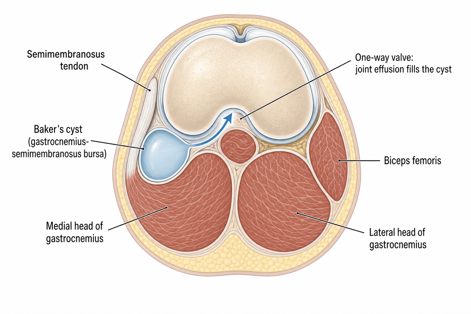
Popliteal fossa anatomy:
The popliteal fossa is a diamond-shaped space posterior to the knee bounded by:
- Superomedially: Semimembranosus and semitendinosus
- Superolaterally: Biceps femoris
- Inferomedially: Medial head of gastrocnemius
- Inferolaterally: Lateral head of gastrocnemius and plantaris
- Floor: Posterior capsule of knee, popliteus muscle, posterior femur and tibia
- Roof: Deep fascia
- Popliteal artery (deepest structure)
- Popliteal vein (superficial to artery)
- Tibial nerve (most superficial)
- Common peroneal nerve (lateral)
- Small saphenous vein (in superficial fascia)
- Popliteal lymph nodes
- Fat
- Located between medial head of gastrocnemius (anteriorly) and semimembranosus tendon (posteriorly)
- Positioned in posteromedial aspect of popliteal fossa
- A communication between the bursa and the knee joint may exist as a normal anatomical variant in some individuals, while in others the bursa arises primarily without a joint connection (Handy 2001, PMID 11590580)
The communication between the bursa and knee joint occurs at the posteromedial capsule, typically between the medial head of gastrocnemius and the joint capsule. A one-way valve mechanism develops, allowing fluid to flow from joint to bursa but not back.
- Flap of tissue acts as one-way valve
- Intra-articular pressure during knee motion forces fluid into bursa
- Bursal pressure cannot overcome valve to return fluid to joint
- Progressive accumulation and distension of bursa
- Can range from 1-2 cm to greater than 10 cm
- May extend proximally along gastrocnemius or semimembranosus
- May extend distally into posterior calf
- Can dissect between muscle planes
- Rarely can extend to ankle or even foot
- Tibial nerve is lateral to the cyst
- Popliteal vessels are deep and lateral to the cyst
- Common peroneal nerve is lateral (around fibular head)
- Large cysts can compress these structures
A basic-science distinction examiners like: a Baker's cyst is a true synovial cyst — its wall is lined by synovium (synovial cells) and it communicates with the joint, so the fluid it contains is genuine synovial fluid. This differs from a ganglion, which has no synovial (or epithelial) lining, contains thick mucinous/gelatinous material, and arises from myxoid degeneration of joint capsule or tendon sheath without a true synovial-lined wall. The practical relevance: it explains why a Baker's cyst reflects intra-articular disease (it is in continuity with the synovial cavity through the one-way valve) and why excised specimens are sent for histology — a synovial lining confirms the diagnosis, whereas atypical wall thickening or solid/nodular tissue should raise concern for synovial proliferative disease (PVNS) or, rarely, a cystic-appearing tumour.
MENISCUSMENISCUS - Common Associated Pathology
Hook:MENISCUS lists the common knee pathologies causing Baker's cyst
Classification
Classification by underlying cause:
- Description
- No underlying knee pathology
- Typical context
- More typical in children
- Clinical Note
- Often resolves spontaneously (paediatric prevalence ~6.3% on MRI; De Maeseneer 1999, PMID 10415188)
- Description
- Associated with intra-articular pathology
- Typical context
- The usual pattern in adults
- Clinical Note
- Requires treatment of underlying condition (older patients usually have coexistent joint pathology; Handy 2001, PMID 11590580)
Associated intra-articular pathology (independent MRI associations; Miller 1996, PMID 8816552):
- Joint effusion
- Meniscal tear
- Degenerative arthropathy (osteoarthritis)
- Inflammatory arthropathy and chronic synovitis (e.g. rheumatoid arthritis), PVNS and chondral injury are also recognised secondary causes
The probability of a cyst rises with the number of these co-existing features. The aetiology classification guides treatment - primary can be observed, secondary requires knee assessment.
Beyond aetiology and size, examiners expect the Rauschning-Lindgren clinical grading, which scores the cyst by symptoms and functional restriction rather than by imaging: Grade 0 — no symptoms; Grade I — slight swelling or fullness with no functional limitation; Grade II — swelling with mild pain and slight restriction of flexion; Grade III — marked swelling with significant pain and substantial restriction of movement. It is useful because it tracks the patient's actual disability and the response to treatment: higher grades (II-III) are the ones that tend to come to aspiration or surgery, whereas Grade 0-I cysts are typically observed while the underlying knee pathology is addressed.
Clinical Presentation and Assessment
History:
- Gradual onset of posterior knee fullness or mass
- Aching discomfort in popliteal fossa
- Worse with prolonged standing or activity
- Tightness or pressure sensation behind knee
- History of knee injury or arthritis
- If ruptured: Acute onset calf pain and swelling
Symptomatic presentation:
- Symptoms
- Painless or mild aching mass
- Key Features
- Gradual onset, worse with activity
- Symptoms
- Acute calf pain, swelling, ecchymosis
- Key Features
- Mimics DVT, crescent sign pathognomonic
- Symptoms
- Paresthesias, numbness in foot
- Key Features
- Tibial or peroneal nerve compression
- Symptoms
- Claudication, swelling
- Key Features
- Rare, popliteal vein or artery compression
Differential diagnosis of a popliteal fossa mass:
- Distinguishing features
- Soft, fluctuant, posteromedial, transilluminates, Foucher sign positive, communicates with joint
- Key investigation
- Ultrasound (anechoic cyst); MRI for joint pathology
- Distinguishing features
- Pulsatile, expansile mass, possible bruit; risk of distal embolisation/thrombosis
- Key investigation
- Duplex ultrasound / CT angiography - do NOT aspirate
- Distinguishing features
- Acute calf pain and swelling; the two can coexist
- Key investigation
- Doppler ultrasound (mandatory before treating either)
- Distinguishing features
- Firm, enlarging, deep mass; may be non-fluctuant; constitutional features
- Key investigation
- MRI with contrast; biopsy via specialist sarcoma unit
- Distinguishing features
- Semimembranosus or pes anserine bursa, peri-articular ganglion
- Key investigation
- Ultrasound / MRI
- Distinguishing features
- Soft, mobile, slow-growing, non-tender
- Key investigation
- Ultrasound / MRI (fat signal)
- Distinguishing features
- Firm nodes; consider infection or malignancy
- Key investigation
- Ultrasound; investigate underlying cause
- Distinguishing features
- Tinel-positive mass, neurological symptoms (tibial/peroneal)
- Key investigation
- MRI; specialist referral
A popliteal artery aneurysm can masquerade as a Baker's cyst. Always assess for pulsatility and a bruit, and confirm any suspicious mass with duplex ultrasound or CT angiography before considering aspiration. Aspirating an aneurysm risks catastrophic haemorrhage and limb-threatening ischaemia.
Physical examination
- Visible fullness in popliteal fossa (better seen in extension)
- Foucher sign: Cyst more prominent with knee in extension, less prominent in flexion
- Compare to contralateral side
- Look for ecchymosis if ruptured
- Soft, fluctuant mass in posteromedial popliteal fossa
- Non-tender (unless ruptured or infected)
- Smooth borders
- Can transilluminate (confirms fluid-filled structure)
- May be compressible
- Check for warmth (suggests inflammation or infection)
Foucher sign is pathognomonic for Baker's cyst. The cyst becomes more prominent and tense with knee extension (fluid pushed posteriorly) and less prominent with knee flexion (gastrocnemius relaxes, space increases). This distinguishes it from solid tumors.
- Usually full ROM
- May have mild flexion discomfort with large cysts
- Assess for signs of underlying knee pathology
- Transillumination: Positive (confirms fluid-filled cyst)
- Compression test: Gentle compression may reduce size temporarily
- Knee examination: Essential to identify underlying pathology
- McMurray test (meniscal tear)
- Joint line tenderness
- Lachman/anterior drawer (ACL)
- Varus/valgus stress (collateral ligaments)
- Crepitus (arthritis)
- Effusion
- Tibial nerve function (ankle plantar flexion, toe flexion, plantar sensation)
- Common peroneal nerve (ankle dorsiflexion, toe extension, first webspace sensation)
- Popliteal and pedal pulses
- Venous examination if suspect DVT
- Acute calf pain and swelling
- Crescent sign: Ecchymosis below medial malleolus (pathognomonic)
- Tenderness in calf
- May have positive Homan's sign (though non-specific)
- Must exclude DVT with Doppler ultrasound
FOUCHERFOUCHER - Clinical Sign
Hook:FOUCHER sign is the key clinical examination finding - cyst more prominent in extension
Investigations

Ultrasound is the first-line imaging modality.
- Non-invasive, no radiation
- Dynamic examination (test Foucher sign)
- Can assess for rupture (fluid tracking into calf)
- Can guide aspiration if needed
- Can assess popliteal vessels (exclude DVT, aneurysm)
- Relatively inexpensive
- Anechoic (fluid-filled) cystic structure
- Located between medial head of gastrocnemius and semimembranosus
- May show neck communicating with joint
- Can measure size accurately
- Doppler can exclude vascular abnormalities
- Limited assessment of intra-articular knee pathology
- Operator dependent
- May miss small meniscal tears
Ultrasound is excellent for confirming the diagnosis and excluding vascular pathology.
Aspiration:
- Can confirm diagnosis (clear, yellow synovial fluid)
- Send for cell count, culture, crystal analysis if concern for infection or inflammatory arthritis
- Provides temporary relief but high recurrence rate
- Not recommended as definitive treatment
Management Algorithm
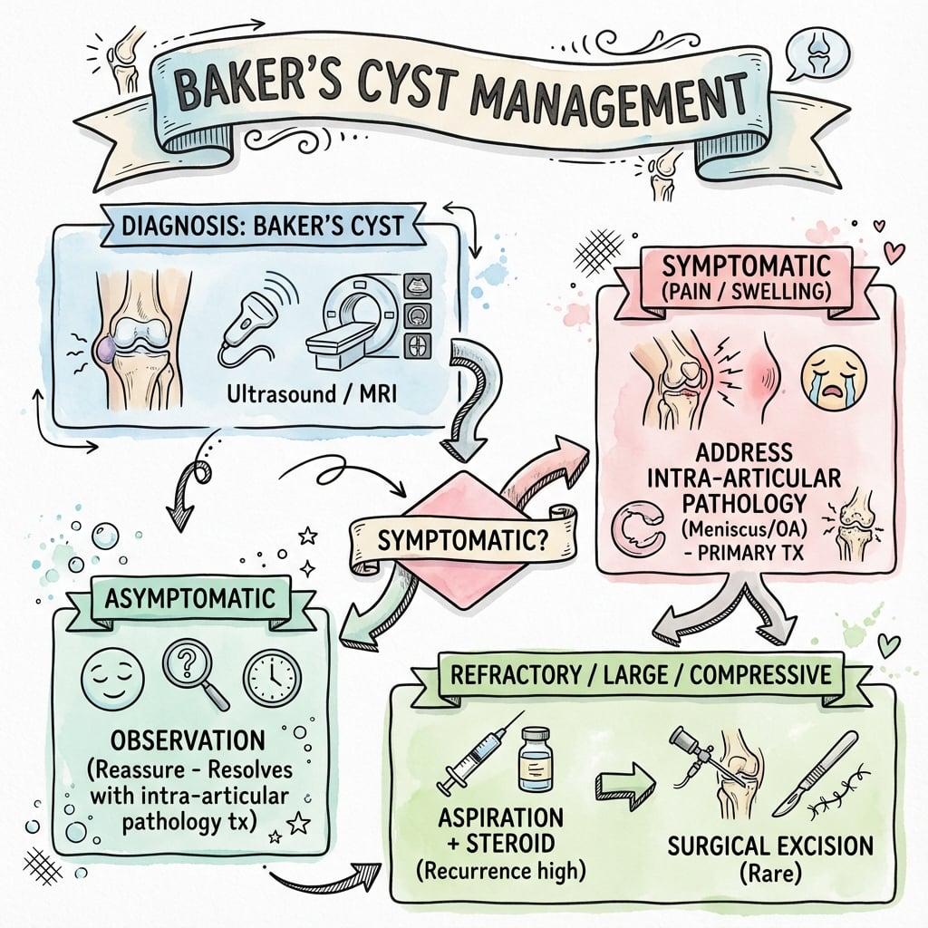
Conservative management is first-line for most Baker's cysts.
- Asymptomatic or minimally symptomatic cysts
- No complications (rupture, compression, infection)
- Patient preference after counseling
- Many cysts are asymptomatic
- May resolve spontaneously if underlying pathology treated
- Monitor size and symptoms
- Avoid activities that increase knee effusion
- Limit prolonged standing
- Low-impact exercise (swimming, cycling)
- Reduce inflammation and synovitis
- May decrease fluid production
- Standard anti-inflammatory doses
- Consider gastroprotection if prolonged use
- Quadriceps strengthening
- Range of motion exercises
- Proprioceptive training
- May help reduce effusion
- This is the most important aspect
- Meniscal tear: consider arthroscopic repair or meniscectomy
- ACL tear: reconstruction if indicated
- Osteoarthritis: optimize medical management, consider injections
- Inflammatory arthritis: optimize DMARD therapy
Simply treating the cyst is inadequate. The cyst is a manifestation of underlying knee pathology. Focus treatment on the intra-articular condition - the cyst often resolves once the effusion is controlled.
Surgical Technique
- Nerve compression with progressive neurological deficit
- Vascular compression with claudication or swelling
- Suspected malignancy (very rare)
- Failed conservative management (6+ months)
- Large cyst (greater than 10 cm) with functional impairment
- Recurrent ruptures
- Patient preference with realistic expectations
- Underlying knee pathology must be addressed (ideally same surgery)
- Patient counseled about recurrence risk
- Conservative measures exhausted
- No active infection
Isolated cyst excision without treating knee pathology is not recommended due to high recurrence.
Complications
- Incidence
- 10-15%
- Prevention/Management
- Exclude DVT with Doppler, treat conservatively
- Incidence
- Less than 5%
- Prevention/Management
- Large cysts - consider excision if symptomatic
- Incidence
- Less than 2%
- Prevention/Management
- Rare, may need urgent excision
- Incidence
- Less than 1%
- Prevention/Management
- Antibiotics, drainage, may need joint washout
- Incidence
- 5-10% of ruptured cysts
- Prevention/Management
- Always exclude with Doppler, anticoagulate if present
- Incidence
- 40-60% (isolated) 10% (combined)
- Prevention/Management
- Address underlying knee pathology
Surgical complications:
- Incidence
- Less than 2%
- Prevention
- Careful dissection, identify and protect nerve
- Incidence
- Less than 1%
- Prevention
- Understand anatomy, meticulous dissection
- Incidence
- 2-5%
- Prevention
- Sterile technique, careful closure, avoid hematoma
- Incidence
- Variable (see above)
- Prevention
- Ligate communication, address knee pathology
- Incidence
- 5-10%
- Prevention
- Early ROM exercises, physiotherapy
- Incidence
- Less than 2%
- Prevention
- DVT prophylaxis, early mobilization
Ruptured Baker's cyst presents identically to DVT with acute calf pain and swelling. The crescent sign (ecchymosis below the medial malleolus) is pathognomonic for ruptured cyst. However, DVT and ruptured cyst can coexist, so always perform Doppler ultrasound to exclude DVT.
Rare complications:
- Compartment syndrome (very rare, from massive dissection)
- Mass effect causing foot drop (peroneal nerve compression)
- Popliteal artery thrombosis (very rare)
Postoperative Care
Post-surgical management following Baker's cyst excision:
- Posterior splint or knee brace at 30 degrees flexion
- Elevation to minimize swelling
- Ice for comfort and inflammation control
- DVT prophylaxis (mechanical compression, pharmacologic based on risk)
- Multimodal pain control (oral analgesics, ice, elevation)
- Wound check at 2 weeks (suture removal if non-absorbable)
- Begin gentle ROM exercises
- Weight bearing as tolerated with crutches for support
- Continue elevation when resting
- Monitor for signs of infection or DVT
- Progress ROM exercises (goal: full range by 6 weeks)
- Begin gentle strengthening of quadriceps and hamstrings
- Wean off crutches as comfort allows
- Return to desk work at 2-3 weeks typically
- Continue physiotherapy
- Full activities as tolerated
- Return to sports at 8-12 weeks (depending on sport demands and underlying knee pathology)
- Follow-up MRI if recurrence suspected or symptoms persist
- Gradual return to high-demand activities
- 2 weeks: Wound check, ensure healing
- 6 weeks: Assess ROM, clinical examination
- 3 months: Ensure resolution, no recurrence
- As needed: If symptoms recur or concerns arise
- Continue treatment of underlying knee pathology
- Report any return of posterior knee swelling
- Maintain healthy knee function (strengthening, weight management)
- Address underlying arthritis or meniscal issues
Outcomes and Prognosis
- Many remain stable or resolve spontaneously
- 30-40% increase in size over time
- 10-15% rupture at some point
- Symptoms vary - many tolerate well
- 80-90% have symptom improvement if underlying knee pathology addressed
- Cyst size may reduce or remain stable
- Recurrence common if pathology not treated
- 50-80% recurrence if underlying pathology untreated
- 20-30% recurrence if knee pathology treated
- Symptom relief usually temporary (weeks to months)
- Recurrence Rate
- 40-60%
- Outcomes
- High recurrence, not recommended
- Recurrence Rate
- 60-70% resolution
- Outcomes
- Many cysts resolve without direct excision
- Recurrence Rate
- Less than 10%
- Outcomes
- Best outcomes, recommended if surgery needed
The prognosis of Baker's cyst depends almost entirely on treatment of the underlying knee pathology. The cyst is a symptom, not the disease. Treat the meniscal tear, arthritis, or synovitis, and the cyst usually resolves or becomes asymptomatic.
- 85-90% good to excellent symptom relief
- Return to full activity at 8-12 weeks
- Low complication rate with experienced surgeon
- Recurrence risk low if knee pathology addressed
- Underlying pathology - treatable causes (meniscal tear) better than degenerative (advanced OA)
- Size - smaller cysts respond better to conservative treatment
- Duration - longstanding cysts more likely to need surgery
- Age - younger patients may be more active and symptomatic
- Treatment of knee - single most important factor
- Primary cysts (no underlying pathology) almost always resolve spontaneously
- Observation is appropriate
- Rarely need any intervention
Guidelines, Registries & Global Practice
Global epidemiology (PubMed-sourced):
- Evidence
- Narrative review
- Source
- Handy, Semin Arthritis Rheum 2001 (PMID 11590580)
- Evidence
- Retrospective cohort
- Source
- Miller et al, Radiology 1996 (PMID 8816552)
- Evidence
- Retrospective cohort
- Source
- De Maeseneer et al, Pediatr Radiol 1999 (PMID 10415188)
- Evidence
- Case-control (210 patients)
- Source
- Han et al, Eur Radiol 2019 (PMID 31811432)
Guidelines and practice variation:
There is no dedicated international society guideline (AAOS, NICE, BOA-BOAST, AO or EFORT) specific to Baker's (popliteal) cyst; management is guided by the evidence above and by guidelines for the underlying knee disorder (e.g. osteoarthritis, meniscal pathology, inflammatory arthritis). Likewise, popliteal cyst is a soft-tissue condition rather than an implant procedure, so it is not tracked by arthroplasty registries (NJR, AJRR, AOANJRR, SHAR, NZJR). Globally consistent principles are:
- Ultrasound first, MRI for the joint - sonography is the imaging method of choice; MRI characterises associated intra-articular pathology (Handy 2001, PMID 11590580; Miller 1996, PMID 8816552).
- Treat the cause, not the cyst - because adult cysts are usually secondary, recurrence is high after isolated excision (~63%; Rauschning & Lindgren 1979, PMID 525326) and far lower when the joint pathology and valve are corrected arthroscopically (~95% good/optimal; Sansone & De Ponti 1999, PMID 10355711).
- Image-guided aspiration/injection is a recognised, lower-morbidity option for symptomatic cysts (Smith et al 2015, PMID 26502415).
- Children differ - paediatric cysts are usually primary and self-limiting, so observation is the default (De Maeseneer 1999, PMID 10415188).
Practice variation: first contact and access to ultrasound/MRI differ by health system (primary-care vs direct specialist referral; publicly funded vs insurance-based imaging), but the diagnostic and treatment hierarchy above is broadly uniform across high-income settings.
Be prepared to discuss the differential diagnosis of popliteal fossa masses (Baker's cyst, popliteal aneurysm, soft-tissue tumour, lymphadenopathy). Know the one-way valve mechanism of communication with the joint. Understand that treatment focuses on the underlying knee pathology, not the cyst itself, and that there is no implant registry or single society guideline for this condition.
MCQ Practice Points
Q: Where is a Baker's cyst located anatomically? A: Between the medial head of gastrocnemius (anteriorly) and the semimembranosus tendon (posteriorly) in the posteromedial popliteal fossa. It represents distension of the gastrocnemius-semimembranosus bursa that communicates with the knee joint.
Q: What percentage of Baker's cysts in adults are associated with underlying intra-articular knee pathology? A: 95% of Baker's cysts in adults are secondary to intra-articular pathology (meniscal tears, OA, inflammatory arthritis). Only 5% are primary. This is opposite to children, where most are primary.
Q: What is Foucher sign? A: The Baker's cyst becomes more prominent and tense with knee extension and less prominent with knee flexion. This occurs because extension pushes fluid posteriorly and tightens the gastrocnemius, while flexion relaxes the muscle and increases space.
Q: What is pseudothrombophlebitis syndrome and what is the pathognomonic sign? A: Pseudothrombophlebitis is acute calf pain and swelling from ruptured Baker's cyst that mimics DVT clinically. The crescent sign (ecchymosis below the medial malleolus) is pathognomonic. DVT must be excluded with Doppler as they can coexist.
Q: What is the recurrence rate after isolated Baker's cyst excision without treating underlying knee pathology? A: High recurrence after isolated cyst excision - a recurrent cyst was found in 63% of knees on follow-up arthrography in Rauschning's classic series (1979). Recurrence falls substantially when the underlying knee pathology and the one-way valve are addressed arthroscopically (optimal/good results in ~95%; Sansone & De Ponti, 1999). The cyst is a symptom, not the disease.
Q: What is the first-line imaging modality for suspected Baker's cyst? A: Ultrasound - confirms fluid-filled cyst, can assess size, exclude vascular pathology, and guide aspiration if needed. MRI is the gold standard for identifying underlying knee pathology and surgical planning.
Clinical Decision Scenarios
Practise clinical reasoning and management decisions out loud
“A 52-year-old woman presents with a painless lump behind her knee that has been gradually increasing in size over 6 months. She has a history of medial knee pain for the past year. On examination, you palpate a soft, fluctuant 4 cm mass in the posteromedial popliteal fossa that becomes more prominent when the knee is extended. What is your diagnosis and management?”
“A 45-year-old man presents to ED with acute onset right calf pain and swelling that started suddenly yesterday. He has a history of right knee pain and had noticed a lump behind his knee previously. On examination, there is calf swelling, tenderness, and you notice some bruising around his medial ankle. The ED team are concerned about DVT. How would you assess and manage?”
“A 55-year-old man with rheumatoid arthritis has a large Baker's cyst that has been present for 18 months. He has tried NSAIDs, had two aspirations (recurred within weeks both times), and is now developing numbness in his foot. MRI shows an 8 cm multiloculated cyst compressing the tibial nerve and some posterior horn medial meniscus degeneration. His rheumatologist has optimized his RA medications. He asks about surgery. What is your approach?”
KEY ANATOMY
- Distension of gastrocnemius-semimembranosus bursa
- Location: posteromedial popliteal fossa
- Between medial head of gastrocnemius and semimembranosus tendon
- Communicates with knee joint via one-way valve (posteromedial capsule)
- Tibial nerve lateral to cyst, popliteal vessels deep and lateral
PATHOPHYSIOLOGY
- 95% secondary to intra-articular knee pathology in adults
- Causes: meniscal tear (40%), OA (30%), RA (15%), ACL tear (10%)
- One-way valve allows fluid to enter bursa but not exit
- Progressive distension from chronic knee effusion
- Primary cysts rare in adults (common in children)
CLINICAL FEATURES
- Gradual onset painless or aching mass behind knee
- Foucher sign: prominent with extension, less with flexion
- Soft, fluctuant, transilluminates
- May have symptoms of underlying knee pathology
- Complications: rupture (10%), nerve compression (less than 5%)
INVESTIGATIONS
- First-line: Ultrasound (confirms cyst, excludes vascular)
- Gold standard: MRI (identifies underlying knee pathology)
- If ruptured: Doppler ultrasound to exclude DVT (mandatory)
- Plain X-rays limited (assess for OA)
- Aspiration: clear yellow synovial fluid
MANAGEMENT
- Conservative first-line: NSAIDs, activity modification, treat knee
- Aspiration ± steroid: temporary relief, 50-80% recurrence
- Surgery indications: nerve/vascular compression, failed conservative
- Isolated excision: 40-60% recurrence
- Combined (arthroscopy + excision): less than 10% recurrence
- Ruptured cyst: exclude DVT, rest, ice, elevation, NSAIDs
SURGICAL TECHNIQUE
- Stage 1: Arthroscopy (treat meniscal tear, synovectomy, loose bodies)
- Stage 2: Posterior approach prone position
- Identify and protect tibial nerve (lateral to cyst)
- Excise cyst completely
- Ligate communication with joint (prevent recurrence)
- Careful hemostasis and layered closure
PEARLS AND TRAPS
- Treat underlying knee pathology, not just cyst (key principle)
- Foucher sign is pathognomonic (prominent in extension)
- Crescent sign (ecchymosis at ankle) = ruptured cyst
- Always exclude DVT in suspected rupture (Doppler)
- Isolated cyst excision not recommended (high recurrence)
- Tibial nerve most at risk during surgery
Evidence Base
- Depending on population and imaging technique, popliteal cysts are seen in approximately 5-32% of knees imaged, with two age peaks (4-7 years and 35-70 years).
- In older patients there is usually coexistent intra-articular joint pathology; pathogenesis depends on a valve-like joint-bursa communication allowing one-way passage of fluid.
- Sonography is the imaging method of choice; cysts extending or rupturing into the calf mimic phlebitis. Most symptomatic cysts respond to intra-articular corticosteroid injection and surgical excision is rarely necessary.
- Review of 400 knee MRI examinations showed significant associations between Baker cyst and joint effusion, meniscal tear, and degenerative arthropathy, each independent of one another.
- No association was found with ACL or medial collateral ligament injury.
- Probability of a Baker cyst rose with the number of co-existing features: approximately 0.08-0.10 with one feature, 0.19-0.21 with two, and 0.38 with all three.
- 40 patients re-examined a mean of 4 years (range 6 months to 15 years) after popliteal cyst excision.
- A recurrent cyst was found in 63% of knees on follow-up arthrography, yet most patients had fewer symptoms than before operation.
- Authors concluded Baker's cysts are usually secondary and should be treated as a manifestation of the underlying joint disorder; isolated excision with tight closure of the communication should be reserved for incurable knee disease with troublesome symptoms.
- 30 adults treated arthroscopically by addressing the intra-articular pathology and correcting the valvular mechanism, with mean follow-up of 32 months.
- A connection between the joint space and the cyst was found in every case, and the popliteal cyst was almost invariably associated with other knee disorders.
- Optimal or good clinical results were achieved in 95% of patients.
- 47 patients had ultrasound-guided aspiration, fenestration, and triamcinolone/bupivacaine injection (UGAFI) as sole treatment.
- Mean WOMAC score improved significantly from 48.55 to 17.15 (p less than 0.0001), with significant gains in pain, stiffness, and physical function.
- 6 patients (12.7%) required re-aspiration for recurrence; there were no infections or other complications.
- Ruptured Baker's cyst is easily misdiagnosed as deep vein thrombosis.
- The crescent sign - ecchymosis appearing around the malleoli/distal calf as synovial fluid dissects distally - helps identify a ruptured cyst.
- Recognising this sign supports rapid, correct diagnosis and avoids unnecessary anticoagulation, though DVT must still be excluded.