Bending Stress | Neutral Axis | Working Length | Plate Mechanics
- Bending moment creates linear stress distribution from maximum tension to maximum compression
- Neutral axis at centroid has zero bending stress - material here does not resist bending
- Stress proportional to distance from neutral axis: σ = My/I where y is distance from neutral axis
- Working length (distance between screws) affects stiffness as L³ - doubling length reduces stiffness 8-fold
- Plate should be placed on tension side for optimal biomechanics
- “Stiffness inversely proportional to working length cubed - critical for bridge plating
- “Section modulus (I/c) determines bending strength - increases with height squared
- “Plate on tension side prevents gap formation and reduces stress shielding
- “Eccentric loading increases bending moment - explains failure in varus/valgus malalignment
Overview and Fundamentals
Bending moment distribution is fundamental to understanding fracture fixation mechanics, and most long bone fractures experience bending loads during physiologic loading. When a long bone or plate is loaded in bending, internal stresses develop that vary linearly from maximum tension on one surface to maximum compression on the opposite surface; the neutral axis, at the centroid of the cross-section, experiences zero bending stress. Understanding that distribution is essential for bridge plating, plate selection and analysing construct failure, because it explains:
- why plates should be on the tension side
- how working length affects construct stiffness
- why eccentrically loaded fractures fail in predictable patterns
- how to optimise screw spacing for biological fixation
The flexure formula. The bending stress at any point is σ = My/I, where M is the bending moment (N·m or N·mm), y is the perpendicular distance from the neutral axis and I is the second moment of area. Maximum stress occurs at the outer fibres, where y is greatest (y = c), giving σ_max = Mc/I. The ratio I/c is called the section modulus and determines bending strength.
Pure and combined bending. In pure bending only a moment acts: the stress distribution is linear and the neutral axis sits at the centroid. Add an axial load and the axial stress shifts the neutral axis, so the distribution becomes asymmetric. Most fractures experience this combined loading, because eccentric axial loads create bending moments (varus/valgus stress), and three-point bending is the most common loading mode in long bones.
Neutral Axis and Stress Distribution
Where it lies. The neutral axis passes through the centroid of the cross-section and is perpendicular to the plane of bending. For symmetric sections (circular, square, I-beam) that is the geometric centre. For a composite section such as plate plus bone, it shifts according to the relative stiffness and area of the two materials.
- Neutral Axis Location
- Centre
- Section Modulus (I/c)
- πd³/32
- Clinical Example
- Intact long bone, IM nail
- Neutral Axis Location
- Centre
- Section Modulus (I/c)
- π(D⁴-d⁴)/32D
- Clinical Example
- Cortical bone, hollow nail
- Neutral Axis Location
- Centre
- Section Modulus (I/c)
- bh²/6
- Clinical Example
- Compression plate
- Neutral Axis Location
- Shifts toward stiffer material
- Section Modulus (I/c)
- Complex calculation
- Clinical Example
- Fixed fracture
The distribution. Stress increases linearly with distance from the neutral axis: maximum tensile stress on one surface, maximum compressive stress on the opposite surface, and σ = 0 at the axis itself, where the material makes no contribution to bending resistance. The sign changes across the neutral axis, from tension to compression, and because the distribution is linear the gradient is constant, dσ/dy = M/I.
When plate and bone share load the neutral axis shifts toward the stiffer material, the steel plate. Bone near the plate then experiences less stress, which is stress shielding, while bone far from the plate experiences more. Proper working length balances that load sharing to promote healing while preventing excessive motion.
Working Length in Fracture Fixation
Working length can be approached from two directions. Beam theory, below, derives it from the mechanics of the loaded plate; implant and fracture biomechanics approaches the same quantity from the construct side — implant material, screw configuration and failure modes. The underlying material behaviour is in stress, strain and modulus, and the lever mechanics in moment arms and levers.
Definition. Working length is the distance between the two innermost screws on either side of a fracture: the holes actually occupied, not the distance between screw holes. Empty holes within the working length are stress concentration points that reduce fatigue life and should be avoided. For a comminuted fracture the plate bridges the entire comminution zone, so the working length runs from the end of the comminution to the first screw. It determines construct stiffness and with it fracture healing, stress shielding and the risk of implant failure.
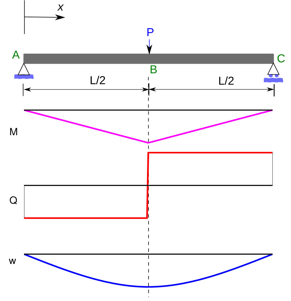
The beam law. Under three-point bending the deflection is δ = FL³/48EI, where F is the load, L the working length, E the elastic modulus and I the area moment of inertia. Stiffness k = F/δ is therefore proportional to 1/L³: doubling the working length reduces stiffness 8-fold, and halving it increases stiffness 8-fold. The other terms matter as well: the modulus E of the plate material (titanium, steel and carbon fibre differ), the plate cross-section I (width × thickness³/12 for a rectangle), and the number and spacing of the screws, which affect load transfer.
- Stiffness
- Very high
- Stress Shielding
- Maximum
- Fracture Motion
- Minimal
- Clinical Application
- Simple fractures, absolute stability
- Stiffness
- Moderate
- Stress Shielding
- Moderate
- Fracture Motion
- Moderate
- Clinical Application
- Most fractures, relative stability
- Stiffness
- Low
- Stress Shielding
- Minimal
- Fracture Motion
- Excessive
- Clinical Application
- Risk of delayed union, implant failure
- Stiffness
- Balanced
- Stress Shielding
- Balanced
- Fracture Motion
- Micromotion only
- Clinical Application
- Modern locking plate technique
Too short. A stiff construct shields the bone excessively and reduces the strain at the fracture site, which may impair healing. All the load passes through the implant, a fatigue risk if the fracture does not heal, and the stress at the screw-bone interface is higher.
Too long. A flexible construct allows excessive fracture motion, which may prevent healing. Implant stress rises, because the bending moment is proportional to L; a cantilever beam effect develops if the fracture does not heal, and the implant may fail before union.
Optimal. For a simple fracture, 3-5 cortices (1.5-2.5 screw holes) on each side; longer for a comminuted fracture. The aim is enough stiffness for healing while still allowing micromotion.
The current principle is fewer screws and a longer working length for biological fixation. The traditional AO teaching of six cortices per fragment is too stiff; modern bridge plating uses 3-4 screws per fragment, with the working length chosen for appropriate flexibility. The exception is the periarticular fracture, which needs a short working length for a stable articular reduction.
Working length and construct flexibility matter because they set the interfragmentary strain — the biological determinant of how a fracture heals. Strain = (change in gap length) / (original gap length). A tissue can only bridge a gap when the local strain is below its tolerance: lamellar/cortical bone tolerates under about 2%, woven bone and callus up to about 10%, and granulation/fibrous tissue more than about 10% (beyond which a nonunion persists). The clinical paradox follows directly: a tiny gap with even small motion generates very high strain and will not heal in a too-flexible construct — so simple fractures need absolute stability (anatomic reduction plus compression, which lowers strain by eliminating the gap). A comminuted fracture distributes the same total motion over many gaps, so the strain at each is low and relative stability (flexible bridge plating, longer working length) promotes abundant callus. This is why an over-stiff construct across a single small gap, and an over-flexible construct across a simple fracture, both fail to heal.
Plate Placement and Tension Band Principle
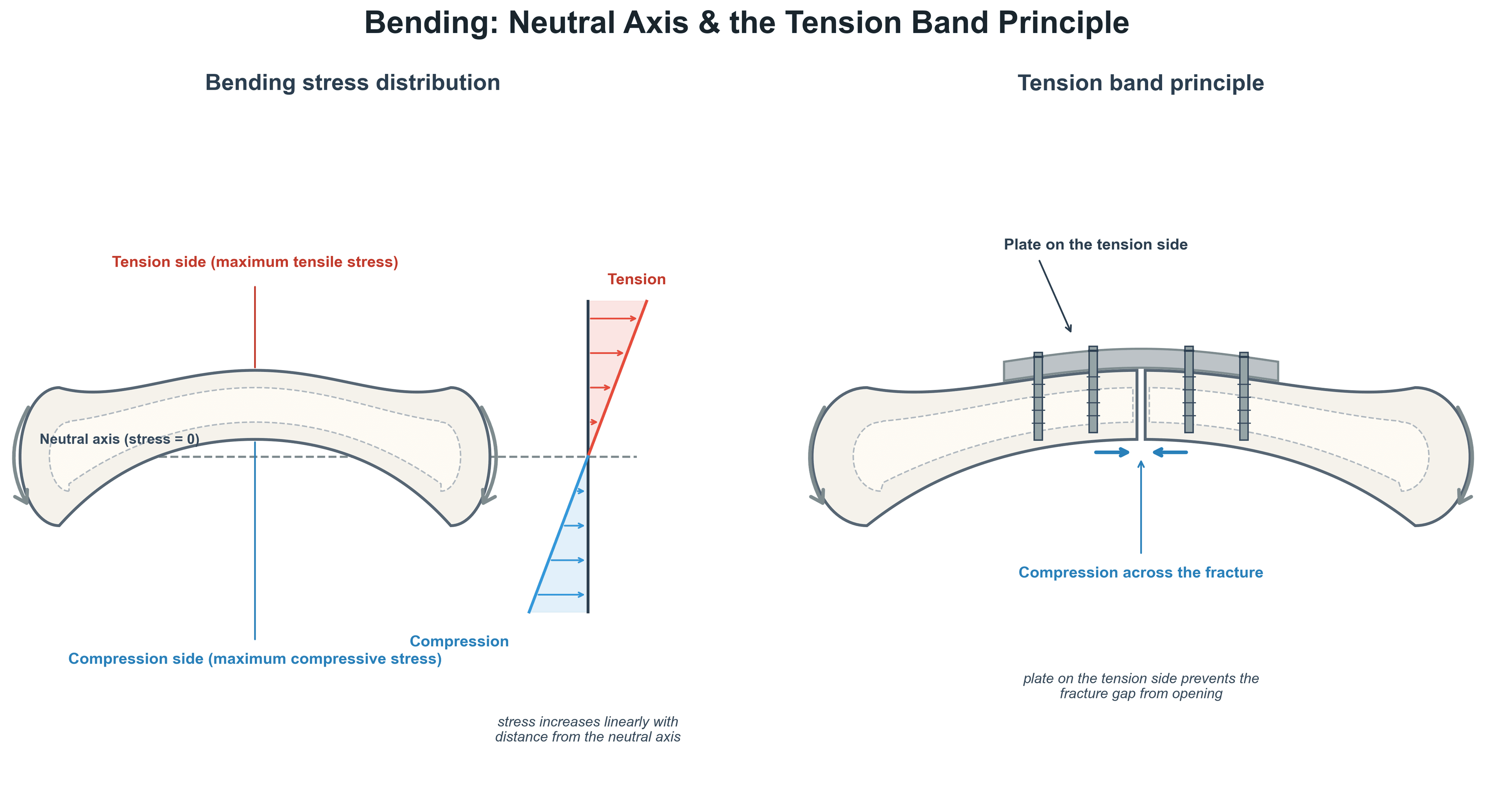
Tension side. Plates should ideally sit on the tension surface of the bone, a principle that follows from the bending moment distribution and composite beam behaviour. The plate resists the tensile stress, so no gap forms on the tension surface, and it creates compression across the fracture (a pre-loading effect). The compression side of the bone tolerates contact and load sharing, and can gap without loss of stability; composite action with the bone reduces the plate stress, and interfragmentary motion is minimised.
Compression side. A plate on the compression side lets the tension surface gap, uncontrolled. The plate carries a higher bending stress, the construct is less stable, and the gap risks delayed union or nonunion.
- Physiologic Tension Surface
- Lateral (tension band of IT band)
- Optimal Plate Position
- Lateral
- Rationale
- Resist varus moment from body weight
- Physiologic Tension Surface
- Anterior or medial
- Optimal Plate Position
- Anteromedial
- Rationale
- Subcutaneous position, resist anterior bow
- Physiologic Tension Surface
- Anterior with forward flexion
- Optimal Plate Position
- Anterolateral
- Rationale
- Accessible, resist AP bending
- Physiologic Tension Surface
- Variable with rotation
- Optimal Plate Position
- Dorsal radius, volar ulna
- Rationale
- Based on anatomy and soft tissue
Eccentric loading. An axial load applied eccentric to the bone's mechanical axis creates a bending moment, M = P × e, where P is the axial force and e is the eccentricity, the perpendicular distance from the load line to the neutral axis.
- A varus knee: medial compartment overload creates a varus bending moment
- The hip joint reaction force, medial to the femoral shaft, creates a varus bending moment in the proximal femur
- The patellar tendon, pulling anterior to the tibial axis, creates an anterior bending moment
This is why a varus malunion of a femoral fracture leads to increased implant stress and failure, why proper alignment is critical to minimise bending moments, and why a plate should resist the direction of bending expected from eccentric loading.
The lateral plate on a distal femur fracture. The hip joint reaction force creates a varus bending moment: with body weight medial to the femoral shaft, the lateral cortex is in tension. A lateral plate resists that tensile stress, prevents a lateral gap and creates medial compression across the fracture. A medial plate would allow a lateral gap and carry higher plate stress.
Beam Theory and Implant Design
Second moment of area. The second moment of area, I, quantifies a cross-section's resistance to bending. It depends on how the material is distributed relative to the neutral axis: material far from the axis contributes more, through the y² term. For a rectangular section I = bh³/12 (b the width, h the height perpendicular to the bending axis), so I is proportional to h³. For a circular section, I = πd⁴/64 when solid and π(D⁴ - d⁴)/64 when hollow.
Hollow tubes. Material near the neutral axis contributes little, so it can be removed while I is maintained by preserving the outer diameter. That is why I-beams and hollow tubes are efficient, with the material concentrated at the extremes, and why a hollow tube is as efficient as a solid rod of the same I/c. It explains the efficiency of cortical bone and the hollow design of intramedullary nails, and it is the reason trabecular bone in the marrow cavity does not resist bending.
Plate thickness. For a plate of width b and thickness h, c = h/2, so the section modulus is I/c = bh²/6 and the maximum bending stress is σ_max = 6M/bh². Doubling the thickness increases I 8-fold (the h³ relationship), increases the section modulus 4-fold (the h² relationship), reduces the stress for the same bending moment 4-fold, and increases stiffness 8-fold. A plate twice as thick is therefore more than twice as strong: a small increase in thickness dramatically improves bending strength and stiffness, though it may increase stress shielding. Modern locked plates can be thinner because they transfer load by a different mechanism, angle stability rather than friction.
A conventional plate transfers load by friction, the plate compressed onto the bone: bending creates the friction force at the plate-bone interface, and the plate requires precise contouring. A locking plate transfers load through the screw-plate construct, which acts as an internal-external fixator and depends less on plate-bone contact. It can use a longer working length and maintain bending stability with fewer screws, which is what allows biological fixation principles.
Bending mechanics predict the dominant mode of implant failure: fatigue. An implant cyclically loaded well below its ultimate strength can still fail after enough cycles, because microcracks initiate at stress concentrations (notably the screw holes, where bending stress peaks) and propagate. The relationship between cyclic stress amplitude and cycles-to-failure is the S-N (Wöhler) curve. Some materials have an endurance (fatigue) limit — a stress below which they survive effectively infinite cycles: stainless steel and titanium alloys have a practical endurance limit, whereas aluminium does not (it always eventually fails). Clinically this sets up a race between fracture union and implant fatigue failure: every load cycle the bone has not yet healed consumes fatigue life, which is why a long-standing nonunion eventually breaks the plate through a screw hole. Lowering the cyclic stress in the implant — load sharing with reduced bone, avoiding empty holes at the fracture, and an appropriate (not excessive) working length — buys fatigue life until the bone heals.
Guidelines, Registries & Global Practice
Bending mechanics is a principle, not a disease, so it is governed by educational and technical standards from the major trauma organisations rather than by NICE/AAOS clinical practice guidelines. The dominant global framework is the AO/OTA synthesis of absolute versus relative stability, with broadly concordant teaching from EFORT (Europe), the British Orthopaedic Association / BOAST standards (UK), the Orthopaedic Trauma Association (North America) and the AOA (Australia). There is little controversy about the underlying physics; practice variation lies in how aggressively surgeons reduce construct stiffness (working length, plate material, far cortical locking) to optimise secondary healing, especially in distal femur fractures.
- Region
- Global
- Position on Bending & Working Length
- Absolute stability (lag screw + compression plate, short working length) for simple/intra-articular; relative stability (bridge plate, longer working length, minimal contact) for comminuted diaphyseal fractures
- Evidence basis
- Expert consensus + biomechanical (Perren, Stoffel)
- Region
- North America
- Position on Bending & Working Length
- Endorses bridge plating with adequate plate span and limited screw density for comminuted shaft fractures
- Evidence basis
- Consensus + cohort/biomechanical
- Region
- UK
- Position on Bending & Working Length
- Stable fixation appropriate to fracture personality; supports biological/relative-stability constructs for comminution
- Evidence basis
- Standard of care guidance
- Region
- Europe
- Position on Bending & Working Length
- Same absolute-vs-relative-stability paradigm; emphasises stiffness modulation to avoid asymmetric callus
- Evidence basis
- Educational consensus
- Region
- Australia / NZ
- Position on Bending & Working Length
- Beam theory, section modulus and working length are core basic-science viva content
- Evidence basis
- Curriculum standard
Global Practice and Variation
The flexure formula (σ = My/I), the L³ working-length relationship and tension-side plating are universal and uncontested. Where practice genuinely varies is stiffness modulation:
- Plate material: Titanium (lower modulus, E ≈ 110 GPa) deflects more than stainless steel (E ≈ 200 GPa) for the same geometry; titanium plating produced 56–76% more callus than steel in distal femur fractures (Lujan 2010).
- Working-length doctrine: Modern teaching favours fewer screws and a longer working length over the older "six cortices per fragment" rule, but periarticular and simple fractures still demand short working lengths and absolute stability.
- Stiffness-reduction devices: Far cortical locking and active/dynamic plates are used selectively (more in North America/Europe) to add controlled axial micromotion (Doornink 2011).
Distal femur locking-plate fixation is the canonical real-world example where over-stiff constructs cause asymmetric callus and nonunion — the strongest signal that bending mechanics is clinically, not just theoretically, important.
MCQ Practice Points
Q: What does the flexure formula σ = My/I represent? A: Bending stress at distance y from neutral axis. M is bending moment, y is perpendicular distance from neutral axis, I is second moment of area. Stress is linear across section, maximum at outer fibers (y = c).
Q: How does doubling the thickness of a plate affect its bending strength? A: Increases strength 4-fold. Section modulus I/c = bh²/6 for rectangular section. Doubling h increases section modulus by factor of 4, thus maximum stress σ_max = M/(I/c) decreases 4-fold for same moment.
Q: If you double the working length in a plated fracture, how does construct stiffness change? A: Stiffness decreases 8-fold. From beam theory, stiffness is inversely proportional to working length cubed: k ∝ 1/L³. Doubling L means new stiffness is 1/(2L)³ = 1/8 of original stiffness.
Q: Why is material at the neutral axis not effective at resisting bending? A: Stress at neutral axis is zero. From σ = My/I, when y = 0 (at neutral axis), σ = 0. Material contributes to bending resistance proportional to its distance from neutral axis. This explains efficiency of hollow tubes.
Q: Why should plates be placed on the tension side of bone? A: Prevents gap formation and reduces plate stress. Plate directly resists tensile stress, creating compression across fracture. Allows load sharing with bone. Plate on compression side would allow gap on tension surface and experience higher stress.
Q: How does varus malalignment affect bending moment in a femoral fracture? A: Increases bending moment via eccentric loading. M = P × e where e is eccentricity. Varus shifts load line medial, increasing distance to lateral cortex, thus increasing varus bending moment and lateral cortex tensile stress.
Exam Viva Scenarios
Practise clinical reasoning and management decisions out loud
“Examiner shows cross-section of a long bone and asks: Explain the stress distribution when this bone is loaded in bending. What is the neutral axis and why is it important?”
“You are treating a comminuted mid-diaphyseal femur fracture with a locked plate. Explain how you would determine the optimal working length and why it matters for fracture healing and construct stability.”
“You are treating a distal femur fracture with lateral locked plate. The examiner asks: Why do we place the plate laterally? What would happen if you placed it medially?”
Bending Stress Fundamentals
- Bending creates LINEAR stress distribution from max tension to max compression
- Flexure formula: σ = My/I (M = moment, y = distance from neutral axis, I = area moment)
- Maximum stress at outer fibers: σ_max = Mc/I where c is distance to extreme fiber
- Neutral axis at centroid: zero stress, no contribution to bending resistance
Section Modulus and Strength
- Section modulus I/c determines maximum bending stress for given moment
- For rectangle: I/c = bh²/6 - proportional to height squared
- Doubling plate thickness increases strength 4x (h² effect) and stiffness 8x (h³ effect)
- Hollow tubes efficient: material at neutral axis removed, outer fibers preserved
Working Length Principles
- Working length = distance between innermost screws on opposite sides of fracture
- Stiffness ∝ 1/L³ - doubling length reduces stiffness 8-fold
- Optimal: 3-5 cortices (1.5-2.5 holes) per fragment for relative stability
- Too short = stress shielding; too long = excessive motion and implant stress
Plate Positioning
- Plate on TENSION side for optimal biomechanics
- Prevents gap formation, creates compression, reduces plate stress
- Femur: lateral plate (varus moment from medial hip reaction force)
- Tibia: anteromedial plate (resist anterior bow, subcutaneous access)
Clinical Applications
- Eccentric loading creates bending moment: M = P × e
- Varus/valgus malalignment increases bending stress and implant failure risk
- Empty screw holes within working length = stress risers (avoid)
- Locked plates allow longer working length than conventional (angle stability)
Key Numbers to Remember
- I/c for rectangle = bh²/6 (width × height squared / 6)
- Deflection ∝ L³ (length cubed relationship)
- Area moment ∝ h³ (height cubed for rectangular section)
- Modern bridge plating: 3-4 screws per fragment (vs old rule of 6 cortices)
Evidence Base
Working Length Controls Stability of the Locked Internal Fixator
- Working length (distance of the first screw to the fracture) was the dominant determinant of both axial and torsional rigidity of the LCP construct
- Omitting one screw hole on either side of the fracture made the construct almost twice as flexible in both compression and torsion
- More than three screws per fragment added little axial stiffness, and four screws did not increase torsional rigidity
- Plate failures invariably occurred through a screw hole where finite element analysis showed peak von Mises stress; greater working length lowered the load to plastic deformation when bone contact was absent
Screw–Bone Interface Modelling and Local Stress Around Locked Screws (FEA)
- In a locked-plate tibial mid-shaft fracture model, the global load–deformation response varied less than 1% between interface modelling strategies
- Interface modelling markedly altered the LOCAL stress–strain field around the screws — the principal determinant of screw loosening, bone damage and stress shielding
- Frictional and tied (bonded) interfaces gave similar peak strains (under 5% difference); the undersized pilot-hole pre-stress model produced the largest peri-screw strains
- Demonstrates that bending stress concentrates at the screw–bone interface, the typical site of construct failure
Scientific Basis of Biological Internal Fixation: Stability vs Biology
- The internal fixator splints rather than compresses; flexible stabilisation induces callus and reliable healing without anatomical reduction (as shown by locked nailing)
- The strain theory defines the tolerable instability — gross instability may still heal by callus, whereas minimal motion across a rigidly fixed small gap can be deleterious
- Extensive plate–bone contact damages periosteal blood supply, causing necrosis and temporary porosity (a biological, not mechanical, cause of so-called stress protection)
- Locked, threaded bolts allow minimally invasive percutaneous osteosynthesis (MIPO) without contouring the splint to the bone
Interfragmentary Strain Governs Healing: Gap Size and Movement
- In a sheep metatarsal osteotomy with an adjustable external fixator, larger interfragmentary movement/strain (≈31% vs ≈7%) stimulated more callus in small gaps (1–2 mm) but not in 6 mm gaps
- Increasing gap size from 1 to 6 mm significantly reduced the bending stiffness of the healed bone
- Healing was inferior once the gap exceeded 2 mm
- Provides the experimental basis for the interfragmentary-strain concept that links construct flexibility, gap size and callus formation
Locked Plating of Distal Femur: Stiffness, Callus and Plate Material
- Across 64 distal femur fractures, deficient periosteal callus (≤20 mm²) was present in 52%, 47% and 37% of fractures at 6, 12 and 24 weeks
- Callus was asymmetric — the medial cortex formed on average 64% more callus than the anterior/posterior cortices, reflecting the stiff lateral construct
- A longer bridge span only minimally improved callus (significant at 6 weeks only)
- More flexible titanium plates produced 56–76% more callus than stainless-steel plates
Far Cortical Locking Reduces Stiffness and Restores Symmetric Motion
- Far cortical locking (FCL) diaphyseal fixation of a periarticular distal femur plate had 81% lower initial stiffness than standard locked plating
- FCL generated nearly five times more interfragmentary motion under one body-weight load (highly significant, p below 0.001)
- FCL produced near-parallel interfragmentary motion, whereas standard locked plates showed 48% less motion at the near cortex than the far cortex (asymmetric, shear-prone)
- Residual strength after 100,000 cycles was equivalent between FCL and standard locked constructs (≈5 kN; p = 0.73)
References
-
Perren SM. Evolution of the internal fixation of long bone fractures: The scientific basis of biological internal fixation: choosing a new balance between stability and biology. J Bone Joint Surg Br. 2002;84(8):1093-1110. PMID: 12463652. doi:10.1302/0301-620x.84b8.13752
-
Stoffel K, Dieter U, Stachowiak G, Gächter A, Kuster MS. Biomechanical testing of the LCP - how can stability in locked internal fixators be controlled? Injury. 2003;34(Suppl 2):B11-B19. PMID: 14580982. doi:10.1016/j.injury.2003.09.021
-
MacLeod AR, Pankaj P, Simpson AHRW. Does screw-bone interface modelling matter in finite element analyses? J Biomech. 2012;45(9):1712-1716. PMID: 22537570. doi:10.1016/j.jbiomech.2012.04.008
-
Gautier E, Sommer C. Guidelines for the clinical application of the LCP. Injury. 2003;34(Suppl 2):B63-B76. doi:10.1016/j.injury.2003.09.026
-
Bottlang M, Doornink J, Lujan TJ, et al. Effects of construct stiffness on healing of fractures stabilized with locking plates. J Bone Joint Surg Am. 2010;92(Suppl 2):12-22. PMID: 21123589. doi:10.2106/JBJS.J.00780
-
Claes L, Augat P, Suger G, Wilke HJ. Influence of size and stability of the osteotomy gap on the success of fracture healing. J Orthop Res. 1997;15(4):577-584. PMID: 9379268. doi:10.1002/jor.1100150414
-
Cordey J, Borgeaud M, Perren SM. Force transfer between the plate and the bone: relative importance of the bending stiffness of the screws and the friction between plate and bone. Injury. 2000;31(Suppl 3):C21-C28. doi:10.1016/s0020-1383(00)80028-5
-
Doornink J, Fitzpatrick DC, Madey SM, Bottlang M. Far cortical locking enables flexible fixation with periarticular locking plates. J Orthop Trauma. 2011;25(Suppl 1):S29-S34. PMID: 21248557. doi:10.1097/BOT.0b013e3182070cda
-
Lujan TJ, Henderson CE, Madey SM, et al. Locked plating of distal femur fractures leads to inconsistent and asymmetric callus formation. J Orthop Trauma. 2010;24(3):156-162. PMID: 20182251. doi:10.1097/BOT.0b013e3181be6720
-
Henderson CE, Lujan TJ, Kuhl LL, et al. 2010 mid-America Orthopaedic Association Physician in Training Award: healing complications are common after locked plating for distal femur fractures. Clin Orthop Relat Res. 2011;469(6):1757-1765. doi:10.1007/s11999-011-1870-6
-
Strauss EJ, Schwarzkopf R, Kummer F, Egol KA. The current status of locked plating: the good, the bad, and the ugly. J Orthop Trauma. 2008;22(7):479-486. doi:10.1097/BOT.0b013e31817996d6
-
Henschel J, Tsai S, Fitzpatrick DC, Marsh JL, Madey SM, Bottlang M. Comparison of 4 methods for dynamization of locking plates: differences in the amount and type of fracture motion. J Orthop Trauma. 2017;31(10):531-537. PMID: 28657927. doi:10.1097/BOT.0000000000000879
Key Biomechanics References
- Hibbeler RC. Mechanics of Materials. 10th ed. Pearson; 2017. (Beam theory and bending stress fundamentals)
- Beer FP, Johnston ER, DeWolf JT, Mazurek DF. Mechanics of Materials. 8th ed. McGraw-Hill; 2020. (Section modulus and stress distribution)
Suggested Reading
- Rüedi TP, Buckley RE, Moran CG. AO Principles of Fracture Management. 3rd ed. Thieme; 2018. Chapter on biomechanics of fracture fixation provides comprehensive review of bending mechanics, working length principles, and plate positioning.