Hierarchical Organisation | 65% Inorganic | Type I Collagen | Lamellar Architecture
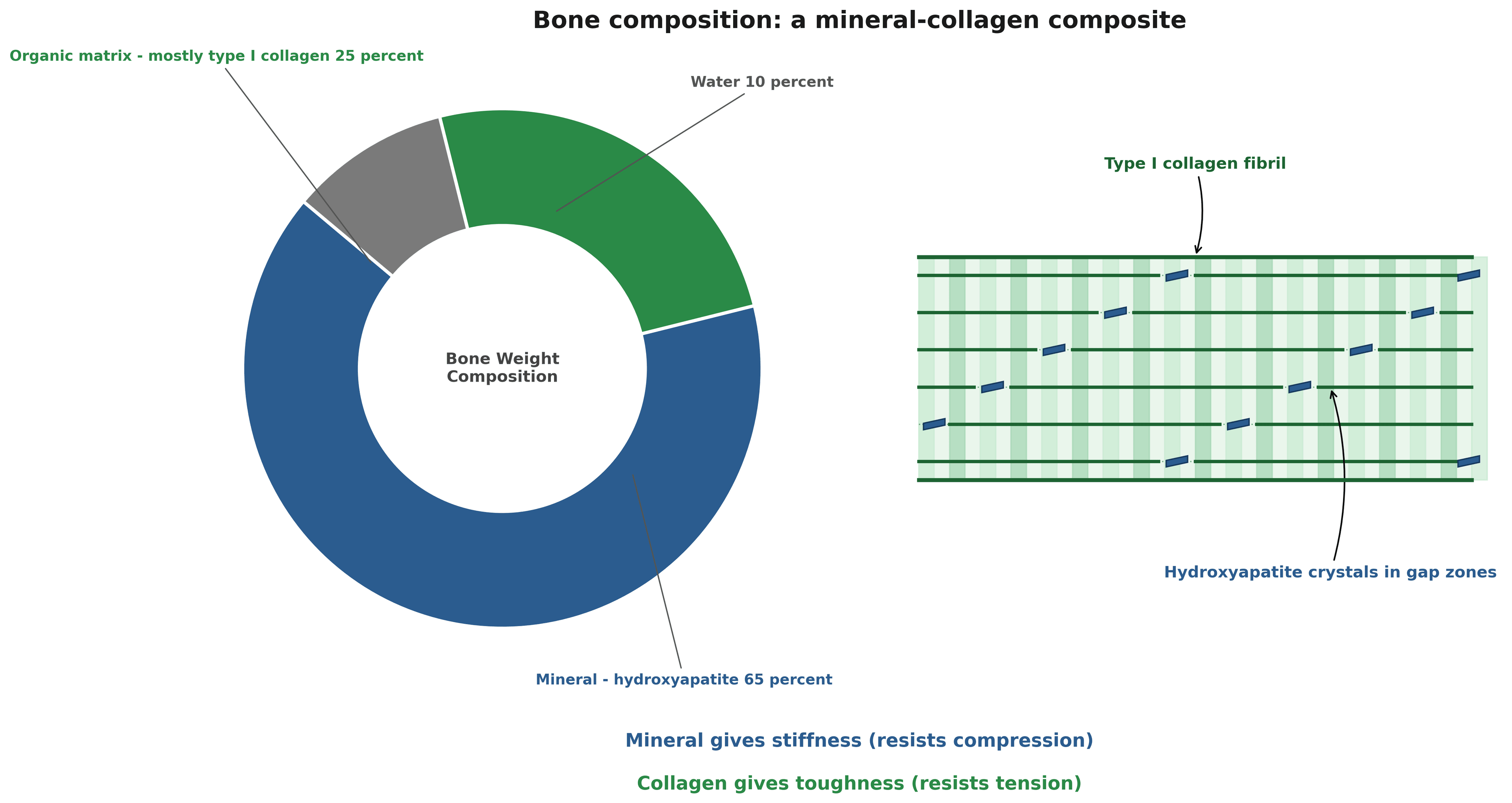
- Bone is a composite material - mineral provides stiffness, collagen provides toughness
- Hydroxyapatite Ca10(PO4)6(OH)2 is the inorganic mineral phase
- Type I collagen (90% of organic matrix) provides tensile strength
- Lamellar bone is organised mature bone, woven bone is immature
- Hierarchical structure: mineral crystals to lamellae to osteons to whole bone
- “Bone mineral density increases with hydroxyapatite deposition
- “Collagen fibril orientation determines mechanical anisotropy
- “Osteoid is unmineralised organic matrix (10-14 day lag before mineralisation)
- “Cortical bone has a lower remodelling rate than cancellous (3-5% vs 20-25% per year)
Overview
Where the rest of this lives. This page is the material - what bone is made of and how it is arranged. The cells that build and remove it have their own pages, osteoblasts and osteoclasts, and the cycle they run together is bone remodeling; the same matrix laid down again after injury is bone healing, and the organic phase in more detail is proteoglycans and collagen. The three diseases that read straight off this composition are worth holding together, because each fails a different component: osteogenesis imperfecta is defective type I collagen - too little normal matrix; osteomalacia is failure to mineralise a normal matrix; and osteoporosis is too little of an otherwise normal, normally mineralised bone. Same composite, three different points of failure.
What bone is. Bone is a specialised connective tissue doing four jobs at once: structural support, protection of vital organs, mineral homeostasis as the calcium and phosphate reservoir, and haematopoiesis in the marrow it encloses.
A composite material. By weight, bone is 65% inorganic mineral (hydroxyapatite), 25% organic matrix and 10% water. The mineral phase provides stiffness and compressive strength; type I collagen, which is 90% of the organic matrix, provides tensile strength and toughness.
The two demonstrations. Take the mineral out with acid and bone becomes flexible, bending like rubber. Take the collagen out with heat and it becomes brittle, shattering like chalk. Both components are needed for normal mechanical properties.
The Mineral Phase
The crystal. The mineral is hydroxyapatite, Ca10(PO4)6(OH)2, in hexagonal crystals of nanoscale dimensions: 50 nm long, 25 nm wide and only 2-3 nm thick. Crystal size and orientation are important to bone biomechanics.
Where the crystals sit. They are deposited within and between the collagen fibrils, aligned with the long axis of the fibril.
Ion substitutions. The apatite lattice tolerates ion exchange, and the substituting ion alters crystallinity and solubility, and through them mineral quality and remodelling dynamics.
- Substitutes For
- Phosphate (PO4)
- Effect
- Decreases crystallinity, increases solubility
- Clinical Example
- Normal bone has 4-8% carbonate
- Substitutes For
- Hydroxyl (OH)
- Effect
- Increases crystallinity, decreases solubility
- Clinical Example
- Fluoride treatment for osteoporosis
- Substitutes For
- Calcium (Ca)
- Effect
- May increase bone formation
- Clinical Example
- Strontium ranelate (historical osteoporosis drug)
- Substitutes For
- Calcium (Ca)
- Effect
- Affects crystal size
- Clinical Example
- Present in normal bone
Fluoride substitutes for hydroxyl groups, creating fluorapatite, which is more resistant to acid dissolution but also more brittle. High-dose fluoride treatment increases bone density but paradoxically increases fracture risk because of the abnormal crystal structure.
Mineralisation of Osteoid
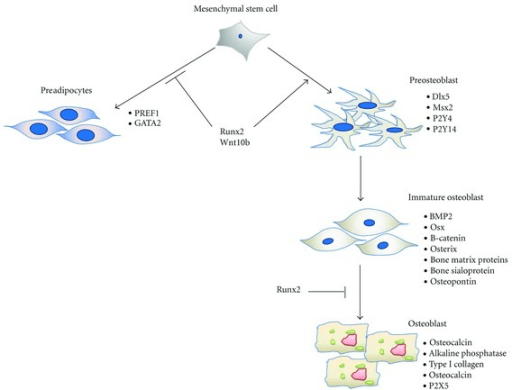
Osteoid comes first. Osteoblasts secrete unmineralised organic matrix - osteoid - and mineral follows it after a lag of 10-14 days. At the mineralisation front that unmineralised layer is 10-15 micrometres thick.
Mineralisation Timeline
Osteoblasts secrete unmineralised organic matrix (osteoid). This layer is 10-15 micrometres thick at the mineralisation front.
Osteoid matures and collagen crosslinks form. Matrix vesicles, containing alkaline phosphatase, bud from the osteoblasts.
Rapid mineral deposition to 70% of final mineral content. Matrix vesicles provide nucleation sites for hydroxyapatite crystals.
Slow increase to 95% of final mineral content. Crystals grow and mature, and bone density increases.
What mineralisation needs. The process is tightly regulated by the osteoblast and requires adequate calcium, phosphate and vitamin D.
Vitamin D deficiency impairs mineralisation, leaving excess unmineralised osteoid. Bone is formed but not properly mineralised, giving soft, deformable bones - rickets in children, osteomalacia in adults.
The Organic Matrix
The collagen molecule. Type I collagen is a triple helix of two alpha-1(I) chains and one alpha-2(I) chain, 300 nm long as the tropocollagen molecule and 1.5 nm in diameter. Its sequence is a Gly-X-Y repeat, with glycine at every third residue.
Assembly. Collagen is built in four steps:
- Intracellular: procollagen synthesis and hydroxylation, which requires vitamin C
- Extracellular: procollagen to tropocollagen, by cleavage of the N- and C-propeptides
- Fibril formation: tropocollagen self-assembles into fibrils with a 67 nm periodicity
- Crosslinking: lysyl oxidase creates covalent crosslinks - pyridinoline and deoxypyridinoline
Osteogenesis imperfecta is caused by mutations in COL1A1 or COL1A2, producing abnormal type I collagen: brittle bones with multiple fractures, blue sclerae and hearing loss. The organic scaffold is defective despite normal mineralisation.
The non-collagenous proteins. The remaining 10% of the organic matrix is a set of proteins that regulate mineralisation and cell-matrix interaction.
- Function
- Binds calcium, regulates mineralisation
- Clinical Significance
- Serum marker of bone formation
- Function
- Cell adhesion, inhibits mineralisation
- Clinical Significance
- Regulates crystal size and growth
- Function
- Binds collagen and mineral
- Clinical Significance
- Links organic and inorganic phases
- Function
- Nucleation of hydroxyapatite
- Clinical Significance
- Initiates mineralisation
- Function
- Inhibits mineralisation
- Clinical Significance
- Prevents ectopic calcification
Osteocalcin is particularly important as a clinical marker. It is secreted by osteoblasts during bone formation and is vitamin K-dependent, through gamma-carboxylation of its glutamate residues.
Vitamin K is required for the gamma-carboxylation of osteocalcin. Deficiency impairs osteocalcin function and may affect bone quality, and warfarin, a vitamin K antagonist, may increase fracture risk with long-term use.
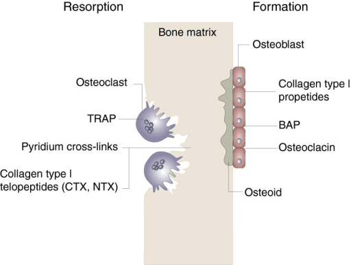
Crosslinks. Crosslink quality affects bone mechanical properties and fracture resistance. Enzymatic crosslinking is the normal route: lysyl oxidase converts lysine to allysine, forming the mature, stable pyridinoline (PYD) and deoxypyridinoline (DPD) crosslinks that increase tensile strength.
Non-enzymatic crosslinks. The advanced glycation end-products (AGEs) accumulate with ageing and with diabetes. They are brittle, they do not contribute to normal strength, and they are associated with reduced bone toughness.
Diabetes mellitus increases AGEs in bone collagen, reducing bone toughness despite normal or high bone mineral density. Diabetic patients have increased fracture risk independent of BMD.
Hierarchical Structure of Bone
Seven levels. The same material is organised on seven scales, from the molecule to the whole bone as an organ.
- Structure
- Tropocollagen + hydroxyapatite crystals
- Size Scale
- 1-100 nm
- Key Feature
- Mineral in collagen gap zones
- Structure
- Mineralised collagen fibrils
- Size Scale
- 100-1000 nm
- Key Feature
- 67 nm periodicity (D-band)
- Structure
- Fibril arrays (lamellae)
- Size Scale
- 1-10 micrometres
- Key Feature
- Parallel fibres in each lamella
- Structure
- Osteons (Haversian systems)
- Size Scale
- 100-300 micrometres
- Key Feature
- Concentric lamellae around central canal
- Structure
- Cortical vs trabecular bone
- Size Scale
- 0.1-1 mm
- Key Feature
- Dense vs porous architecture
- Structure
- Whole bone regions
- Size Scale
- 1-10 mm
- Key Feature
- Diaphysis, metaphysis, epiphysis
- Structure
- Whole bone
- Size Scale
- 10-100 mm
- Key Feature
- Integrated mechanical structure
Bone strength depends on integrity at all hierarchical levels. Osteoporosis (trabecular thinning at the mesostructure level), osteogenesis imperfecta (collagen defect at the molecular level) and osteomalacia (mineralisation defect at the nanoscale) all compromise strength, each by a different mechanism.
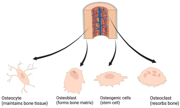
The structure adapts to load. The hierarchy is not static, and the osteocyte is the cell that makes it adapt. Osteocytes sit in lacunae connected by canaliculi - the lacunocanalicular network - and mechanical loading drives interstitial fluid flow and shear stress over their processes, which they transduce into biochemical signals, notably modulation of sclerostin, the SOST-gene product that normally inhibits bone formation.
Wolff's law and the mechanostat. This is the cellular basis of Wolff's law - form follows function, trabeculae aligning along principal stress trajectories - and of Frost's mechanostat. Below a lower strain threshold (disuse) net resorption occurs; within a physiological window bone mass is maintained; above an upper threshold (overload) modelling adds bone, while pathological overload causes microdamage.
What that framework explains. Disuse osteopenia, the periprosthetic bone loss of stress shielding, and exercise-induced bone gain are all consequences of the same osteocyte-driven feedback acting on the structures described here.
Cortical and Trabecular Bone
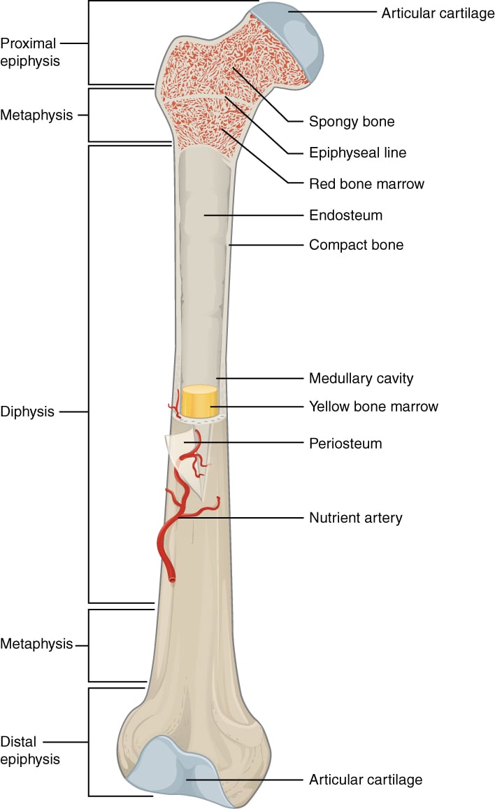
Cortical (compact) bone is dense lamellar bone: 80% of skeletal mass but only 20% of the bone surface area, with a porosity of 5-10% and a remodelling rate of 3-5% per year. It forms the diaphyses of the long bones and the outer shell of every bone, and it is the tissue that provides mechanical strength and protection.
The osteon. Its structural unit is the osteon, or Haversian system, a cylinder 200-300 micrometres in diameter:
- A central Haversian canal carrying blood vessels and nerves
- 4-20 concentric lamellae wrapped around it
- Osteocyte lacunae and canaliculi between the lamellae
- A cement line at the outer boundary
Secondary osteons are the ones produced by remodelling, and each is surrounded by a cement line, also called a reversal line.
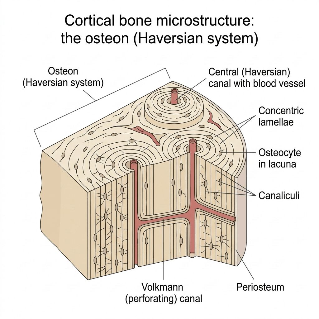
Cement lines mark the boundary of remodelling cycles. They are hypermineralised and weaker than the surrounding bone, which makes them sites for crack initiation but also for crack deflection - a toughening mechanism.
Trabecular (cancellous, spongy) bone is the other 20% of skeletal mass, and it carries 80% of the bone surface area, at a porosity of 50-90% and a remodelling rate of 20-25% per year. It fills the vertebrae, the pelvis and the metaphyses of the long bones.
The struts. A network of interconnected trabeculae, each 50-300 micrometres thick, oriented along the lines of stress (Wolff's law), with marrow in the spaces between them.
How it behaves. Trabecular bone has a lower modulus than cortical bone and is anisotropic, its strength depending on the direction of loading. Its high surface-to-volume ratio makes it the more metabolically active tissue: it is the metabolic reserve, and it distributes the loads handed to it.
- Cortical Bone
- 80%
- Trabecular Bone
- 20%
- Cortical Bone
- 20%
- Trabecular Bone
- 80%
- Cortical Bone
- 5-10%
- Trabecular Bone
- 50-90%
- Cortical Bone
- 3-5%
- Trabecular Bone
- 20-25%
- Cortical Bone
- Diaphyses, outer shell
- Trabecular Bone
- Metaphyses, vertebrae
- Cortical Bone
- Mechanical strength
- Trabecular Bone
- Metabolic reserve
Why the turnover rates matter. The higher remodelling rate of trabecular bone makes it more responsive to metabolic change, and also more vulnerable to resorptive disease.
Osteoporosis affects trabecular bone earlier and more severely than cortical bone, because of its higher surface area and remodelling rate. Vertebral and hip fractures reflect trabecular bone loss.
The stress-strain curve. The composite composition produces the mechanical behaviour examiners ask about, starting with the curve itself: an initial linear elastic region, whose slope is the Young's modulus, then a yield point, then a plastic region before failure; the area under the curve is the energy absorbed, which is toughness.
The numbers. Cortical bone has a Young's modulus of roughly 17-20 GPa, far below steel at about 200 GPa. Trabecular bone is much lower, roughly 0.1-2 GPa, and depends steeply on density.
Anisotropy. Bone is strongest in compression and along the long (osteon) axis, weaker in tension and weakest in shear, which is why the skeleton is loaded preferentially in compression.
Viscoelasticity. Bone is strain-rate dependent: stiffer, stronger, and storing more energy at high loading rates. High-energy trauma therefore releases more energy and drives more comminution, while low-rate loading produces simpler patterns. In those terms, removing the mineral leaves a ductile, low-modulus material, and removing the collagen a stiff but brittle one.
Lamellar versus Woven Bone
- Lamellar Bone (Mature)
- Highly organised, parallel fibres
- Woven Bone (Immature)
- Random, disorganised fibres
- Lamellar Bone (Mature)
- Slow (1-2 micrometres/day)
- Woven Bone (Immature)
- Rapid (4-6 micrometres/day)
- Lamellar Bone (Mature)
- High
- Woven Bone (Immature)
- Low
- Lamellar Bone (Mature)
- Low
- Woven Bone (Immature)
- High
- Lamellar Bone (Mature)
- Normal adult bone, remodelling
- Woven Bone (Immature)
- Fracture callus, fetal bone, Paget disease
Lamellar bone is the mature form: collagen fibres parallel within each lamella, and the orientation alternating between lamellae like plywood. The slow deposition is what allows that organisation.
Woven bone is laid down rapidly, with disorganised collagen orientation, and it is weaker and more flexible than lamellar bone. It is the bone of the fracture callus, and it is remodelled to lamellar bone over months.
Paget disease produces bone with both woven and lamellar patterns together - the mosaic pattern, a jigsaw-puzzle appearance on histology. The rapid, disorganised remodelling produces weak bone despite increased density.
Clinical Relevance and Applications
Why composition explains fracture patterns. High-energy trauma overcomes both components at once, the mineral that carries compression and the collagen that carries tension. Osteoporotic bone has adequate mineral but poor microarchitecture.
- Disease/Condition
- Osteomalacia/Rickets
- Clinical Implication
- Vitamin D supplementation, correct underlying cause
- Disease/Condition
- Osteogenesis imperfecta
- Clinical Implication
- Bisphosphonates, fracture prevention, genetic counselling
- Disease/Condition
- Osteoporosis
- Clinical Implication
- Antiresorptive therapy, fracture risk assessment
- Disease/Condition
- Age-related bone loss
- Clinical Implication
- Monitoring with DXA, fall prevention
- Disease/Condition
- Paget disease
- Clinical Implication
- Bisphosphonates to normalise remodelling
Bone quality affects implant fixation. Osteoporotic bone has reduced holding power for screws, because trabecular loss reduces the surface area available for fixation, and cortical bone gives better screw purchase than cancellous bone. Consider cement augmentation or alternative fixation strategies in poor quality bone.
Fracture healing. Woven bone forms first in the callus and is remodelled to lamellar bone over 12-18 months. Three things get in the way: smoking impairs collagen synthesis, vitamin D deficiency delays mineralisation, and NSAIDs may inhibit bone healing.
Surgical planning. Assess bone quality on the preoperative imaging, then read it into the construct:
- Cortical thickness guides plate selection
- Trabecular pattern influences screw trajectory
- Bone density affects implant choice
- Consider bone grafting for defects
Differentiating Bone Composition Disorders
A favourite examiner manoeuvre is to ask which structural component is at fault in a given metabolic bone disease. Mapping each disease to the affected component — mineral phase, organic (collagen) phase, mineralisation process, or architecture/remodelling — makes the differential systematic rather than a list to be memorised.
- Component at Fault
- Architecture (quantity, microarchitecture)
- Mineral/Matrix Pattern
- Normal composition, reduced bone quantity and connectivity
- Discriminating Clue
- Low BMD with normal calcium, phosphate, ALP; trabecular thinning
- Component at Fault
- Mineralisation process
- Mineral/Matrix Pattern
- Excess unmineralised osteoid, normal collagen
- Discriminating Clue
- Low/normal calcium and phosphate, high ALP, low vitamin D; Looser zones
- Component at Fault
- Organic phase (type I collagen)
- Mineral/Matrix Pattern
- Defective collagen scaffold, mineralisation ongoing
- Discriminating Clue
- Blue sclerae, dentinogenesis imperfecta, hearing loss, family history
- Component at Fault
- Remodelling (architecture)
- Mineral/Matrix Pattern
- Disorganised woven-plus-lamellar mosaic, high turnover
- Discriminating Clue
- Markedly raised ALP, bone pain/deformity, mosaic cement lines on histology
- Component at Fault
- Organic phase (collagen crosslink quality)
- Mineral/Matrix Pattern
- Normal/high mineral, AGE-crosslinked brittle collagen
- Discriminating Clue
- Fracture despite normal or high BMD; raised pentosidine
- Component at Fault
- Mineralisation + remodelling
- Mineral/Matrix Pattern
- Mixed: high-turnover (osteitis fibrosa) or low-turnover (adynamic)
- Discriminating Clue
- CKD, abnormal PTH, calcium, phosphate; bone biopsy for turnover
Osteoporosis = too little bone of normal quality. Osteomalacia = enough matrix but not enough mineral. Osteogenesis imperfecta = defective collagen scaffold. Paget = disorganised over-remodelling. Diabetic bone = normal mineral, poor collagen. Biochemistry (Ca, PO₄, ALP, PTH, vitamin D) plus histology localises the lesion to a structural component.
Guidelines, Registries & Global Practice
Bone composition and structure is a basic-science topic that underpins the way every major examining body and society frames metabolic bone disease, fracture risk and implant fixation. The principles are universal; the way bone quality is assessed and acted upon varies by guideline and by resource setting.
Global Epidemiology of Bone Strength Disorders
- Osteoporosis affects an estimated 1 in 3 women and 1 in 5 men over age 50 worldwide for a fragility fracture in their remaining lifetime, reflecting trabecular microarchitectural deterioration superimposed on age-related collagen and mineral changes.
- Fragility fractures are projected to rise sharply as populations age, with the largest absolute increases anticipated in Asia.
- Osteogenesis imperfecta (primary collagen/organic-matrix defect) has a birth prevalence of roughly 1 in 15,000-20,000, mostly autosomal dominant COL1A1/COL1A2 mutations.
- Vitamin D insufficiency, the dominant driver of impaired mineralization (osteomalacia/rickets), is highly prevalent across all latitudes, with higher rates at high latitudes, in people with darker skin pigmentation, and where cultural dress limits sun exposure.
- Diabetes mellitus increasingly contributes to fracture burden via accumulation of advanced-glycation-end-product collagen crosslinks that reduce bone toughness independent of mineral density.
Controversies and Areas of Uncertainty
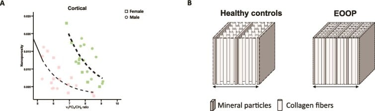
Intrafibrillar versus extrafibrillar mineral. The relative contribution of mineral located within versus around the collagen fibrils to whole-bone mechanics remains debated. Atomistic modelling indicates that intrafibrillar mineral alone cannot account for whole-bone stiffness, which implies that extrafibrillar mineral is essential, but the exact partition and its biological control are unsettled.
Bone quality beyond BMD. DXA-measured BMD explains only part of fracture risk. How best to capture the "quality" contribution - collagen crosslinks, crystallinity, microdamage, microarchitecture - in routine practice is unresolved; the Trabecular Bone Score and HR-pQCT are surrogates, not direct measures.
Fluoride and strontium. Agents that alter the mineral phase increase measured density but historically gave disappointing or paradoxical fracture outcomes, strontium ranelate having been withdrawn in many regions over cardiovascular concerns. More mineral does not equal stronger bone.
Osteocalcin's systemic role. Beyond its established role as a bone-formation marker, undercarboxylated osteocalcin has been proposed as a hormone influencing glucose metabolism and other systems. The clinical significance in humans remains an area of active investigation and is not yet exam-established fact.
MCQ Practice Points
Q: What is the approximate composition of bone by weight?
A: 65% inorganic mineral (hydroxyapatite), 25% organic matrix (90% Type I collagen), and 10% water. The inorganic phase provides stiffness and compressive strength, while the organic matrix provides toughness and tensile strength.
Q: What is the chemical formula for hydroxyapatite, the primary mineral in bone?
A: Ca₁₀(PO₄)₆(OH)₂ - calcium phosphate hydroxide. Calcium and phosphate ions can be substituted (e.g., carbonate for phosphate, fluoride for hydroxyl) which affects bone quality. Fluoride increases density but also increases brittleness.
Q: What is the key difference between cortical and cancellous bone in terms of remodeling rate?
A: Cortical bone: 3-5% annual turnover rate, comprises 80% of skeletal mass Cancellous bone: 20-25% annual turnover rate, comprises only 20% of mass but 80% of surface area
The higher surface area of cancellous bone explains its faster turnover and greater susceptibility to metabolic bone diseases.
Q: What is the typical lag time between osteoid deposition and mineralization?
A: 10-14 days. Osteoid is unmineralized organic matrix secreted by osteoblasts. The mineralization lag time is clinically relevant - increased lag indicates osteomalacia, while decreased lag may indicate impaired matrix maturation.
Basic Science Viva Scenarios
Practise clinical reasoning and management decisions out loud
“Describe the composition of bone and explain how each component contributes to its mechanical properties.”
“Explain the hierarchical organization of bone from the molecular level to whole bone. How does this relate to fracture risk in osteoporosis?”
“A 72-year-old diabetic woman sustains a low-energy distal femoral fracture. Her DXA bone mineral density is in the normal range. Using your knowledge of bone composition and structure, explain why she fractured and how bone quality affects your fixation strategy.”
Composition by Weight
- 65% inorganic (hydroxyapatite mineral)
- 25% organic (90% Type I collagen + 10% non-collagenous proteins)
- 10% water
- Composite material: mineral = stiffness, collagen = toughness
Hydroxyapatite Mineral
- Ca10(PO4)6(OH)2 chemical formula
- Crystal size: 50nm × 25nm × 2-3nm (nanoscale)
- Deposits in collagen gap zones (67 nm periodicity)
- Ion substitutions: carbonate, fluoride, strontium affect properties
Collagen and Organic Matrix
- Type I collagen = 90% of organic matrix
- Triple helix (two alpha-1, one alpha-2 chain), 300 nm length
- Crosslinks: pyridinoline and deoxypyridinoline (enzymatic)
- Non-collagenous: osteocalcin, osteopontin, osteonectin, BSP
Hierarchical Levels
- Level 1-2: Tropocollagen + crystals → mineralized fibrils (67 nm)
- Level 3-4: Lamellae → osteons (200-300 micrometers)
- Level 5: Cortical (dense, 80% mass) vs trabecular (porous, 80% surface)
- Levels 6-7: Whole bone regions and organ
Cortical vs Trabecular
- Cortical: 80% mass, 5-10% porosity, 3-5% remodeling/year
- Trabecular: 20% mass, 80% surface, 50-90% porosity, 20-25% remodeling/year
- Trabecular affected first in osteoporosis (higher surface area)
- Cortical provides strength, trabecular provides metabolic reserve
Key Clinical Correlations
- Osteogenesis imperfecta: Type I collagen defect (brittle bones)
- Osteomalacia: Mineralization defect (soft bones, excess osteoid)
- Osteoporosis: Multi-level failure (trabecular loss, cortical porosity)
- Paget disease: Woven bone mosaic pattern (weak despite high density)
Evidence Base
Hierarchical Structure of Bone
- Surveyed bone mechanical data across the full hierarchy from nanoscale to whole bone
- Mechanical properties emerge from structural organization at each level
- Simple composite rule-of-mixtures formulae only partly predict bone behaviour
- Load transfer between organic and inorganic subunits is incompletely understood
Intrafibrillar Mineralization and Compressive Strength
- Full-atomistic simulation of the three-dimensional mineralized collagen fibril
- Compressive modulus rises monotonically as intrafibrillar mineral density increases
- Intrafibrillar mineral alone gives a modulus an order of magnitude below whole bone
- Extrafibrillar mineralization is therefore mandatory for bone's load-bearing capacity