Anatomic Reduction Required | Plate Fixation Standard | Compartment Syndrome Risk
- Anatomic reduction essential - malunion impairs rotation
- Plate both bones - 3.5mm LC-DCP or locking plates
- Minimum 6 cortices each side of fracture (3 screws)
- Restore radial bow - critical for supination
- Compartment syndrome risk - high energy mechanism
- “Radial bow is at junction of proximal and middle thirds - must restore
- “Plate radius on volar (Thompson) surface - less symptomatic
- “Plate ulna on dorsal/tension surface
- “Loss of just 10 degrees of rotation = significant functional deficit
Non-anatomic reduction impairs rotation. Unlike other long bone fractures, the forearm requires anatomic reduction to restore rotation. Even 10° angulation or 10° rotation malalignment significantly reduces supination/pronation.
Restore the radial bow - the normal curve of the radius is critical for supination. Maximum bow at junction of proximal and middle thirds. Loss of bow = loss of rotation, especially supination.
Both bones require plating in adults. Standard is 3.5mm LC-DCP or locking plates. Minimum 6 cortices (3 screws) each side of fracture. May need larger construct for comminution.
High risk with both-bone fractures, especially high-energy. Maintain high index of suspicion. Pain with passive finger extension, tense compartments = emergency fasciotomy.
- Key Consideration
- Standard displaced fracture
- Treatment
- ORIF both bones with 3.5mm plates
- Key Consideration
- Compression achievable
- Treatment
- LC-DCP with lag and compression
- Key Consideration
- Cannot compress
- Treatment
- Bridge plating, may need longer construct
- Key Consideration
- Soft tissue compromise
- Treatment
- Urgent debridement, internal or external fixation
- Key Consideration
- Multiple fractures one bone
- Treatment
- Long plate spanning all fractures
- Key Consideration
- Floating elbow/wrist
- Treatment
- Prioritize forearm stability
- Key Consideration
- Growth plate considerations
- Treatment
- Often closed reduction, casting acceptable
FOREARMFOREARM - Fixation Principles
Hook:FOREARM fixation requires attention to every detail
BOWBOW - Radial Bow Restoration
Hook:The radial BOW is essential for supination
PLATESPLATES - Placement Principles
Hook:PLATES positioned correctly prevent complications
COMPARTMENTSCOMPARTMENTS - Syndrome Recognition
Hook:COMPARTMENTS syndrome is a clinical diagnosis requiring immediate fasciotomy
Overview and Epidemiology
Both-bone forearm fractures in adults are typically high-energy injuries requiring operative fixation. The forearm is unique because it functions as a paired bone articulated joint - both bones rotate around each other, and any disruption of anatomy impairs this function.
Mechanism of injury:
- Direct trauma - blow to forearm
- Often comminuted at impact site
- Industrial injuries, assault
- Indirect trauma - fall on outstretched hand
- Rotational component common
- Motor vehicle accidents
- High-energy mechanisms predominate - MVA, falls from height, sport
The forearm functions as a paired rotational joint - the radius rotates around the ulna through approximately 150° arc. This requires intact proximal radioulnar joint (PRUJ), interosseous membrane (IOM), and distal radioulnar joint (DRUJ). Both-bone fractures disrupt this mechanism.
Associated injuries:
- Compartment syndrome (high risk)
- Neurovascular injury
- Ipsilateral elbow injury (floating elbow)
- Ipsilateral wrist injury (floating wrist)
- Associated hand injuries
Anatomy and Biomechanics
- Proximal Radius (Supinator/Biceps): Supinated and flexed
- Middle Radius (Pronator Teres): Pronated (if distal to insertion)
- Distal Radius (Pronator Quadratus): Pronated
- Radial head - articulates with capitellum and PRUJ
- Radial tuberosity - biceps insertion
- Radial bow - maximum curvature at junction proximal/middle thirds
- Lateral surface - covered by wrist extensors
- Volar surface - FPL origin, plate placement area
- Olecranon - triceps insertion
- Coronoid - anterior buttress
- Shaft - relatively straight, triangular cross-section
- Subcutaneous border - medial, palpable throughout
- Dorsal surface - ECU groove, plate placement area
- Interosseous membrane (IOM) - connects radius to ulna
- PRUJ - proximal radioulnar joint
- DRUJ - distal radioulnar joint
- Interosseous space - maintained for rotation
The radial bow is the normal lateral convexity of the radius. Maximum bow is at the junction of proximal and middle thirds. Loss of even 10% of radial bow can result in loss of supination. Always compare to contralateral side and use pre-contoured plates.
- Normal supination/pronation: 75°/75° approximately
- Radius rotates around relatively fixed ulna
- Rotation occurs at PRUJ, DRUJ, and through IOM
- Loss of 10° rotation malalignment = significant functional loss
- Thompson (dorsolateral radius) - between ECRB and EPL
- Henry (volar radius) - between BR and PT/FCR
- Ulnar approach - along subcutaneous border
Classification Systems
Location Classification
- Radius
- Near radial head/tuberosity
- Ulna
- Near olecranon/coronoid
- Radius
- Shaft - most common
- Ulna
- Shaft
- Radius
- Near wrist
- Ulna
- Near DRUJ
Considerations by location:
- Proximal: risk of PIN, need different approach
- Middle: standard Thompson + ulnar approach
- Distal: consider associated DRUJ/wrist injury
Location determines surgical approach and implant selection.
Clinical Presentation and Assessment
History:
- Mechanism (high vs low energy)
- Time of injury
- Open wound (assume open until proven otherwise)
- Associated injuries
- Hand dominance, occupation
Physical examination:
- Significance
- Displaced fracture
- Action
- Document, assess NV, reduce
- Significance
- Open fracture
- Action
- Antibiotics, urgent surgery
- Significance
- Compartment syndrome
- Action
- Emergency fasciotomy
- Significance
- Impending compartment syndrome
- Action
- Serial monitoring or fasciotomy
- Significance
- Arterial injury
- Action
- Urgent reduction, angiography/repair
- Significance
- Nerve injury/compression
- Action
- Document pre-op, may resolve with reduction
- Significance
- Floating segment
- Action
- Address all injuries
Compartment syndrome assessment:
The classic 5 Ps (Pain, Pallor, Pulselessness, Paralysis, Paresthesia) are late signs. Early compartment syndrome: Pain out of proportion and pain with passive stretch of fingers. Don't wait for all 5 Ps - fasciotomy needed urgently.
Key examination points:
- Compartment assessment - volar and dorsal
- Neurovascular status - PIN, median, ulnar nerves; radial and ulnar pulses
- Skin integrity - open wound assessment
- Associated injuries - elbow, wrist, hand
- Soft tissue swelling - predictor of complications
The quick-guide lists "floating elbow/wrist" as a reason to prioritise forearm stability, but the floating elbow is a defined, high-stakes entity worth stating explicitly:
- Definition: ipsilateral fractures of the forearm (one or both bones) AND the humerus, so the elbow joint is "floating" - disconnected from both the proximal and distal skeletal segments. (The analogous floating wrist is an ipsilateral forearm + distal radius/carpal injury.)
- Why it matters: it marks a high-energy limb and carries a markedly increased rate of compartment syndrome, open injury, neurovascular injury and polytrauma - have a very low threshold for fasciotomy and a full secondary survey.
- Management principle: stabilise both fractures operatively (fix the humerus and the forearm) so the elbow can be mobilised early - a key reason floating-elbow injuries are an operative indication even where an isolated humeral shaft fracture might be braced. Non-operative management of either component leaves the elbow unstable and stiff.
- Sequence: commonly fix proximal-to-distal (humerus then forearm) or address the more accessible/urgent injury first, but the goal is a stable construct top-to-bottom permitting early elbow motion.
Exam point: "floating elbow" = ipsilateral humerus + forearm fracture, a high-energy injury with a high compartment-syndrome rate that converts an otherwise brace-able humeral fracture into an operative both-segment fixation to allow early elbow mobilisation.
Investigations
Radiographic assessment:
Standard views:
- AP forearm - entire forearm including elbow and wrist
- Lateral forearm - true lateral
- Dedicated elbow views - AP and lateral if proximal injury
- Dedicated wrist views - AP and lateral if distal injury

Always image the joint above and below. A forearm fracture series must include adequate views of the elbow and wrist to exclude Monteggia (proximal) or Galeazzi (distal) variants.
- Fracture location and pattern (both bones)
- Degree of comminution
- Angulation and displacement
- Radial head congruent with capitellum?
- DRUJ congruent?
- Shortening present?
- Rarely indicated for shaft fractures
- May help for complex articular extension
- 3D reconstruction for difficult patterns
- If clinical suspicion
- Absolute pressure greater than 30mmHg concerning
- Delta pressure (Diastolic - Compartment) less than 30mmHg = fasciotomy
Differential Diagnosis
A "both-bone forearm fracture" on a single film can be a fracture-dislocation in disguise. Distinguishing these patterns changes the operation.
- Key Feature
- Fractures of radial AND ulnar shafts; PRUJ and DRUJ congruent
- Pitfall / Distinguishing Test
- Confirm radial head aligns with capitellum and DRUJ is reduced
- Key Feature
- Proximal/mid ulna fracture with radial head dislocation
- Pitfall / Distinguishing Test
- Radiocapitellar line must pass through capitellum on every view
- Key Feature
- Distal-third radius fracture with DRUJ disruption
- Pitfall / Distinguishing Test
- Assess DRUJ widening/instability; the 'fracture of necessity' needing ORIF
- Key Feature
- Radial head fracture + IOM rupture + DRUJ disruption
- Pitfall / Distinguishing Test
- Check wrist pain/DRUJ in any radial head fracture; longitudinal instability
- Key Feature
- Single ulnar shaft fracture, no radial injury
- Pitfall / Distinguishing Test
- Exclude radial head dislocation before calling it isolated
- Key Feature
- Bowing or incomplete cortical break
- Pitfall / Distinguishing Test
- Different management - often closed reduction; not adult ORIF principles
Management Algorithm
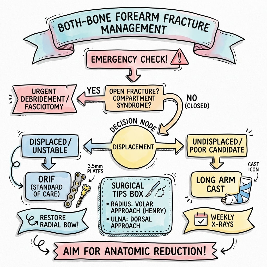
- Exclude emergencies first - compartment syndrome (clinical diagnosis), open wound, vascular injury. Treat these before definitive fixation.
- Image the joint above and below to exclude Monteggia (proximal) and Galeazzi (distal) variants.
- Default to ORIF of both bones with 3.5mm plates; conservative management is reserved for the rare truly undisplaced fracture or the patient unfit for surgery.
- Restore length, rotation and the radial bow, then confirm smooth pronation/supination intra-operatively.
- Plan soft-tissue cover with plastics input for high-grade open injuries.
Definitive Management
Non-operative management:
- Isolated, undisplaced fractures (rare)
- Non-ambulatory patients
- Significant medical comorbidities precluding surgery
- Patient refusal
- Long arm cast, elbow 90°, neutral rotation
- Weekly X-rays for first 3 weeks
- Any displacement = convert to operative
- Expect prolonged immobilization and stiffness
Non-operative treatment is rarely indicated for adult both-bone forearm fractures. Non-anatomic healing leads to loss of rotation and poor function. Operative fixation is the standard of care.
Surgical Technique
Surgical Approaches
- Incision: lateral epicondyle to Lister's tubercle
- Interval: ECRB and EPL (proximal), EPB/APL (distal)
- Danger: PIN crosses at level of radial head
- Pros: excellent visualization, less symptomatic plate
- Incision: lateral to biceps tendon to radial styloid
- Interval: BR/PT and FCR/radial artery
- Pros: safer for proximal third (PIN protected)
- Cons: plate may be more prominent
- Incision along subcutaneous border
- Directly onto bone (ECU/FCU interval)
- Minimal soft tissue dissection
- Plate on dorsal tension surface
Thompson (dorsolateral) is preferred for middle and distal radius - plate less symptomatic, good exposure. Henry (volar) is safer for proximal third - PIN is protected as it crosses deeper in the supinator. Choose approach based on fracture level.

Complications
- Incidence
- 2-5%
- Management
- Emergency fasciotomy
- Incidence
- 5-10%
- Management
- Corrective osteotomy if symptomatic
- Incidence
- 2-5%
- Management
- Revision fixation, bone graft
- Incidence
- 3-5%
- Management
- Excision if mature, interposition
- Incidence
- 1-3% closed, higher open
- Management
- Debridement, antibiotics
- Incidence
- 2-5%
- Management
- Usually neurapraxia, observation
- Incidence
- 2-3%
- Management
- Revision with bone graft
- Incidence
- 5-10%
- Management
- Protected activity, avoid early removal
- Most critical early complication
- High-energy injuries at highest risk
- Pain with passive finger extension is early sign
- Emergency fasciotomy - both volar and dorsal compartments
- Loss of rotation most functionally limiting
- Greater than 10° angulation or rotation = significant deficit
- Corrective osteotomy with bone graft if symptomatic
- Abnormal bone bridge between radius and ulna
- Risk factors: high energy, single incision, head injury
- Prevention: orthogonal plating (90° apart), avoid IOM damage
- Treatment: excision when mature (greater than 1 year), interposition material
Refracture can occur after plate removal, especially if removed early. Recommend leaving plates in place unless symptomatic. If removal needed, wait 18-24 months post-op, and protect with splint and activity restriction for 6-8 weeks after removal.
The topic covers how to prevent synostosis (orthogonal plating, separate incisions) but an examiner will expect the Vince and Miller classification of established post-traumatic radioulnar synostosis, which is by location and guides resection risk and prognosis:
- Type I - distal (intra-articular at the DRUJ): synostosis at the distal third/DRUJ; resection risks the distal radioulnar articulation.
- Type II - middle third (diaphyseal): the commonest type, in the mid-shaft region; generally the best prognosis for resection.
- Type III - proximal third (at or near the PRUJ/bicipital tuberosity): the worst prognosis and highest recurrence; resection here is hazardous because of proximity to the PIN and the PRUJ.
Management principles: excise only mature synostosis (clinically quiescent, normalised bone scan/markers - commonly delayed to about a year), restore the interosseous space, interpose tissue/material where used, protect the PIN (especially Type III), and consider single-dose radiotherapy or indomethacin prophylaxis against recurrence.
Exam point: grade established synostosis by Vince and Miller (I distal/DRUJ, II diaphyseal - best prognosis, III proximal/PRUJ - worst and PIN-threatening), and operate only on mature lesions with PIN protection and recurrence prophylaxis.
Postoperative Care and Rehabilitation
Postoperative protocol:
- Posterior splint for comfort
- Elevation
- Finger motion immediately
- Monitor for compartment syndrome
- Wound check day 2 and 5-7
- Begin active ROM elbow and wrist
- Gentle active supination/pronation
- Wean from splint
- Light functional use
- Progressive active ROM
- Expect near full ROM by 6 weeks
- No resistance/lifting
- Light activities of daily living
- Begin gentle strengthening
- Progressive loading
- Return to light work
- X-ray at 6 weeks to confirm union
- Full strengthening
- Return to sport/heavy work
- Full union expected
- Final ROM assessment
Key rehabilitation principles:
- Early motion critical - prevents stiffness
- Stable fixation allows early motion
- Focus on rotation (supination/pronation)
- Progressive loading after union confirmed
- Final ROM assessment at 6 months
Both-bone forearm fractures typically unite in 12-16 weeks. Confirm radiographic union before allowing heavy activities. Delayed union more common with comminution, open fractures, and smoking.
Outcomes and Prognosis
Expected outcomes:
- Expectation
- 90-95% primary union
- Expectation
- Less than 10-20° loss supination/pronation
- Expectation
- 85-95% of contralateral
- Expectation
- DASH scores typically excellent
- Expectation
- 3-6 months most patients
Prognostic factors:
- Open vs closed (open = worse outcomes)
- Energy of injury
- Quality of reduction
- Bone quality
- Patient compliance
- Smoking status
Most activities of daily living require approximately 50° supination and 50° pronation. Loss of rotation up to 20° is often well-tolerated functionally. Greater loss typically symptomatic and may warrant corrective surgery.
Guidelines, Registries & Global Practice
Global epidemiology:
- Forearm shaft fractures account for roughly 1-2% of all adult fractures; both-bone patterns are the high-energy subset.
- Bimodal distribution: young men from high-energy trauma (road traffic, especially motorcycles, industrial machinery, falls from height, contact and wheeled sport) and older adults from lower-energy falls.
- Male predominance in the high-energy group; rising motorcycle use makes these injuries disproportionately common in low- and middle-income countries.
Guideline and society positions (areas of genuine agreement and difference):
- Position on adult both-bone forearm fractures
- Anatomic open reduction and compression/neutralisation plating (3.5mm) of both bones is the reference standard; bridge plating for comminution.
- Position on adult both-bone forearm fractures
- Operative fixation for displaced adult diaphyseal forearm fractures; plate fixation favoured over intramedullary nailing for restoring rotation and bow.
- Position on adult both-bone forearm fractures
- Open fractures follow BOAST open-fracture standards: IV antibiotics within 1 hour, combined ortho-plastic care, early definitive fixation with soft-tissue cover.
- Position on adult both-bone forearm fractures
- Endorse anatomic plate fixation; recognise locking plates mainly for osteoporotic or comminuted bone rather than routine use.
- Unlike arthroplasty, forearm shaft fractures are not tracked by a dedicated implant registry; the evidence base is institutional cohorts and trauma databases rather than national joint registries.
- Across cohorts, plate fixation delivers union rates of roughly 90-98%, with locking and conventional 3.5mm constructs performing comparably.
- Well-resourced settings: routine ORIF with 3.5mm plates, intra-operative imaging, on-site plastic surgery for Gustilo IIIB/C, and compartment-pressure monitoring where the diagnosis is equivocal.
- Limited-resource settings: square or Hackethal intramedullary nailing and external fixation are used more often when plating implants or image intensifiers are unavailable; transfer pathways may be limited, so compartment syndrome is frequently managed at the presenting hospital.
Be prepared to discuss surgical approaches, fixation principles (especially radial bow restoration), compartment syndrome recognition and management, and open fracture protocols. Both-bone fractures are common viva topics testing fundamental principles.
Controversies and Areas of Uncertainty
- Locking vs conventional plates: Locking plates have not shown superior union or function over conventional 3.5mm DCP/LC-DCP in standard diaphyseal bone (Iacobellis & Biz; Hertel). Their clearest role is osteoporotic bone and bridge plating of comminution, not routine use.
- Plate vs intramedullary nailing: Modern locked forearm nails achieve good union, but restoring the radial bow and rotation is harder than with plates. Nailing is most attractive in segmental fractures, poor soft tissues, or limited-resource settings; plating remains the default where anatomic reduction is the priority.
- Routine implant removal: Removing plates exposes the patient to refracture (around 4% in long-term cohorts) and a second operation. Most surgeons leave asymptomatic plates in situ; if removal is undertaken, delay it (commonly 18-24 months) and protect the limb afterwards.
- Acceptable malalignment: "Anatomic" is the goal, but the precise threshold beyond which rotation is meaningfully lost is debated; radial bow restoration matters more than absolute angular numbers.
- Compartment pressure thresholds: The diagnosis remains clinical. Absolute and delta-pressure cut-offs (delta less than 30mmHg) guide equivocal cases but should never delay fasciotomy when the clinical picture is clear.
- Single vs separate incisions: A single incision for both bones increases synostosis risk; orthogonal plating through separate approaches is preferred, though high-energy injury itself is the dominant non-modifiable risk factor.
MCQ Practice Points
Q: Why is anatomic reduction essential in both-bone forearm fractures? A: The forearm is a paired rotational joint - radius rotates around ulna. Even 10° of angulation or rotational malunion significantly reduces supination and pronation. Unlike other diaphyseal fractures, the forearm does not tolerate malunion.
Q: What is the minimum fixation for both-bone forearm fractures? A: 3.5mm plates with minimum 6 cortices (3 screws) each side of the fracture for each bone. Eight cortices (4 screws) provides more security and is often preferred.
Q: Where is the maximum radial bow and why is it important? A: Maximum bow is at the junction of proximal and middle thirds of the radius. The bow is critical for supination - loss of radial bow results in loss of supination. Must restore with contoured plate.
Q: What is the danger with Thompson (dorsolateral) approach to the radius? A: The posterior interosseous nerve (PIN) crosses at the level of the radial head (approximately 4cm from lateral epicondyle). Protect by supinating forearm and staying anterior to supinator. Consider Henry approach for very proximal fractures.
Q: How do you reduce the risk of radioulnar synostosis? A: Use orthogonal plating (plates 90° apart), avoid single incision for both bones, meticulous soft tissue handling, preserve interosseous membrane, and avoid high-energy mechanisms (not modifiable).
Clinical Decision Scenarios
Practise clinical reasoning and management decisions out loud
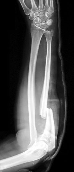
“A 35-year-old man falls from a motorcycle at low speed. He has a closed both-bone forearm fracture in the mid-shaft. X-rays show transverse fractures of both radius and ulna with 100% displacement. How would you manage this injury?”
“A 28-year-old construction worker has his forearm caught in machinery. He has a Gustilo Type II open both-bone forearm fracture with a 3cm wound over the volar forearm. The fractures are at different levels. What is your management?”
“Six hours after plating a both-bone forearm fracture, the nurse calls you because the patient is in severe pain despite adequate analgesia. His fingers are swollen and he has severe pain with passive finger extension. What do you do?”
KEY PRINCIPLES
- Anatomic reduction essential - non-anatomic = loss of rotation
- Plate both bones in adults
- Restore radial bow (max at proximal-middle junction)
- Orthogonal plating (90° apart) reduces synostosis
FIXATION STANDARD
- 3.5mm LC-DCP or locking plates
- Minimum 6 cortices (3 screws) each side
- 8 cortices (4 screws) preferred
- Bicortical screw purchase
SURGICAL APPROACHES
- Radius: Thompson (dorsolateral) for mid/distal
- Radius: Henry (volar) safer for proximal (PIN protected)
- Ulna: Direct over subcutaneous border
- PIN danger zone: 4cm from lateral epicondyle
PLATE POSITIONING
- Radius: volar (Thompson) surface - less symptomatic
- Ulna: dorsal tension surface
- 90° apart to reduce synostosis risk
- Contour plate to restore radial bow
COMPARTMENT SYNDROME
- Pain with passive finger stretch = early sign
- 5 Ps are LATE signs - don't wait
- Emergency fasciotomy if clinical suspicion
- Release volar AND dorsal compartments
OPEN FRACTURES
- Antibiotics within 1 hour
- Thorough debridement
- Type I/II: internal fixation appropriate
- Type III: consider external fixation initially
Evidence Base
Compression-plate fixation of forearm diaphyseal fractures
- 330 acute diaphyseal radius/ulna fractures (244 patients) treated with ASIF compression plates; union rate 97.9% radius and 96.3% ulna over up to 9 years follow-up.
- Established compression plating as the benchmark for adult forearm shaft fractures with restoration of function.
Effect of malunion (radial bow) on functional outcome after plating
- 55 adults followed mean 6 years; restoration of the normal amount and location of the maximum radial bow correlated with good functional rotation (more than 80% of normal) and grip strength recovery.
- Failure to restore the radial bow was associated with loss of forearm rotation.
Immediate internal fixation of open forearm diaphyseal fractures
- 50 open forearm fractures (Gustilo I-III) with immediate plate fixation; excellent/good function in 85%, deep infection in only 2 patients and 6 non-unions.
- Outcomes tracked the severity of the initial soft-tissue injury and surgical technique.
Locking compression plate (LCP) fixation of forearm shaft fractures
- 47 patients (64 segments) with 3.5mm titanium LCPs; union 91.5%, non-union 4 segments, 37 excellent/6 satisfactory by Anderson criteria, mean DASH 13.5.
- Locking plates reproduce the high union rates of conventional compression plates when technique is meticulous.
Long-term outcome and refracture after plate osteosynthesis of forearm shafts
- 133 patients, 3.5mm DCP, mean 10.2-year follow-up; 96.2% united before 6 months, one superficial infection in closed fractures.
- After plate removal in 70 patients there were 3 refractures (4.3%), occurring a mean of 8.7 months later.