The Most Common Newborn Foot Posture
- It is postural, not structural — supple and passively correctable to neutral and beyond
- Dorsiflexed + everted — the dorsum of the foot can touch the front of the tibia
- Exclude CVT — congenital vertical talus is RIGID with a fixed rocker-bottom sole
- Exclude posteromedial bowing of the tibia — palpate the tibia; a bowed bone means a leg-length problem to monitor
- Check the hips — 6.1% of calcaneovalgus feet had sonographic DDH in the largest cohort, the highest of any foot deformity; a shared packaging cause, so a marker rather than a proven independent risk factor
- “Calcaneovalgus is a deformity of POSITION (soft tissue), vertical talus is a deformity of BONE (talus)
- “The convexity in calcaneovalgus is dorsal/lateral and reducible; in CVT the convexity is plantar and rigid
- “Always run your thumb down the subcutaneous border of the tibia to feel for a posteromedial bow
- “First-line treatment for the isolated foot is parental reassurance plus gentle passive stretching
- “If it does not correct passively, think structural disease — re-examine and image
Overview/Epidemiology
Calcaneovalgus foot (talipes calcaneovalgus) is a postural deformity of the newborn in which the foot is held in excessive dorsiflexion and eversion. In its classic form the dorsum of the foot lies against the anterolateral surface of the tibia and the sole faces outward (laterally).
Position, not structure. It is a supple, passively correctable posture caused by intrauterine moulding rather than a fixed bony abnormality. The bones and joints are normally formed; it is the soft tissues that temporarily hold the foot up against the shin.
Reviews of the newborn foot group calcaneovalgus with metatarsus adductus and pes supinatus as benign malpositions with an excellent prognosis, distinct from the true malformations of clubfoot and vertical talus (Delpont et al, Arch Pediatr 2014). That single idea, position versus structure, drives the whole topic.
Who gets it. Along with metatarsus adductus, it is one of the most common foot postures seen at birth. It is more frequent in first-borns and after oligohydramnios or breech positioning, all features of a tight intrauterine environment, which is why it is called a "packaging" deformity. It is often unilateral and commonly accompanied by other moulding signs.
Why a benign foot still matters. Calcaneovalgus is a diagnosis of exclusion, and three conditions around it are not benign:
- The rigid rocker-bottom of congenital vertical talus (CVT) must be ruled out. If the everted foot does not correct passively, it is not calcaneovalgus.
- The identical posture can sit on top of a posteromedial bow of the tibia, which produces a leg-length discrepancy that needs monitoring to skeletal maturity.
- There is a real and actionable association with DDH, 6.1% in the largest cohort, so every calcaneovalgus foot gets a hip assessment. The word is association, not independent risk factor, for the reason given under Pathophysiology.
Anatomy/Biomechanics
The normal newborn foot. At birth the foot has a wide range of passive motion. The talus, calcaneus and navicular are normally aligned, the talar head is supported by the spring (plantar calcaneonavicular) ligament, and the talonavicular joint is congruent.
The calcaneovalgus foot. Every element of the deformity is reducible, and the bones and joints are structurally normal:
- Ankle: marked dorsiflexion, with the anterior ankle and dorsal soft tissues tight and the Achilles and posterior structures stretched, the opposite of clubfoot
- Hindfoot: valgus (everted), from lateral soft-tissue tightness
- Forefoot: abducted and dorsiflexed
- Talus: normally located, with a congruent talonavicular joint that is not dislocated
The contrast with vertical talus. In congenital vertical talus the talus is plantarflexed and vertical and the navicular is dislocated onto its dorsum, a structural, irreducible problem. Calcaneovalgus is all soft tissue on normal bone, and recognising that is what makes it benign.
The mirror image of clubfoot. Clubfoot (talipes equinovarus) points the foot down and in, equinus with varus; calcaneovalgus points it up and out, calcaneus (dorsiflexion) with valgus. The two sit at opposite ends of the equinus-calcaneus axis, which is why calcaneovalgus is sometimes called the opposite of clubfoot.
Pathophysiology
Calcaneovalgus is a mechanical, in-utero soft-tissue deformity, not a developmental error of the foot skeleton. It develops in sequence:
- Intrauterine constraint. A crowded uterus (oligohydramnios, a large fetus, breech, a tight primigravid uterus) holds the foot pressed against the front of the leg.
- Sustained dorsiflexion and eversion. The anterior and dorsal capsule shortens, so the foot "sets" in the up-and-out position.
- A normal skeleton. Because the joints are congruent and the talus is correctly placed, the foot remains fully reducible.
- Postnatal remodelling. Once the constraining force is removed at birth, normal kicking and growth allow the soft tissues to recover, which is why most resolve spontaneously.
The structural mimics behave differently. In CVT the deformity is a fixed talonavicular dislocation: no amount of stretching reduces it, so it does not self-correct and needs casting then surgery. In posteromedial bowing of the tibia the everted foot posture is a marker of the same intrauterine moulding, but the tibia underneath is bowed.
The DDH link. The same crowded intrauterine environment that produces the foot posture also predisposes to hip instability, which is the mechanistic basis for the documented association between calcaneovalgus and DDH (Haberg et al, Bone Joint J 2020). That shared cause is also the reason the association cannot be called independent. Breech presentation and oligohydramnios each produce both the foot posture and the unstable hip, and the cohort reporting the 6.1% figure did not adjust for them, so part of the excess is confounding rather than a link between foot and hip.
What that changes. Nothing you do: the hip is assessed either way, and a marker does not need to be causal to be useful. It does mean you should not teach the foot as a cause of the hip problem, and you should not expect correcting the foot to alter hip risk.
Classification Systems
There is no single eponymous classification of calcaneovalgus. The clinically useful framework places the everted newborn foot on a supple-to-rigid spectrum, and separates isolated postural deformity from structural disease and from underlying bone or neuromuscular pathology.
- What it is
- Isolated, supple, idiopathic packaging deformity on normal bone
- How it behaves
- Resolves spontaneously
- What it is
- The same supple foot posture over a bowed tibia
- How it behaves
- A leg-length discrepancy develops
- What it is
- Rigid rocker-bottom with a dislocated talonavicular joint
- How it behaves
- Structural; needs treatment
- What it is
- Over-pull of the dorsiflexors / weak plantarflexors, in spina bifida, sacral agenesis or polio
- How it behaves
- Progressive and does not self-correct; heel-walking risk
- What it is
- Seen in conditions with generalised ligamentous laxity
- How it behaves
Rigidity is the decisive split. The supple foot of calcaneovalgus is passively correctable past neutral into plantarflexion and is benign. A rigid foot, fixed with a plantar convexity that will not reduce, should make you think of CVT. This single bedside test, "can I correct it?", does most of the diagnostic work.
Directions of Congenital Tibial Bowing
The everted foot can overlie a bowed tibia, and the direction of the apex tells you both the diagnosis and the prognosis. This is the framework behind the calcaneovalgus differential, and noting the direction of the apex when you palpate the tibia is part of every calcaneovalgus assessment.
- Underlying diagnosis
- Congenital posteromedial bowing of the tibia (the calcaneovalgus association)
- Prognosis / significance
- Benign: the bow remodels substantially; the durable problem is a leg-length discrepancy to monitor
- Underlying diagnosis
- Congenital pseudarthrosis of the tibia, strongly linked to neurofibromatosis type 1
- Prognosis / significance
- Dangerous: progresses to fracture and pseudarthrosis
- Underlying diagnosis
- Fibular hemimelia (postaxial longitudinal deficiency)
- Prognosis / significance
- Structural limb deficiency with leg-length discrepancy and foot/ankle anomaly
The single most important safety point is to separate the benign posteromedial bow that travels with calcaneovalgus from the sinister anterolateral bow of NF1 and congenital pseudarthrosis. The anterolateral bow is protected and braced; osteotomising an apparently cosmetic bow can precipitate a pseudarthrosis.
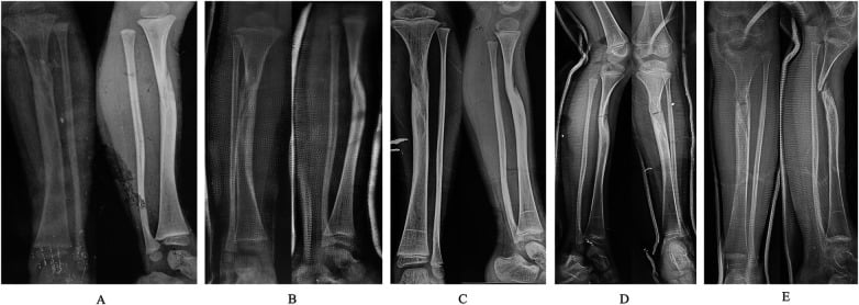
Each entity is developed in its own topic: posteromedial-bow-tibia, congenital-pseudarthrosis-of-the-tibia / neurofibromatosis, and fibular-hemimelia.
Clinical Assessment
History. The foot is noted at birth, and parents describe it as "folded up" against the shin. Ask about the pregnancy and delivery for the packaging clues (oligohydramnios, breech, a first pregnancy, twins), and about a family history of DDH or foot deformity.
Inspection. Beyond the posture itself, look for the other moulding signs: torticollis, plagiocephaly, and metatarsus adductus of the other foot.
Passive correction: the defining manoeuvre. Gently bring the foot into plantarflexion and inversion. Calcaneovalgus corrects easily past neutral, and that supple reducibility is the diagnosis. If it will not reduce, abandon the benign diagnosis and reassess for CVT.
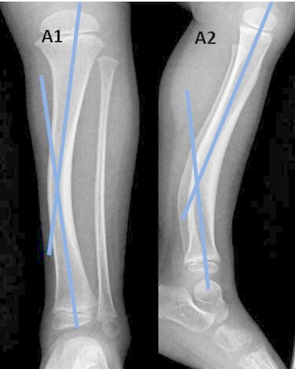
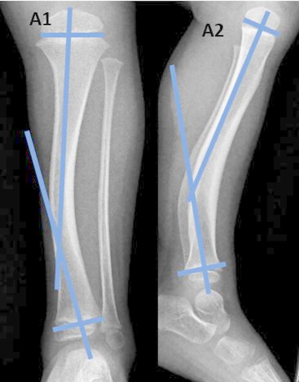
Palpate the tibia, and do not skip it. Run a thumb along the subcutaneous medial border of the tibia. A posteromedial bow (concavity facing anterolaterally) signals posteromedial bowing of the tibia, which carries a different prognosis and a leg-length problem.
Examine the hips; this is mandatory. Perform the Barlow and Ortolani tests and assess for limited abduction and leg-length difference (Galeazzi), then arrange hip ultrasound as described under Investigations.
Screen neurologically. Assess spontaneous movement, tone and the spine (sacral dimple, hairy patch) to exclude a neuromuscular calcaneus from spina bifida.
CVTRigid vs Supple: the key split
Hook:If you cannot Correct it, think Vertical talus; always check the Tibia.
Investigations
Calcaneovalgus is a clinical diagnosis. Investigations are used to exclude the mimics, not to confirm a benign postural foot, and a supple, passively correctable foot in an otherwise normal newborn needs no foot radiographs.
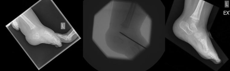
When to image the foot. Radiographs are for the rigid foot, or when doubt remains, to exclude CVT. Failure of a presumed calcaneovalgus foot to passively plantarflex beyond neutral should trigger imaging for vertical talus, coalition or neuromuscular deformity. The navicular is not ossified in the newborn, so talonavicular position is inferred from the talar and metatarsal axes rather than seen directly.
The three lateral views each answer a different question:
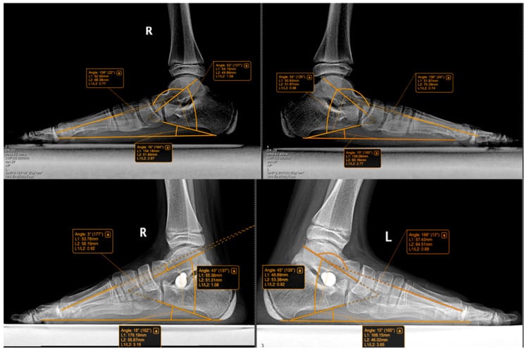
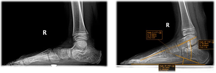
- Lateral weight-bearing or simulated standing. In calcaneovalgus Meary's (talo-first-metatarsal) angle stays essentially co-linear or only mildly disrupted, because the bones are normally aligned. In CVT the talus is vertical and the line is grossly broken, with a plantar apex.
- Forced plantarflexion lateral, "the money view". The single most useful film to separate supple from rigid. The calcaneovalgus or normal foot reduces and its alignment restores; in CVT the talonavicular joint stays dislocated.
- Forced dorsiflexion lateral. Assesses hindfoot equinus, a feature of CVT that is absent in calcaneovalgus.
The oblique talus. This is the entity between the two, and the reason the money view has three outcomes rather than two. The talonavicular joint is dorsally subluxed on the standing lateral but reduces on forced plantarflexion, so the money view looks like the benign result while the standing film looks like CVT. The abnormality persisting on the standing lateral distinguishes it from postural calcaneovalgus, and reducibility distinguishes it from CVT.
Most oblique tali are observed or treated with stretching and casting rather than open reduction. They need follow-up rather than reassurance, because a minority behave like CVT.
The tibia, if a bow is palpable. AP and lateral radiographs of the tibia and fibula confirm posteromedial bowing and provide a baseline for monitoring angular remodelling and leg-length discrepancy over time.
The hips. Given the association, arrange hip ultrasound (Graf) in the newborn period as part of DDH screening, per the local screening pathway.
Management Algorithm
Test passive correction first. A supple foot goes to reassurance and stretching, a rigid one to imaging and structural treatment, and in every case the hips are checked and the tibia palpated.
The supple foot: reassurance and stretching. This is the treatment for isolated, supple, passively correctable calcaneovalgus, the great majority:
- Reassure the parents that it is a benign positional posture that almost always resolves with growth.
- Teach passive stretching: gentle plantarflexion and inversion stretches at each nappy change.
- Review to confirm resolution, and re-examine the hips.
The outcome is excellent and most need nothing more.
The persistent foot. When the deformity has not settled over the first weeks despite stretching, continue and reinforce the stretching technique. Serial casting or splinting is occasionally used for a stubborn supple foot, holding it in the corrected plantarflexed-inverted position, though true postural calcaneovalgus rarely needs casting. Failure to correct should prompt reassessment for CVT, and the next section sets out what else to look for.
When it is not simple calcaneovalgus. Treatment follows the true diagnosis:
- Rigid rocker-bottom (CVT): treat as vertical talus, with reverse Ponseti serial casting, percutaneous Achilles tenotomy, talonavicular reduction and pinning, and later surgery if needed.
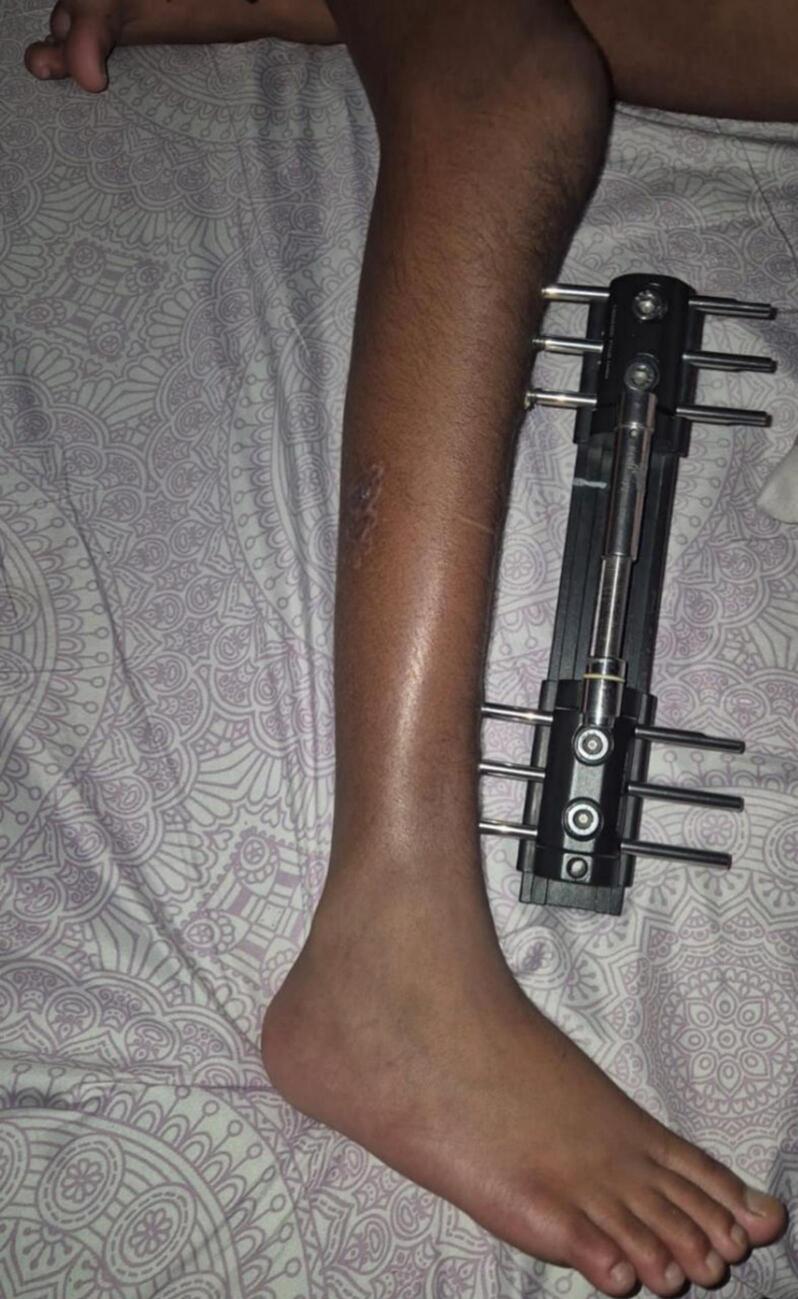
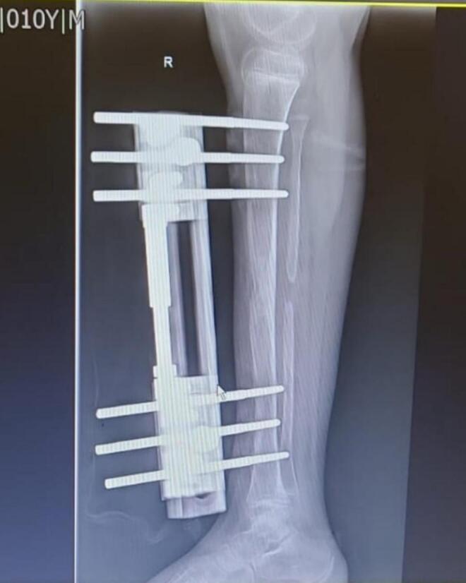
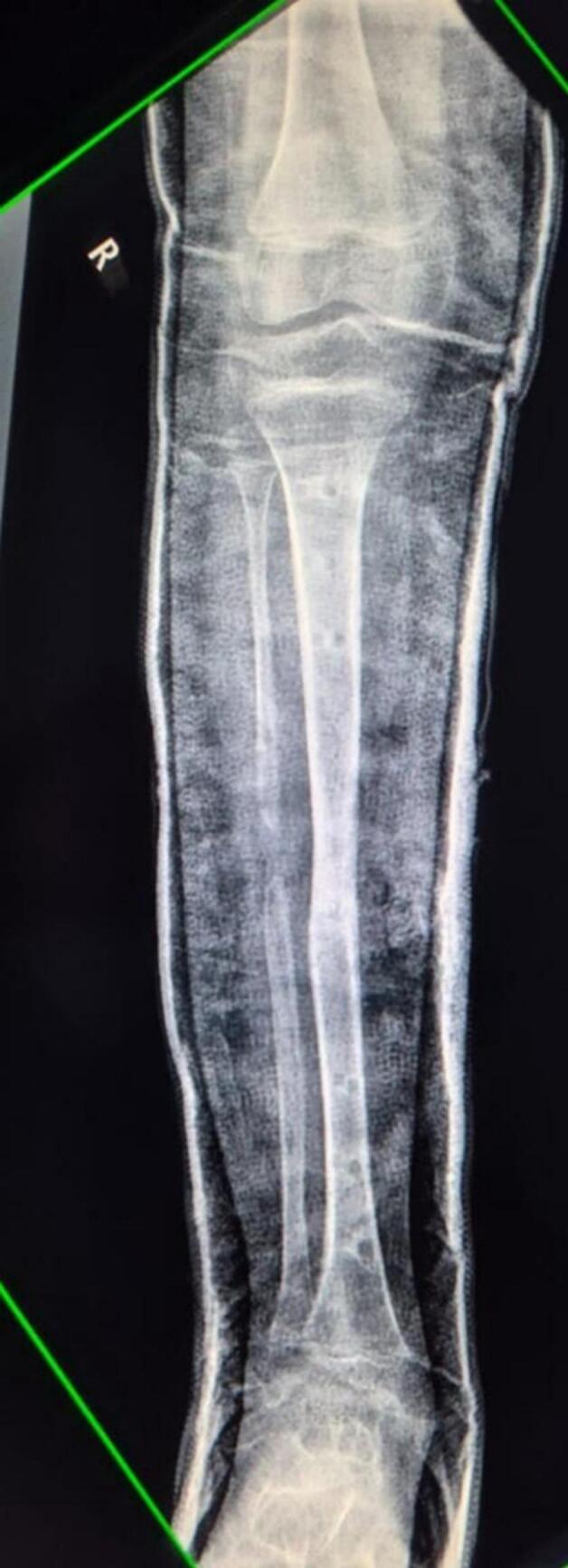
- Posteromedial bowing of the tibia: the foot posture is observed and stretched, but the focus shifts to monitoring angular remodelling and leg-length discrepancy. Significant discrepancy (commonly more than 40 mm at maturity) is managed with limb lengthening (e.g. Ilizarov); epiphysiodesis of the long side is an option for smaller discrepancies.
- Neuromuscular calcaneus: treat the underlying disorder, with tendon balancing/transfer (e.g. of the over-pulling dorsiflexors) for a paralytic calcaneus.
The Resistant Calcaneovalgus Foot
Almost every calcaneovalgus foot corrects easily, so a supple-looking foot that is unusually resistant to correction is a red flag. Do not accept "resistant calcaneovalgus"; look harder before labelling the foot benign:
- Re-test rigidity. A foot that will not reduce is not calcaneovalgus; reassess for congenital vertical talus.
- If it is still reducible but stubborn or slow, look for an associated lesion. The classic association is congenital dislocation of the peroneal tendons, in which the tendons lie displaced over the lateral malleolus, the superior peroneal retinaculum being the key stabilising structure.
- Reconsider the cause. Think of a neuromuscular calcaneus (spina bifida, sacral agenesis) if the pattern is paralytic, and a syndromic or ligamentous-laxity cause where there is generalised laxity.
What Purnell showed. Feet with a congenital peroneal tendon dislocation needed more prolonged, aggressive treatment than an isolated calcaneovalgus foot, and the children usually carried other stigmata of intrauterine packaging or oligohydramnios.
Complications
- Cause / Context
- Mistaking a rigid rocker-bottom for benign calcaneovalgus; the outcome is poor because delay worsens the structural deformity
- Prevention / Management
- Test passive correction; image the rigid foot; treat CVT early
- Cause / Context
- Failure to examine the hips in a packaging deformity; the foot is benign but a missed DDH is not
- Prevention / Management
- Barlow/Ortolani + ultrasound as part of screening
- Cause / Context
- Not palpating the tibia
- Prevention / Management
- Palpate the tibial border; baseline radiograph; monitor leg lengths to maturity
- Cause / Context
- Treating a benign foot aggressively
- Prevention / Management
- Reassurance; avoid unnecessary casting/surgery for a supple foot
Outcomes/Prognosis
The foot. The natural history is excellent: isolated postural calcaneovalgus resolves spontaneously in the great majority within the first weeks to months of life. A minority retain a flexible flatfoot posture into childhood, from residual hindfoot valgus; it is usually asymptomatic and managed expectantly, with orthotics only if painful.
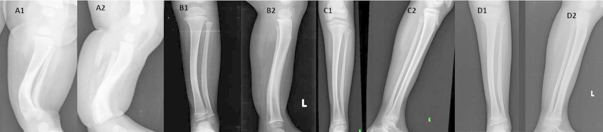
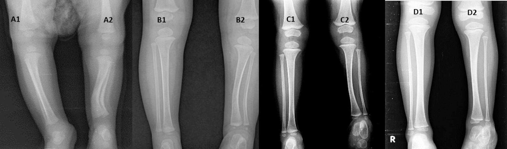
The bowed tibia. The bow remodels substantially: in one long-term series more than half of children needed no surgery for the deformity or leg length. A measurable leg-length discrepancy persists, from growth disturbance of the bowed tibia, and around 40 percent in that series ultimately required lengthening (Sevencan et al, Jt Dis Relat Surg 2022).
A residual discrepancy is the rule, so monitoring leg lengths to skeletal maturity is essential whenever a bow is present. Counsel families that the foot posture and the bow improve earlier than the final limb-length discrepancy becomes predictable.
Guidelines, Registries & Global Practice
Global epidemiology
- Calcaneovalgus and metatarsus adductus are the most frequently observed newborn foot postures worldwide; both are associated with first pregnancies, breech presentation and oligohydramnios (intrauterine packaging).
- There is no dedicated registry for calcaneovalgus; the strongest population-level data come from national newborn screening cohorts (e.g. the Norwegian cohort linking calcaneovalgus to DDH).
Side-by-side guidance
- Position on isolated supple calcaneovalgus
- Benign postural deformity; reassure and stretch
- Key action
- Examine hips for DDH
- Position on isolated supple calcaneovalgus
- Reassure; spontaneous resolution expected
- Key action
- Selective hip ultrasound per screening pathway
- Position on isolated supple calcaneovalgus
- Distinguish postural malposition from structural CVT
- Key action
- Image only the rigid or atypical foot
- Position on isolated supple calcaneovalgus
- Foot deformity is a flagged DDH risk factor
- Key action
- Trigger deliberate hip assessment
The consistent global message: treat the supple foot with reassurance, exclude the rigid mimic (CVT), and use the foot as a prompt to examine the hips and tibia.
High- versus limited-resource practice
- Well-resourced settings: ready access to hip ultrasound and tibial radiographs; structured newborn screening pathways capture the DDH association.
- Limited-resource settings: diagnosis rests on the bedside passive-correction test and clinical hip examination; the priority is not to miss a rigid CVT or an associated dislocatable hip, both of which carry far greater long-term cost than the benign foot itself.
Controversies & Areas of Uncertainty
- How aggressively to treat a slow-resolving supple foot: most authorities favour reassurance and stretching, but the role and threshold for serial casting/splinting in stubborn (yet still supple) feet is not standardised.
- Screening intensity for DDH: calcaneovalgus is an established DDH risk factor, but whether every case warrants formal ultrasound versus careful clinical examination depends on local screening pathways.
- Calcaneovalgus and later flexible flatfoot: whether infantile calcaneovalgus predisposes to symptomatic flexible flatfoot in childhood is debated; most residual flatfeet are asymptomatic.
- Timing of lengthening in posteromedial bowing: when, and at what discrepancy, to lengthen versus perform contralateral epiphysiodesis remains a judgement call, as remodelling potential is considerable but variable.
MCQ Practice Points
Q: What is the position of the foot in calcaneovalgus? A: Dorsiflexed and everted — the dorsum lies toward the shin (the mirror image of the equinovarus clubfoot).
Q: Is calcaneovalgus a structural or postural deformity? A: Postural (intrauterine packaging). The bones are normal and it is passively correctable.
Q: Which foot deformity carries the highest associated risk of DDH? A: Talipes calcaneovalgus (about 6 percent), higher than clubfoot or metatarsus adductus.
Q: Which radiograph best separates calcaneovalgus from congenital vertical talus? A: Forced plantarflexion lateral — the supple foot reduces (Meary's line restores); CVT stays dislocated.
Q: First-line treatment of isolated supple calcaneovalgus? A: Reassurance plus gentle passive stretching — most resolve spontaneously; no foot radiographs needed.
Q: A calcaneovalgus-looking foot with a palpable tibial bow suggests what? A: Posteromedial bowing of the tibia — observe the foot but monitor for leg-length discrepancy.
Additional Quiz Questions
Viva Scenarios
Practise clinical reasoning and management decisions out loud
“A midwife asks you to review a newborn whose left foot is folded up against the front of the leg. What is your approach?”
“Two newborns both have an everted, dorsiflexed-looking foot. How do you tell calcaneovalgus from congenital vertical talus, clinically and radiologically?”
“A newborn has a calcaneovalgus-looking foot, but when you palpate the leg the tibia feels bowed. What is going on and how does it change management?”
Key Features
- Dorsiflexed + everted
- Supple / correctable
- Postural (packaging)
- Resolves spontaneously
Must Exclude
- Vertical talus (rigid)
- Posteromedial tibial bow
- DDH (examine hips)
- Neuromuscular calcaneus
Investigations
- Clinical diagnosis
- No X-ray if supple
- Forced PF lateral if rigid
- Hip ultrasound (DDH)
Management
- Reassurance
- Passive stretching
- Monitor LLD (if bow)
- Treat CVT if rigid
Evidence Base
Sankar, Weiss & Skaggs — Orthopaedic Conditions in the Newborn
- Frames calcaneovalgus deformity among the core newborn foot conditions alongside metatarsus adductus, clubfoot and congenital vertical talus
- Emphasises careful physical examination with newborn-specific physiology to reach the correct diagnosis, since contractures that spontaneously improve are normal in the newborn
- Stresses that conditions such as DDH must be actively identified and treated early to prevent long-term morbidity
Haberg et al — Is Foot Deformity Associated with DDH?
- Population cohort of 60,844 Norwegian children with prospectively registered risk factors and selective ultrasound screening
- Children with a congenital foot deformity had a significantly higher rate of DDH than the general population (p less than 0.001)
- Risk of DDH was HIGHEST for talipes calcaneovalgus at 6.1 percent, exceeding clubfoot (3.5 percent) and metatarsus adductus (1.5 percent)
Sevencan et al — Evolution of Posteromedial Bowing of the Tibia Over Time
- 22 children with congenital posteromedial bowing of the tibia followed from birth to skeletal maturity
- Posteromedial bow remodelled satisfactorily in 13 of 22 (59 percent), who needed no surgery for deformity or leg-length discrepancy within their first decade
- 9 of 22 (41 percent) required Ilizarov lengthening for leg-length discrepancy (mean residual discrepancy 21 mm in the non-operated group)
Purnell et al — Congenital Dislocation of Peroneal Tendons in the Calcaneovalgus Foot
- Four neonates with congenital peroneal tendon dislocation, each associated with a calcaneovalgus foot and other stigmata of intrauterine malposition / oligohydramnios
- The calcaneovalgus foot in these cases was more resistant to correction and needed more prolonged, aggressive treatment than an isolated calcaneovalgus foot
- Identifies the superior peroneal retinaculum as the critical stabilising structure for the peroneal tendons