Retain a sound distal cement mantle and place a new stem into a prepared proximal mantle
- The retained mantle is part of the fixation construct. If it is loose, fractured, infected or malpositioned, cement-in-cement is contraindicated.
- The old stem must be removed without destabilising the mantle. A polished taper may be extracted with axial slap or controlled trephine techniques; do not lever against a thin mantle.
- The mantle is inspected and prepared circumferentially. Remove fibrous tissue, debris and loose cement while preserving the distal plug and stable distal cement.
- The new stem must be compatible with the cement-in-cement technique and fit the prepared mantle; a random stem inserted into a residual mantle is not a cement-in-cement revision.
- The technique saves distal bone but does not correct a poorly positioned mantle. Version, offset, leg length, cement mantle thickness and proximal support remain surgeon responsibilities.
- “The indication is a well-fixed, well-aligned cement mantle with a failed stem, not simply any cemented revision.
- “A polished taper and a roughened or prepared cement interface behave differently; follow the implant system's validated technique.
- “Complete cement removal is safer than retaining a mantle with an occult distal fracture or infection.
- “The common technical failure is a new stem that is too small, malrotated, underfilled or unsupported proximally.
A retained cement mantle can harbour biofilm. A sinus, positive aspiration, purulence or multiple positive deep samples requires an infection strategy, usually complete removal rather than cement-in-cement.
A mantle that looks intact on one AP film may be cracked posteriorly or distally. Open the proximal mantle enough to assess circumferential continuity and use intraoperative imaging when the distal end is uncertain.
Mark the old version, trial the new stem and control rotation before cement cures. A stem that is too anteverted or retroverted can cause instability and impingement even when radiographs look centred.
Prepare flexible reamers, cement restrictor tools, endofemoral or transfemoral extraction options and a long revision stem. The operation must not depend on a mantle that proves unsalvageable.
Definition, Indications and Contraindications
Cement-in-cement revision removes a failed femoral stem while retaining a well-fixed distal polymethylmethacrylate mantle. The proximal mantle is opened or revised, the old cement surface is cleaned and roughened or otherwise prepared according to the validated system, and a new cemented stem is inserted into the existing mantle with fresh cement. It is a bone-preserving revision strategy.
Indications
- Aseptic loosening, fracture or wear of a cemented femoral stem with a well-fixed distal mantle.
- A well-positioned mantle with sufficient length, diameter and circumferential integrity for the planned new stem.
- A patient in whom preserving femoral bone and avoiding extensive cement extraction reduces fracture or perforation risk.
- A revision in which stem offset, length, version and proximal support can be restored within the existing cement geometry.
Contraindications
- active or incompletely investigated periprosthetic joint infection;
- a sinus tract, purulence, infected cement or positive evidence that cannot be safely explained away;
- gross distal mantle loosening, a circumferential fracture or a distal cement defect;
- severe mantle malposition causing unacceptable version, leg length or offset;
- a mantle that is too narrow, too short or too thin for the new stem;
- distal femoral fracture, perforation or severe osteolysis that requires a different revision construct;
- an implant or cement system without a validated cement-in-cement compatibility and preparation method.
A mantle may be retained only after clinical, laboratory, radiographic and intraoperative assessment. The threshold for complete cement removal should be low when the patient has infection risk, unexplained pain or a mantle that cannot be inspected reliably.
Anatomy and Technical Hazards
The femoral canal is surrounded by a thin cortex in the proximal femur and a stronger diaphysis. A retained mantle may be eccentric, tapered, cracked or bonded to the cortex. The operation is performed close to the greater trochanter, abductors, calcar, femoral vessels and sciatic nerve.
- The greater trochanter and abductors control exposure and postoperative stability. Excessive trochanteric stripping weakens the hip and may require an osteotomy.
- The calcar and medial cortex are common sites of proximal bone loss and fracture. A new stem with inadequate medial support can subside or cantilever.
- The lateral femoral cortex may be thinned by old windows, cerclage or cement extrusion. Broaching or stem insertion can propagate a crack.
- The femoral nutrient vessels and endosteal blood supply are reduced by circumferential stripping and repeated cement removal. Preserve cortex and periosteal attachments.
- The sciatic nerve lies posterior to the hip and is at risk from excessive traction, leg lengthening, posterior retractors and cement extrusion.
- The femoral canal may be bowed. A straight stem or instrument can perforate the cortex when the retained mantle is eccentric.
The safest operation is controlled, visualised and reversible: expose enough to inspect, use flexible tools, recheck imaging and have a plan for a fracture or a mantle that must be removed.
Mantle Classification for the Operation
A practical operative classification is more useful than a label that does not change the plan:
- Radiographic and intraoperative features
- Continuous circumferential mantle, no distal lucency or fracture, acceptable axis and length
- Plan
- Cement-in-cement revision is reasonable
- Radiographic and intraoperative features
- Small proximal defect with sound distal fixation and a reproducible repair or augment technique
- Plan
- Retain only if the repair and new stem achieve a validated stable construct
- Radiographic and intraoperative features
- Motion, distal debonding, circumferential crack, missing plug or major radiolucency
- Plan
- Remove all cement and use a full femoral revision strategy
- Radiographic and intraoperative features
- Sinus, purulence, positive evidence or an uninspectable mantle
- Plan
- Stage infection and remove contaminated cement
Before proceeding, use a long thin instrument or a controlled tap to assess whether the distal mantle is fixed. Do not accept a false sense of security from a proximal collar that is merely held by scar.
Planning the New Stem
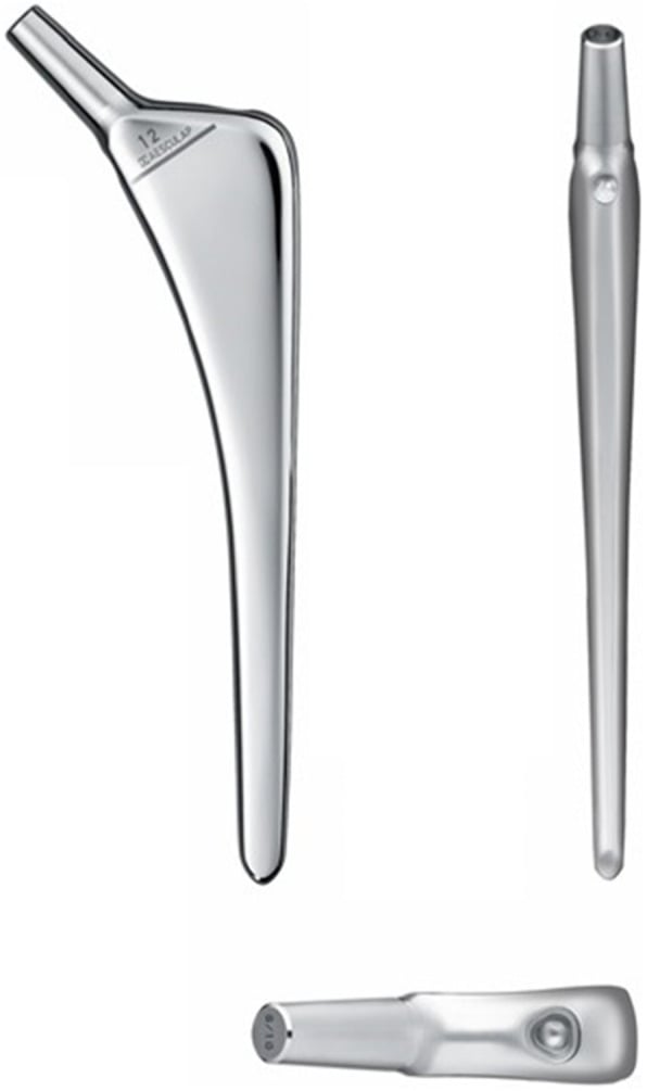
The new stem should fit the retained mantle after preparation, not the old stem's dimensions. Template the stem on AP and lateral femur:
- maintain or intentionally change the hip centre;
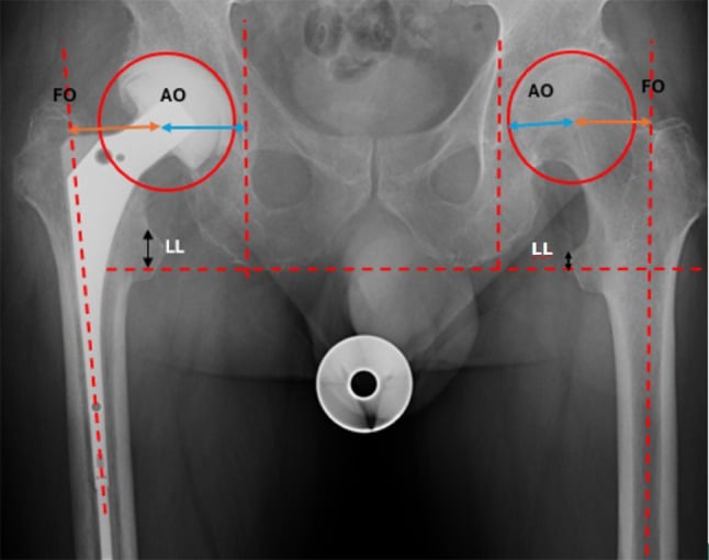
- restore offset and leg length without excessive lengthening;
- select an appropriate distal diameter and length within the mantle;
- achieve the planned version relative to the femoral neck, lesser trochanter and contralateral hip;
- provide proximal support and a cement mantle that surrounds the stem without a gross void;
- confirm compatibility of the stem geometry, cement, centraliser and restrictor with the cement-in-cement technique.
A polished tapered stem may rely on controlled subsidence within a cement mantle, whereas a composite beam or rough stem follows different mechanics. Do not transpose the target mantle thickness, centralisation method or cement handling from one system to another. Read the device-specific technique and use the stem family for which the cement-in-cement evidence exists.
Assessment and Imaging
History and examination
Clarify the index stem design, cement type if known, date of implantation, prior revision, instability, fracture, infection, dislocation, leg-length change and the patient's functional goals. Examine gait, abductor strength, hip flexion and rotation, scars and sciatic function. Document pre-existing foot drop or sensory changes before surgery.
Look for infection: wound drainage, a sinus, warmth, rest pain, night pain and systemic symptoms. Aseptic loosening and low-grade infection overlap clinically; the investigation must be composite.
Infection work-up
Obtain serum inflammatory markers and aspirate the hip when feasible before antibiotics. Send fluid for cell count with differential and culture according to the laboratory protocol. At revision, obtain multiple separately labelled deep tissue samples and histology before the therapeutic antibiotic dose when the patient is stable. A negative culture does not prove an aseptic failure in a patient with a sinus or other major criteria.
Radiographs
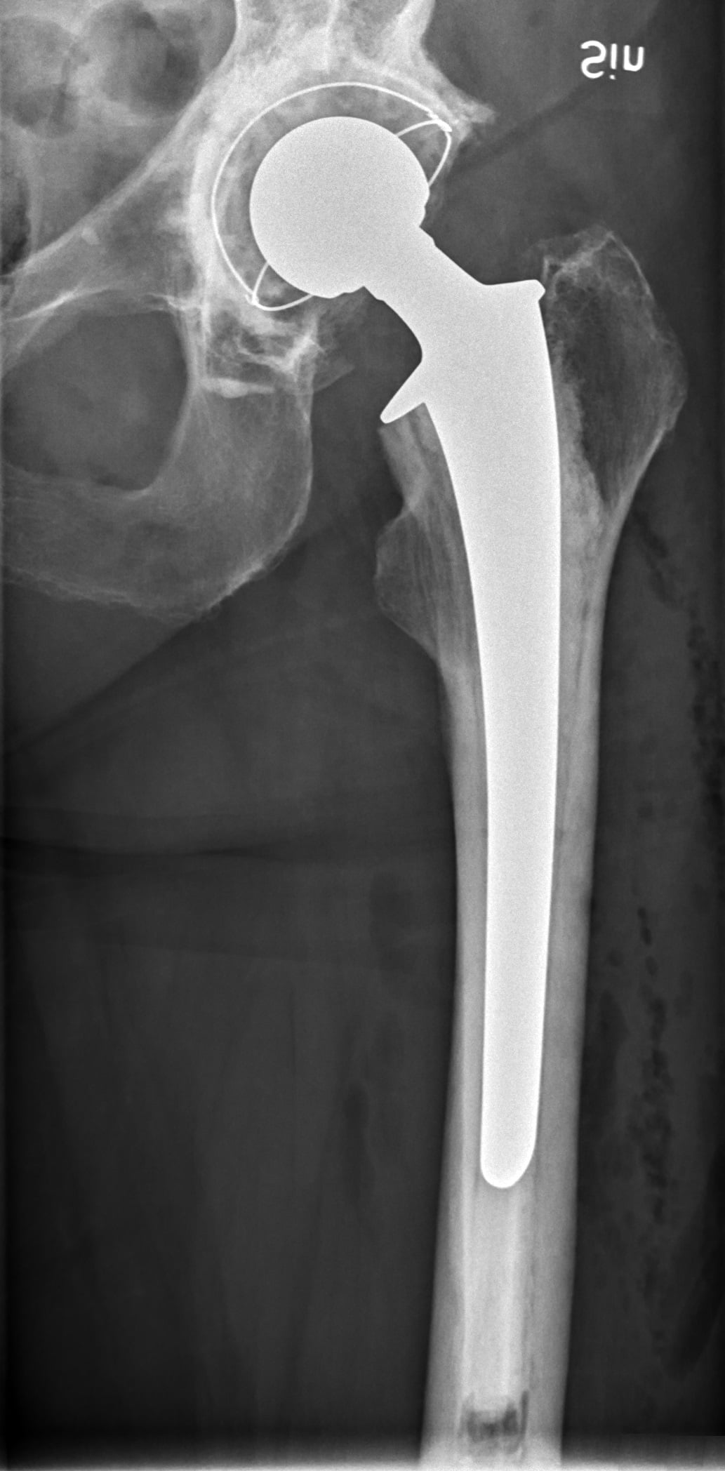
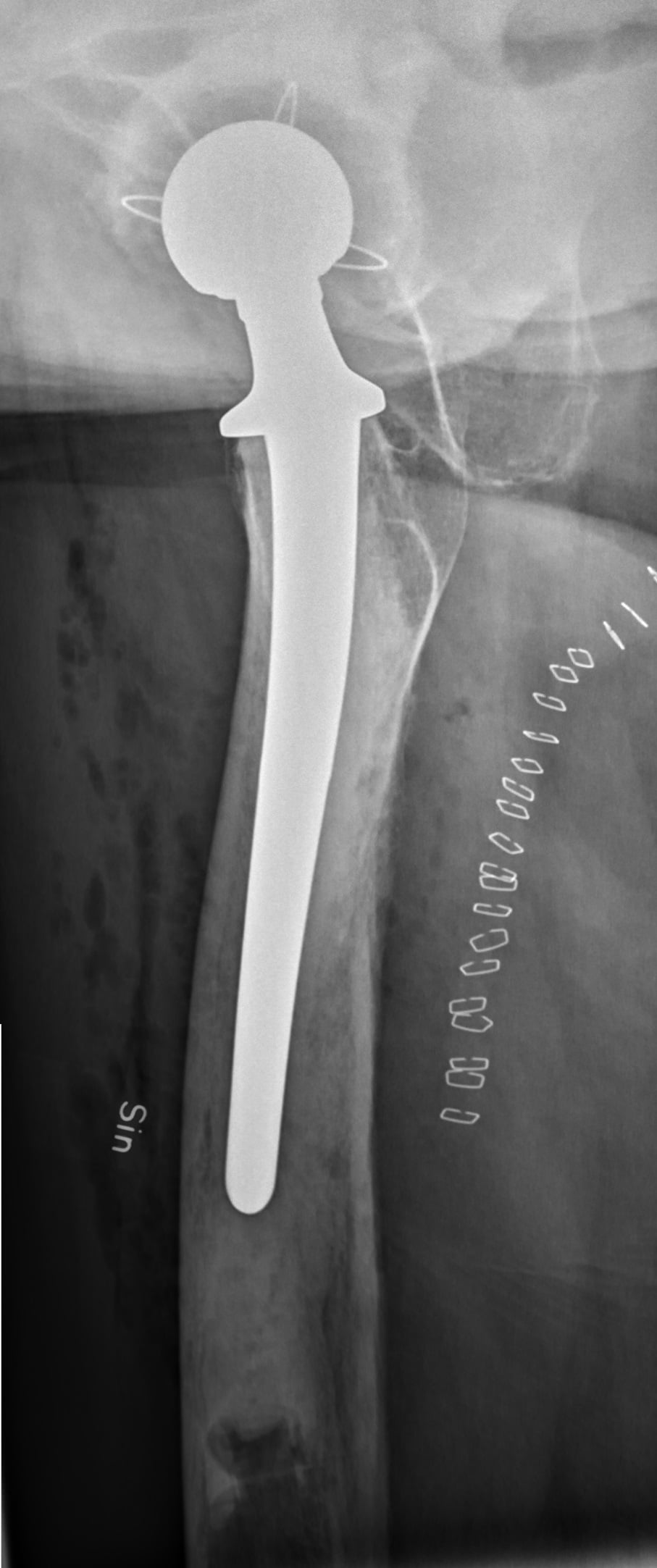
Obtain AP pelvis, AP and lateral femur including the whole cement mantle, and views of the hip and knee when the planned stem will span a distal defect. Assess:
- radiolucent lines in each Gruen zone;
- mantle fracture, distal pedestal, cement extrusion and endosteal scalloping;
- stem subsidence, varus or valgus, version clues and proximal calcar loss;
- the cement mantle's length, thickness and relation to the femoral cortex;
- retained cerclage, cables, wires and previous cortical windows;
- hip centre, offset, leg length and the opposite femur for stem templating.
Radiographs can underestimate a posterior or distal mantle defect. CT is useful for complex osteolysis, cortical perforation, an uncertain distal cement plug or a suspected fracture, but metal artefact may limit detail. Compare with serial images; progressive subsidence and new radiolucency are stronger than a single borderline line.
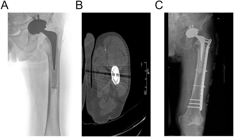
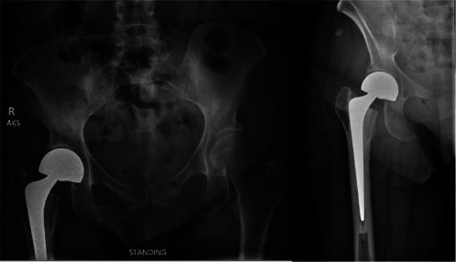
Operative Technique: PIPADRAW
Cement-in-cement femoral revision sequence
- Use the lateral decubitus position for a posterior or direct lateral approach, or supine for an anterior approach, with the pelvis secured and the entire femur accessible to fluoroscopy.
- Prep from the iliac crest to the foot. Keep the opposite hip accessible for comparing leg length and rotation.
- Protect pressure points and plan for blood loss, cell salvage and a possible extended exposure.
- Confirm AP and lateral imaging of the whole femur. Have flexible osteotomes, stem extraction tools, cement drills, long guidewires, trephines and a full-removal revision stem ready.
- Compare the implant label and stem design with the preoperative template. Do not assume that an apparently similar stem is compatible with the retained mantle.
- Keep cerclage cables, plates and a strut graft available for an intraoperative femoral fracture.
- Administer antibiotics after deep cultures when an infection assessment is required and the patient is stable; use a therapeutic regimen when infection is established.
- Confirm the preoperative aspiration, imaging, mantle classification and rescue plan in the team briefing.
- Use a tourniquet only if it does not compromise soft tissue or obscure the assessment of bleeding bone.
- Reuse the safest previous incision when feasible and preserve viable skin bridges. Identify the abductor and capsule according to the chosen approach.
- Expose the proximal femur enough to see the stem, collar, calcar and mantle; avoid stripping the entire femur.
- If an extended trochanteric osteotomy or cortical window is required, plan it before levering the stem.
- Clear the collar and proximal cement, then attach the extraction device in line with the stem.
- Apply controlled axial blows or the validated extraction method. Avoid levering against the proximal cortex or retained mantle.
- Stop if the mantle moves with the stem. Reassess whether the distal mantle is actually fixed and convert to complete cement removal if necessary.
- Examine the mantle circumferentially for cracks, distal debonding, fibrous tissue, cement fragments and a retained distal plug.
- Remove loose proximal cement and membrane. Prepare or roughen the interface exactly as specified by the implant system; do not gouge through the distal mantle.
- Pass a flexible instrument to confirm the length and central continuity of the mantle. Use fluoroscopy if the distal endpoint is uncertain.
- Trial the new stem or broach inside the prepared mantle and reduce the hip.
- Check hip centre, offset, leg length, version, abductor tension and stability through flexion, adduction and rotation.
- Compare the femoral version and foot progression with the other side. A mantle that forces malrotation is not a successful salvage.
- Maintain the femur in line with the canal and avoid eccentric reaming or stem insertion.
- Protect the lateral cortex around old windows and cables; use cerclage before a crack propagates when appropriate.
- Avoid excessive traction or lengthening that can injure the sciatic nerve.
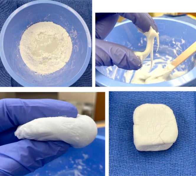
- Irrigate and dry the mantle according to the cement system. Apply fresh low-viscosity or high-viscosity cement as specified, with a controlled retrograde technique and appropriate vacuum or pressurisation when possible.
- Insert the new stem in the planned version and depth, maintaining axial control until the cement cures. Use the specified centraliser, restrictor and cement mantle preparation.
- Remove excess cement before it hardens, especially posteriorly and around the calcar, while protecting the sciatic nerve and soft tissue.
- Obtain AP and lateral images of the whole femur before closure.
- Check stem depth, version clues, cement mantle continuity, cortical cracks, offset and leg length. Image the knee if the stem tip or old hardware is near it.
- If a fracture or mantle defect is identified, stabilise it before closure and revise the plan rather than accepting a compromised construct.
- Repair capsule, abductors, vastus and fascia according to the approach. Close the skin without tension and protect the soft tissue.
- Use approach-specific hip precautions and weight bearing according to stem stability, fracture risk and any osteotomy or graft.
- Begin thrombosis prevention, antibiotics as indicated, ankle pumps and physiotherapy with a clear plan for transfers and gait aids.
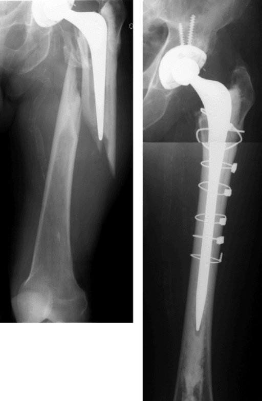
Complications and Conversion to Full Revision
- Clues
- Stem and cement move together, crack or distal plug displacement
- Immediate response
- Stop levering, obtain fluoroscopy and protect the femur
- Salvage
- Complete cement removal, fracture fixation and a long revision stem or staged reconstruction
- Clues
- Persistent drainage, pain, fever, sinus or positive cultures
- Immediate response
- Re-open the diagnosis and obtain cultures
- Salvage
- Remove contaminated cement and stage; do not simply exchange the stem in place
- Clues
- Early shortening, changing offset or progressive radiolucency
- Immediate response
- Protect weight bearing and obtain full femoral imaging
- Salvage
- Assess mantle and stem compatibility; revise with a stable cement or uncemented construct
- Clues
- Crack at the stem tip or old window, new pain or deformity
- Immediate response
- Stabilise and image the full femur
- Salvage
- Bypass the fracture with a long revision stem, cable-plate or strut graft as indicated
- Clues
- Early instability, poor offset or malversion
- Immediate response
- Reduction and sciatic assessment
- Salvage
- Correct the mechanical cause; do not rely on a liner exchange alone if the stem position is wrong
- Clues
- Cement in soft tissue, nerve symptoms or poor mantle pressurisation
- Immediate response
- Remove accessible excess before cure and assess nerve/vascular status
- Salvage
- Debride or revise if compressive, infected or mechanically compromising
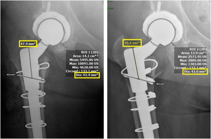
Postoperative Care and Outcomes
Weight bearing depends on stem stability, bone quality, the retained mantle, any osteotomy and the presence of a fracture. A straightforward cement-in-cement revision may permit earlier loading than a full femoral reconstruction, but an old or thin mantle does not justify unrestricted loading without radiographic and clinical review.
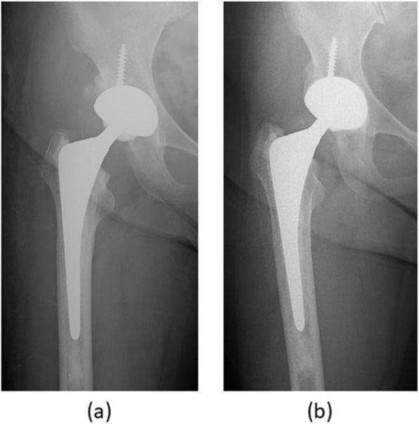
Follow the wound, sciatic function, leg length, hip stability and pain. Obtain AP and lateral femur radiographs at planned intervals and compare with the immediate postoperative films. Look for stem subsidence, cement mantle fracture, progressive radiolucency, new osteolysis, periprosthetic fracture and dislocation. A small controlled subsidence may be expected for some polished taper systems; progressive migration with pain is not.
Guidelines, Registries & Global Practice
Global evidence. Cement-in-cement revision is a niche, bone-preserving technique supported by cohort studies and systematic reviews rather than randomised trials. Systematic reviews generally report encouraging survivorship in carefully selected cases, but the evidence is affected by selection of well-fixed mantles, variable stem designs and inconsistent definitions of radiographic failure.
Practice principles:
- Use a composite infection assessment before retaining cement. The technique is not an infection-control strategy.
- Retain only a mantle that is well fixed, intact, adequately positioned and compatible with the new stem and cement system.
- Preserve femoral bone and blood supply, but convert to complete cement removal when the mantle is loose, fractured, malpositioned or impossible to inspect.
- Restore version, offset and leg length; a retained mantle does not excuse malalignment.
- Use a full-removal rescue plan and counsel about fracture, stem subsidence, dislocation, infection and later revision.
Registries. National arthroplasty registries report outcomes of cemented and uncemented femoral revisions but usually do not identify cement-in-cement as a separate, adequately powered category. Centres should record mantle status, stem design, cement system, reason for revision, infection status, fracture, reoperation and radiographic migration.
Global practice. Where validated cement-in-cement implant systems and extraction tools are available, the technique can reduce operative time and distal femoral damage. Where compatibility data or intraoperative imaging are limited, complete cement removal with a standard revision strategy may be safer. The safe choice is the one the team can execute and revise, not the one that appears shortest on paper.
MCQ Practice Points
Q: When is cement-in-cement revision indicated? A: A failed cemented femoral stem with a well-fixed, intact, acceptably positioned distal cement mantle. A large defect or a cemented stem alone is not enough.
Q: When must the retained cement mantle be removed instead? A: Active or uncertain infection, distal loosening, a circumferential fracture, major cement defect, severe malposition or an uninspectable mantle. Retaining a bad mantle converts a controlled problem into a hidden mechanical or biological failure.
Q: What must be checked before the cement cures? A: Stem depth, version, hip centre, offset, leg length, stability, mantle fill and protection of the sciatic nerve. Version cannot be corrected after the cement has set without another revision.
Q: Why use the cement-in-cement technique? A: It preserves distal femoral bone, reduces cement extraction trauma and can shorten a selected revision. It does not necessarily reduce the overall biological or mechanical risk of revision arthroplasty.
Q: What is the commonest intraoperative conversion, and to what? A: An intraoperative mantle fracture or evidence of distal loosening requires conversion to complete cement removal and a full femoral revision plan, often with a long stem and fracture fixation.
In-cement technique for revision hip arthroplasty
- Consecutive series of 54 in-cement femoral revisions in 51 patients (mean age 70.3 years) retaining the original cement-bone interface
- 42 procedures followed for a mean of 29.2 months (6 to 51) with clinical and radiological assessment
- No radiological evidence of loosening at follow-up
- The retained mantle technique is biomechanically stronger than recementing after complete mantle removal
The cement-in-cement technique is a reliable option in hip arthroplasty revision surgery: a systematic review
- PRISMA systematic review of 16 non-comparative studies: 1899 hips in 1856 patients (mean age 72.2, 37% male), mean follow-up 7.2 years
- Intraoperative femoral or acetabular fracture 5.3%, manageable without relevant sequelae
- Dislocation 2.8% (1.6% requiring re-revision); aseptic-loosening failure of the revised component only 2%
- Re-revision rate 9.3%; functional patient-reported outcomes improved above the minimal clinically important difference
Exam Viva Scenarios
Practise clinical reasoning and management decisions out loud
“A 78-year-old has painful aseptic loosening of a polished cemented femoral stem. The AP and lateral films show a continuous distal mantle without radiolucency or fracture, acceptable version and a stable cement plug. How do you plan the revision?”
“During extraction, the old stem and the distal cement mantle move together. Preoperative films had suggested a fixed mantle. What is the correct response?”
“A patient planned for cement-in-cement revision has normal inflammatory markers and a dry aspiration. At surgery there is purulent-appearing proximal membrane and two deep samples later grow the same organism. The new stem has not yet been cemented. What do you do?”
Indication
- Failed cemented femoral stem with a well-fixed, intact, adequately positioned distal mantle
- Usually aseptic loosening or stem failure, after a composite infection assessment
- Compatible new stem, cement system and sufficient mantle length and diameter required
- Preserves distal bone and avoids extensive cement extraction
Contraindication
- Active, suspected or incompletely investigated infection
- Loose, fractured, distal or uninspectable mantle
- Severe malposition, inadequate length or an incompatible implant system
- Convert to complete cement removal and a full revision plan when doubt remains
PIPADRAW
- Position and image the whole femur; prepare extraction and rescue tools
- Approach through safe scar, remove the stem axially and inspect the mantle circumferentially
- Prepare the retained cement according to the validated system; protect cortex and sciatic nerve
- Trial length, offset, version and stability; cement the new stem under axial control
- Image, close, protect weight bearing and monitor migration, fracture, infection and dislocation
Failure
- Mantle moves or fractures: stop, protect femur and remove all cement
- Positive cultures or purulence: abandon definitive revision and stage infection
- Subsidence: exclude infection and mantle failure, then revise with stable fixation
- Dislocation: correct the cause; do not simply exchange the liner
- Always have a full-removal and fracture-bypass plan
References
- Haddad FS, Masri BA, Garbuz DS, Duncan CP. Femoral bone loss in total hip arthroplasty: classification and preoperative planning. Instr Course Lect. 2000;49:83-96. PMID: 10829164.
- Lieberman JR, Moeckel BH, Evans BG, Salvati EA, Ranawat CS. Cement-within-cement revision hip arthroplasty. J Bone Joint Surg Br. 1993;75(6):869-871. PMID: 8245073.
- Lie SA, Havelin LI, Furnes ON, Engesaeter LB, Vollset SE. Failure rates for 4762 revision total hip arthroplasties in the Norwegian Arthroplasty Register. J Bone Joint Surg Br. 2004;86(4):504-509. PMID: 15174543.
- Mandziak DG, Howie DW, Neale SD, McGee MA. Cement-within-cement stem exchange using the collarless polished double-taper stem. J Arthroplasty. 2007;22(7):1000-1006. PMID: 17920472.
- Cnudde PHJ, Kärrholm J, Rolfson O, Timperley AJ, Mohaddes M. Cement-in-cement revision of the femoral stem: analysis of 1179 first-time revisions in the Swedish Hip Arthroplasty Register. Bone Joint J. 2017;99-B(4 Supple B):27-32. PMID: 28363891.
- Xará-Leite F, Pereira AD, Andrade R, et al. The cement-in-cement technique is a reliable option in hip arthroplasty revision surgery: a systematic review. Eur J Orthop Surg Traumatol. 2021;31(1):7-22. PMID: 32666308.
- Gie GA, Linder L, Ling RS, et al. Impacted cancellous allografts and cement for revision total hip arthroplasty. J Bone Joint Surg Br. 1993;75(1):14-21. PMID: 8421012. DOI: 10.1302/0301-620X.75B1.8421012.
- Keeling P, Prendergast PJ, Lennon AB, Kenny PJ. Cement-in-cement revision hip arthroplasty: an analysis of clinical and biomechanical literature. Arch Orthop Trauma Surg. 2008;128(10):1193-1199. PMID: 17940780.
- Stefanovich-Lawbuary NS, Parry MC, Whitehouse MR, Blom AW. Cement in cement revision of the femoral component using a collarless triple taper: a midterm clinical and radiographic assessment. J Arthroplasty. 2014;29(10):2002-2006. PMID: 24927866.
- Ling RS, Timperley AJ, Linder L. Histology of cancellous impaction grafting in the femur. A case report. J Bone Joint Surg Br. 1993;75(5):693-696. PMID: 8376422. DOI: 10.1302/0301-620X.75B5.8376422.