A revision-like arthroplasty operation with fracture, infection, bone-loss and abductor risks
- Conversion THA has higher infection, fracture, dislocation, abductor and revision risk than routine primary THA.
- Obtain full-length femoral imaging and plan every screw, nail, cement fragment and stress riser before choosing the stem.
- A sinus or suspicious nonunion requires an infection pathway; a normal CRP does not prove aseptic failure.
- The femoral neck, greater trochanter and abductor mechanism may be absent or distorted, so a standard primary stem and direct anterior plan may be unsafe.
- Counsel about possible osteotomy, long stem, trochanteric fixation, staged surgery, leg-length correction and postoperative restrictions.
- “The key difference from primary THA is altered anatomy plus retained hardware and stress risers.
- “Remove a cephalomedullary nail or plate in the planned sequence; do not create a femoral fracture by extracting a broken screw or nail blindly.
- “A long revision stem should bypass screw holes, osteotomy and fracture by a sufficient length and obtain distal fixation.
- “If infection is found, stop the conversion and stage the reconstruction rather than cementing through a contaminated femur.
Failed fracture fixation can harbour low-grade infection. Use serum markers, aspiration when feasible, preoperative planning for multiple deep cultures and histology, and a staged strategy when infection is established.
Radiographs and CT show screw trajectories, nail locking bolts, cement, cortical windows, nail bow and distal stress risers. Extraction is a separate operation within the arthroplasty.
Greater trochanter nonunion, lateral plate removal and gluteal scarring increase limp and dislocation risk. Plan trochanteric fixation or a soft-tissue reconstruction.
Prepare cerclage, a long revision stem, plate, cables, strut graft, flexible extraction tools and an osteotomy option. A broken screw can become a cortical fracture if forced.
Definition and Indications
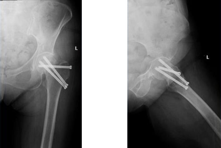
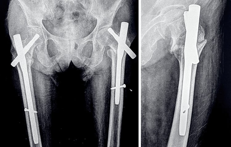
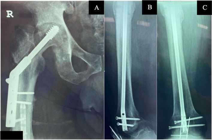
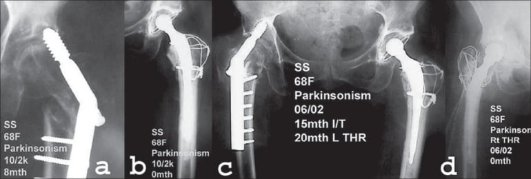
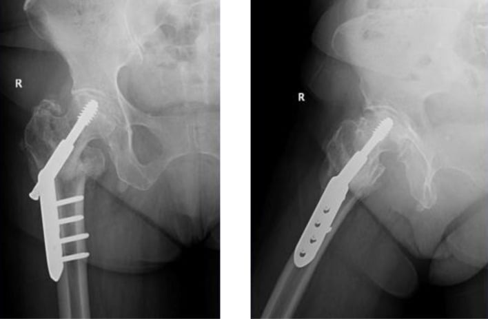
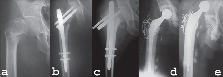
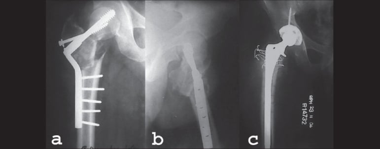
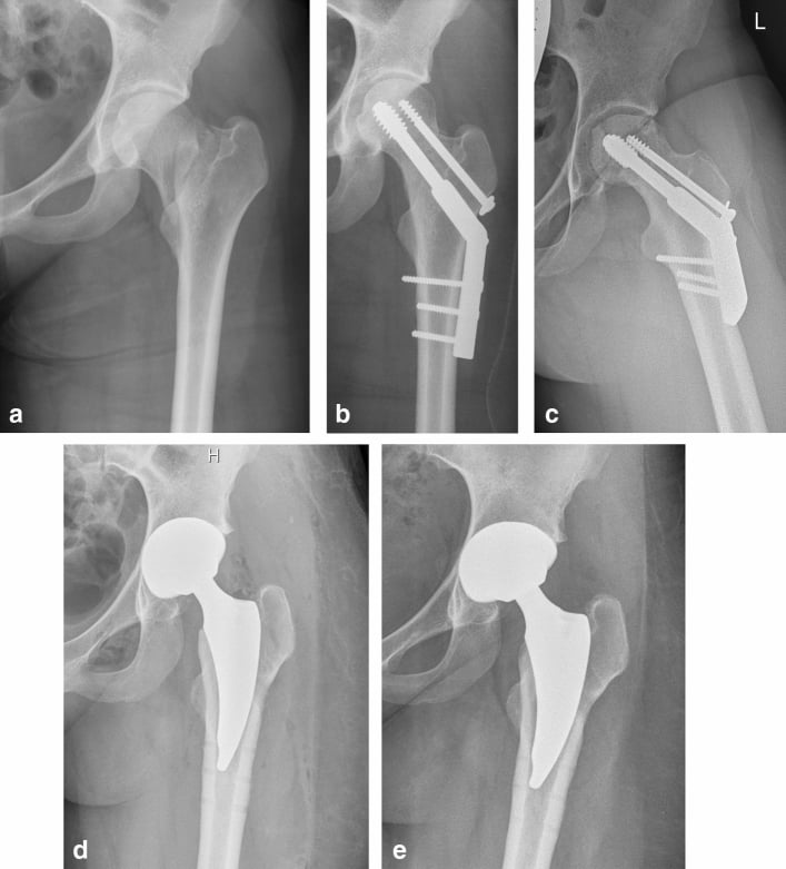
Conversion THA is total hip arthroplasty performed after failed internal fixation of a proximal femoral or acetabular fracture. Failure may be nonunion, malunion, collapse, avascular necrosis, post-traumatic arthritis, implant cut-out, broken hardware, infection or painful hardware with a non-reconstructable hip.
Common indications
- Femoral neck nonunion or avascular necrosis after screws, a dynamic hip screw or other fixation.
- Intertrochanteric or subtrochanteric nonunion with varus collapse, loss of the femoral neck or failed cephalomedullary nail.
- Post-traumatic acetabular or femoral-head degeneration with retained fixation that cannot be safely left in place.
- Cut-out, implant breakage, migration or a painful malunion that prevents useful hip reconstruction.
- Selected infection-controlled cases after staged debridement and fracture-healing assessment.
The indication should explain why THA is preferred over revision fixation, valgus/proximal femoral osteotomy, hemiarthroplasty, fusion or resection. A patient with a reconstructable nonunion and preserved joint may not need conversion THA.
Anatomy and Hazards After Fixation
- Femoral neck and head: collapse shortens the limb and changes the centre; screws may have penetrated the joint or damaged the head.
- Greater trochanter and abductors: lateral plates, cables, nonunion and previous approaches can disrupt the abductor lever arm.
- Proximal femoral canal: nails and cement may have enlarged, bowed or blocked the canal; a primary stem can perforate or fail to bypass stress risers.
- Old screw holes and locking bolts: each is a stress riser and must be bypassed or protected by the revision stem.
- Sciatic nerve: posterior scars, length restoration and retractors create risk. Document baseline function and avoid excessive acute lengthening.
- Femoral vessels and soft tissues: previous surgery may make the iliopsoas, femoral vessels and capsule adherent, particularly with anterior hardware.
- Acetabulum: post-traumatic bone loss, screws and protrusio can make the acetabular side a revision problem too.
Assessment and Examination
History
Record fracture pattern, original reduction, implant type, every revision, weight-bearing history, infection or wound drainage, antibiotics, falls, head collapse, pain location, leg-length change and pre-injury function. Ask about current aids, home stairs, hip and spine disease and expectations.
Examination
Inspect scars, sinus, trochanteric prominence and limb shortening. Observe gait, Trendelenburg sign and transfers. Palpate the greater trochanter, fracture site, hardware and groin. Measure hip flexion, extension, abduction and rotation; assess fixed flexion or external rotation deformity.
Document sciatic, femoral and peroneal function, pulses and skin perfusion. Examine the opposite hip, knee and ankle. Test the abductor mechanism and note whether a greater trochanteric fragment moves independently.
Infection
Obtain serum inflammatory markers and aspirate the hip or nonunion when feasible. Plan multiple deep tissue and membrane samples and histology at surgery. A draining sinus or purulent nonunion is infection until proven otherwise. A negative aspiration does not override a strong clinical or radiographic suspicion.
Imaging and Templating
Obtain AP pelvis, AP and lateral views of the full femur including the knee, and long-leg alignment when shortening or deformity is substantial. Compare with old films to understand implant migration and fracture healing.
CT with metal artefact reduction is useful for:
- nonunion geometry and malunion;
- femoral head collapse, intra-articular screw penetration and acetabular bone loss;
- nail bow, distal locking bolts, cement, cortical windows and stress risers;
- canal patency and distal fixation length;
- a suspected infection cavity or sequestrum;
- vessel or organ proximity when hardware has migrated.
Template the conversion from the femur upward:
- plan the order of hardware removal and whether any implant can be left temporarily;
- choose a stem that bypasses every screw hole, osteotomy and fracture;
- restore hip centre, offset, version and length without sciatic over-lengthening;
- plan calcar replacement, modular proximal body, cable-plate or trochanteric fixation;
- have extraction tools for broken screws and a full-removal rescue strategy.
Approach and Reconstruction Choice
The approach depends on scars, hardware, deformity, the acetabular side and the need for an osteotomy. A posterior or lateral approach may facilitate nail removal and trochanteric reconstruction; an anterior approach may be possible in selected femoral neck failures but does not eliminate the need to map posterior hardware and distal stress risers.
- Preferred strategy
- Cementless or cemented THA with planned hardware removal
- Important modification
- Check abductor and calcar; use a modular or standard stem only if the canal is safe
- Preferred strategy
- Long revision stem or proximal femoral reconstruction
- Important modification
- Fix trochanteric fragment and bypass the nonunion and screw holes
- Preferred strategy
- Long diaphyseal-engaging stem with cables/plate or osteotomy
- Important modification
- Map nail extraction and bypass the fracture by reliable distal fixation
- Preferred strategy
- Revision acetabular shell, augment, cage or other reconstruction
- Important modification
- Plan both sides as a revision THA, not an isolated femoral conversion
- Preferred strategy
- Staged removal, debridement and temporary antibiotic construct
- Important modification
- Delay definitive THA until infection and coverage are controlled
Operative Technique: PIPADRAW
Conversion THA sequence
- Use lateral or supine positioning according to the approach, secure the pelvis and keep the entire femur and foot accessible.
- Prep from iliac crest to foot; include old lateral or distal locking incisions and potential osteotomy/flap fields.
- Protect pressure points and coordinate blood conservation, cell salvage and nerve monitoring when deformity or lengthening is substantial.
- Confirm AP and lateral images of the hip, full femur and knee. Have broken-screw extraction tools, flexible reamers, nail extractors, cerclage, cables, plates and revision stems ready.
- Verify implant type, locking-bolt positions, cement, canal bow and planned stem bypass before incision.
- Keep a temporary spacer or staged pathway available if infection is encountered.
- Administer antibiotics after deep samples when the patient is stable and infection must be characterised; treat sepsis immediately.
- Confirm the hardware-removal sequence, acetabular plan, stem plan, trochanteric fixation and postoperative weight-bearing restrictions.
- Prepare the limb and skin so every sinus can be excised and the final closure remains viable.
- Use the safest previous incision or an extensile approach with preserved skin bridges. Excise sinus tracts en bloc.
- Expose the greater trochanter, fracture/nonunion and hardware enough to control extraction. Protect abductors and the sciatic nerve.
- Do not force the hip into dislocation when screws or nail fragments can be removed through a planned separate window.
- Remove locking bolts and proximal hardware before extracting a nail; remove plates and screws with the planned sequence, using fluoroscopy for buried or broken components.
- If a screw is cold-welded or broken, use a trephine or extraction system rather than excessive torque against thin cortex.
- Obtain multiple deep samples and histology before antibiotics when appropriate. Debride infected membrane and non-viable tissue.
- Remove the femoral head or failed neck segment and prepare the acetabulum. Treat acetabular bone loss with the appropriate revision strategy.
- Identify the true femoral canal, remove cement or debris, and ream along the canal while protecting the cortex.
- Freshen the nonunion only to viable bone and prepare the greater trochanter for cable, plate or abductor repair.
- Trial the stem and head, checking hip centre, offset, length, version, abductor tension and sciatic nerve tension.
- Compare the foot progression and knee/ankle clearance with the opposite limb. A long-standing shortened hip may not tolerate complete anatomical length restoration.
- Test stability through flexion, adduction and rotation; assess the trochanteric fragment and soft-tissue repair.
- Bypass every screw hole, cortical window, fracture and nonunion with the planned revision stem; use cerclage before stem insertion if a crack is likely.
- Avoid excessive traction or acute lengthening that can injure the sciatic nerve. Protect the femoral vessels during anterior or medial dissection.
- Preserve viable abductors and soft tissue; a primary approach that destroys the only functioning abductor is not a safe shortcut.
- Insert the selected cemented or uncemented stem with stable distal fixation and planned version. Use a long stem or modular body when proximal bone is deficient.
- Fix the greater trochanter or nonunion with cables, plate, graft or a strut as indicated. Reconstruct the capsule and abductors.
- Place the acetabular shell, liner and femoral head, then repeat stability, length and offset checks.
- Obtain AP and lateral images of the pelvis and full femur including all old hardware sites.
- Check stem bypass, cortical cracks, trochanteric fixation, cup position, head centre, length, offset and absence of retained loose metal.
- If a fracture or unstable trochanter is found, stabilise it before closure and adjust the postoperative loading plan.
- Repair abductors, capsule and fascia without tension; close old hardware windows and protect skin bridges.
- Use hip precautions and protected weight bearing according to stem, fracture and trochanteric fixation. Begin transfer training and thrombosis prevention.
- Continue antibiotics according to cultures and monitor for early infection, fracture, dislocation and nerve dysfunction.
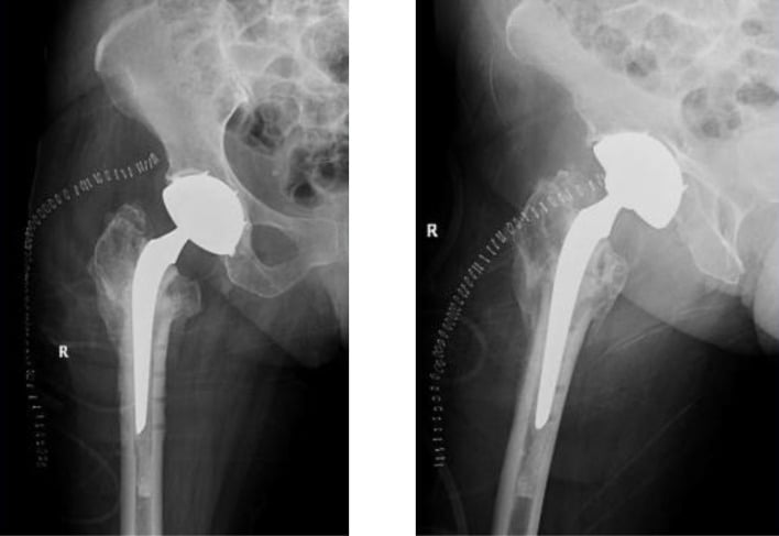
Complications and Salvage
- Clues
- Drainage, sinus, fever, positive cultures or persistent nonunion pain
- Response
- Cultures, imaging and infection-team review
- Salvage
- Debridement and staged implant removal/reconstruction
- Clues
- Cortical crack, loss of stem stability or deformity
- Response
- Reduce and cable/plate; image full femur
- Salvage
- Long stem bypass, fracture fixation and protected loading
- Clues
- Early instability, abductor deficiency or malversion
- Response
- Reduction, sciatic exam and mechanical imaging
- Salvage
- Correct stem/cup/offset/soft tissue; use dual mobility or constraint selectively
- Clues
- Foot drop, sensory loss or severe pain
- Response
- Check length, retractors, haematoma and hardware
- Salvage
- Remove causative compression or revise excessive lengthening; rehabilitate
- Clues
- Trendelenburg gait, lateral pain or fragment migration
- Response
- Assess fixation and abductor continuity
- Salvage
- Revision cable-plate/graft and abductor repair
- Clues
- Progressive shortening, thigh pain or radiolucency
- Response
- Exclude infection and image full femur
- Salvage
- Revise with stable distal fixation and bypass stress risers
Postoperative Care and Outcomes
Weight bearing depends on the weakest element: stem fixation, trochanteric repair, nonunion, fracture or acetabular reconstruction. A simple femoral neck conversion may progress faster than a subtrochanteric nonunion with an extended osteotomy. Use a brace or abduction pillow when the soft tissues require it.
Serial radiographs should include the whole femur. Look for stem subsidence, periprosthetic fracture, retained metal, trochanteric union, cup migration, dislocation and progressive osteolysis. New or persistent pain requires infection, fracture, loosening and abductor assessment; it is not automatically postoperative adaptation.
Guidelines, Registries & Global Practice
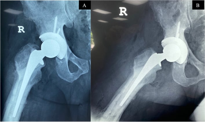
Global evidence. Conversion THA after failed fixation has higher complication risk than primary THA, especially after intertrochanteric and subtrochanteric failure. Evidence comes from comparative cohorts and systematic reviews, with outcomes influenced by fracture pattern, implant, timing, infection, deformity and surgeon experience.
Practice principles:
- Treat the case as a revision arthroplasty with an infection and fracture work-up.
- Map and remove hardware in a controlled sequence; protect the femur from stress-riser fracture.
- Use a stem that bypasses holes, nonunion and cortical windows and obtains real distal fixation.
- Restore hip mechanics without excessive sciatic lengthening and reconstruct abductors/trochanter.
- Obtain multiple cultures and stage infection rather than converting a contaminated nonunion directly to THA.
Registries. Hip arthroplasty registries report conversion THA incompletely because the index fracture fixation and indication may not be captured in the same record. Centres should record fracture type, fixation, nonunion, infection, hardware, stem, osteotomy, dislocation, fracture and reoperation.
Global practice. The operation may be performed with standard or modular stems, cemented reconstruction or staged revision depending on resources. A safe plan includes extraction tools, imaging, fracture fixation, cultures and a postoperative rehabilitation pathway wherever it is performed.
MCQ Practice Points
Q: Why does conversion THA after failed fixation carry higher risk than primary THA? A: Retained hardware, distorted anatomy, occult infection, nonunion, abductor damage, bone loss, limb shortening and femoral stress risers make it a revision-like operation.
Q: What must be imaged before conversion THA? A: The pelvis and the entire femur including the knee. Plan every screw, locking bolt, cement fragment, canal bow and stress riser.
Q: What is the stem rule in conversion THA? A: Bypass every screw hole, osteotomy, fracture and nonunion with a stem that obtains reliable distal fixation. A primary stem may not bypass the risk.
Q: What intraoperative finding changes the plan immediately? A: A sinus, purulence, positive deep cultures or an unsafe tissue envelope. Stop definitive conversion and stage infection control.
Q: What is the commonest soft-tissue failure after conversion? A: Trochanteric nonunion or abductor deficiency causing limp and dislocation. Plan trochanteric fixation and abductor repair rather than accepting a bare or mobile fragment.
Total hip arthroplasty after failed internal fixation of proximal femoral fractures
- 102 conversion THAs (1987-2008) after failed internal fixation of hip fracture: 39 intertrochanteric, 63 femoral neck
- Failure aetiology: osteonecrosis 35, arthritis 32, early fixation failure 25, nonunion 10
- Early surgical complications in 12 of 102 (11.8%): 5 dislocations (4.9%), 4 periprosthetic fractures (3.9%), 2 haematomas, 1 infection
- Complication rates exceed those of primary arthroplasty - the revision-like risk profile is the core message
Complications in conversion arthroplasty methods for failed intertrochanteric fracture fixation: a systematic review and meta-analysis
- Meta-analysis of five studies: 138 conversion arthroplasties with 49 complications; mean age 73 (32-96)
- Mean interval from fixation failure to conversion 11 months; mean operative time 132 minutes
- Complication distribution did not differ by method: cemented THA 4.1%, uncemented THA 81.6%, hybrid 2.0%, hemiarthroplasty 12.2% (all non-significant)
- The choice of conversion method did not influence the complication rate
Exam Viva Scenarios
Practise clinical reasoning and management decisions out loud
“A 71-year-old has a broken cephalomedullary nail, varus subtrochanteric nonunion and severe shortening. The hip has secondary arthritis. There is no sinus, but CRP is mildly raised. How do you plan conversion THA?”
“A 58-year-old has painful femoral neck nonunion, head collapse and retained cannulated screws. The greater trochanter and abductor mechanism are intact. What is your operation?”
“During conversion THA for an intertrochanteric nonunion, the tissue is friable and two deep samples grow the same organism. A long stem is already trialled but not cemented. What is your next step?”
Assess
- Fracture pattern, nonunion/malunion, head collapse, infection, hardware, shortening and abductors
- Serum markers, aspiration when feasible, multiple deep cultures and histology
- AP pelvis plus AP/lateral full femur including knee; CT for canal and hardware
- Document sciatic/femoral/peroneal function and vascularity
Plan
- Hardware extraction sequence and broken-screw/nail tools
- Stem bypass of all holes, nonunion, osteotomy and stress risers
- Hip centre, offset, length, version, trochanter and abductor repair
- Long/modular stem, cerclage, plate, graft, staged infection and fracture rescue
PIPADRAW
- Position and image the complete femur; prepare cultures and extraction
- Approach through scars, remove hardware in sequence and protect abductors
- Debride/culture, prepare acetabulum and true femoral canal
- Trial mechanics, insert bypass stem, fix trochanter, image and protect loading
Failure
- Infection: stop definitive conversion and stage
- Fracture: cable/plate and bypass with long stem
- Dislocation: analyse cup, stem, offset, version and abductors
- Nerve palsy: assess length, retractors, haematoma and hardware
References
- Archibeck MJ, Carothers JT, Tripuraneni KR, et al. Total hip arthroplasty after failed internal fixation of proximal femoral fractures. J Arthroplasty. 2013;28(1):168-171. PMID: 22682040. DOI: 10.1016/j.arth.2012.04.003.
- Mortazavi SMJ, Greenky M, Bican O, et al. Total hip arthroplasty after prior surgical treatment of hip fracture: is it always challenging? J Arthroplasty. 2012;27(1):31-36. PMID: 21798695.
- Haidukewych GJ, Berry DJ. Hip arthroplasty for salvage of failed treatment of intertrochanteric hip fractures. J Bone Joint Surg Am. 2003;85(5):899-904. PMID: 12728042. DOI: 10.2106/00004623-200305000-00019.
- Tabsh I, Waddell JP, Morton J. Total hip arthroplasty for complications of proximal femoral fractures. J Orthop Trauma. 1997;11(3):166-169. PMID: 9181498. DOI: 10.1097/00005131-199704000-00005.
- Moon NH, Shin WC, Kim HJ, et al. Cementless total hip arthroplasty following failed internal fixation for femoral neck and intertrochanteric fractures. Injury. 2019;50(3):713-719. PMID: 30670321.
- Laffosse JM, Molinier F, Tricoire JL, Bonnevialle N, Chiron P, Puget J. Cementless modular hip arthroplasty as a salvage operation for failed internal fixation of trochanteric fractures in elderly patients. Acta Orthop Belg. 2007;73(6):729-736. PMID: 18260485.
- Della Valle CJ, Berger RA, Rosenberg AG, Jacobs JJ, Sheinkop MB, Paprosky WG. Extended trochanteric osteotomy in complex primary total hip arthroplasty. A brief note. J Bone Joint Surg Am. 2003;85(12):2385-2390. PMID: 14668509.
- Paprosky WG, Weeden SH, Bowling JW Jr. Component removal in revision total hip arthroplasty. Clin Orthop Relat Res. 2001;(393):181-193. PMID: 11764348. DOI: 10.1097/00003086-200112000-00021.
- Parvizi J, Zmistowski B, Berbari EF, et al. New definition of periprosthetic joint infection: from the Workgroup of the Musculoskeletal Infection Society. Clin Orthop Relat Res. 2011;469:2992-2994. PMCID: PMC3183178. PMID: 21938532. DOI: 10.1007/s11999-011-2102-9.
- Hsieh PH, Shih CH, Chang YH, Lee MS, Shih HN, Yang WE. Two-stage revision hip arthroplasty for infection: comparison between the interim use of antibiotic-loaded cement beads and a spacer prosthesis. J Bone Joint Surg Am. 2004;86-A(9):1989-1997. PMID: 15342762.