Exercise-Induced | ICP Measurement | Fasciotomy | Athletes
- Aching pain during exercise that resolves with rest - distinguishes from acute compartment syndrome
- ICP measurement is gold standard - pre-exercise, 1 min post, 5 min post
- Diagnostic thresholds: over 15mmHg rest, over 30mmHg at 1 min, over 20mmHg at 5 min
- Fasciotomy is definitive treatment - excellent outcomes in most
- Rule out other causes: stress fracture, MTSS, popliteal entrapment, nerve entrapment
- “Anterior compartment most common (45%), often with lateral (35%)
- “Symptoms reproducible with specific exercise intensity and duration
- “Neurological symptoms (paresthesias, foot drop) often present
- “Bilateral in 85% - if unilateral, reconsider diagnosis
CECS: Aching during exercise, relieved by rest, reproducible, no tissue necrosis risk. Acute CS: Severe pain at rest, progressive, emergency, tissue death imminent.
Pedowitz criteria: Resting over 15mmHg OR 1 min post over 30mmHg OR 5 min post over 20mmHg. Any ONE criterion positive = diagnostic.
85% are bilateral - if truly unilateral, strongly reconsider differential diagnosis. May need to measure and release both legs.
Must rule out: stress fracture (bone scan), MTSS (diffuse pain), popliteal artery entrapment (ABI), nerve entrapment (EMG), vascular claudication.
- Key Finding
- Reproducible, tight compartments
- Action
- ICP measurement pre/post exercise
- Key Finding
- Over 30mmHg at 1 min post
- Action
- Diagnose CECS, consider fasciotomy
- Key Finding
- Positive bone scan
- Action
- Think stress fracture, not CECS
- Key Finding
- Single leg affected
- Action
- Reconsider diagnosis, exclude other causes
- Key Finding
- Anterior compartment CECS
- Action
- Indicates nerve involvement, needs release
- Key Finding
- Activity modification, orthotics failed
- Action
- Proceed to fasciotomy
15-30-20ICP Measurement Thresholds
Hook:15-30-20: The Pedowitz criteria numbers in sequence!
ALDSLeg Compartments
Hook:ALDS compartments - Anterior and Lateral most common in CECS!
ACHINGSymptoms Pattern
Hook:ACHING during exercise that gets better with rest = CECS pattern!
STAMPSDifferential Diagnosis
Hook:STAMPS out the differential diagnosis for exercise leg pain!
Overview and Epidemiology
What is CECS?
Chronic Exertional Compartment Syndrome (CECS) is a condition where:
- Exercise induces elevated intracompartmental pressure
- Muscles swell within non-compliant fascial boundaries
- Blood flow is impaired during activity
- Symptoms develop predictably with specific exercise
- Symptoms resolve with rest (no tissue necrosis)
CECS: Reversible, chronic, no tissue necrosis, not an emergency
Acute CS: Progressive, irreversible without treatment, tissue death, EMERGENCY
Anatomy and Compartments
Leg Compartment Anatomy
- Contents
- TA, EHL, EDL, peroneus tertius
- Nerve
- Deep peroneal
- Function Lost if Affected
- Dorsiflexion, toe extension
- Contents
- Peroneus longus and brevis
- Nerve
- Superficial peroneal
- Function Lost if Affected
- Eversion, sensory first web
- Contents
- TP, FHL, FDL, popliteus
- Nerve
- Tibial nerve
- Function Lost if Affected
- Toe flexion, inversion
- Contents
- Gastrocnemius, soleus, plantaris
- Nerve
- Sural nerve (sensory)
- Function Lost if Affected
- Plantarflexion
Pathophysiology
The Pressure-Ischemia Cycle
CECS develops through a predictable sequence of events during exercise:
Step 1: Muscle Expansion
- Exercise increases muscle blood flow by up to 10-fold
- Active muscle volume increases by 20% due to hyperemia
- Metabolic demands require increased tissue perfusion
Step 2: Fascial Constraint
- The fascia surrounding leg compartments is non-compliant
- Cannot stretch to accommodate increased muscle volume
- Creates a closed space with rising pressure
Step 3: Pressure Rise
- Normal resting pressure: 8-10 mmHg
- Exercise increases pressure to 30-80+ mmHg in CECS patients
- Critical threshold: when pressure exceeds capillary perfusion pressure
Step 4: Ischemia
- Elevated tissue pressure compresses capillaries
- Arterial inflow maintained but venous outflow impaired
- Relative tissue ischemia develops
- Pain and neurological symptoms ensue
Step 5: Recovery
- Cessation of exercise reduces metabolic demand
- Muscle volume decreases over 10-15 minutes
- Pressure normalizes and symptoms resolve
- No permanent tissue damage (unlike acute compartment syndrome)
Why Anterior Compartment Most Common
The anterior compartment is affected in 45% of CECS cases because:
- It has the smallest fascial envelope relative to muscle mass
- Contains muscles with high activity during running (tibialis anterior)
- Experiences the greatest percentage volume change with exercise
- Less fascial compliance than other compartments
Classification Systems
Compartment-Based Classification
- Frequency
- 45%
- Key Features
- Most common, dorsiflexion weakness
- Nerve at Risk
- Deep peroneal
- Frequency
- 35%
- Key Features
- Often combined with anterior
- Nerve at Risk
- Superficial peroneal
- Frequency
- 15%
- Key Features
- Medial symptoms, harder to diagnose
- Nerve at Risk
- Tibial nerve
- Frequency
- 5%
- Key Features
- Rare, calf cramping
- Nerve at Risk
- Sural nerve
- Frequency
- 60%+
- Key Features
- Multiple compartments involved
- Nerve at Risk
- Multiple nerves
Anterior and lateral compartments are often affected together. When releasing one, always assess the other. Over 60% of patients have multiple compartment involvement.
History
History Taking
- Aching, cramping pain with exercise
- Develops after predictable duration/intensity
- Relieved within minutes of rest
- Bilateral in 85% of cases
- "How long into exercise does pain start?"
- "How quickly does it resolve with rest?"
- "Is it the same every time?"
- "Any numbness or tingling?"
- "Any weakness (foot drop)?"
- Pain at rest
- Pain that doesn't resolve with rest
- Point tenderness (stress fracture)
- Night pain
Proper technique and attention to detail ensure optimal outcomes.
Examination
Physical Examination
- Compartments soft
- Normal neurology
- No tenderness typically
- Compartment tightness/firmness
- May have temporary neurological deficit
- Muscle herniation possible through fascial defects
- Symptoms reproducible
- Assess motor function per compartment
- Sensory assessment (first web space = deep peroneal)
- Pulses (usually normal, but check)
Proper technique and attention to detail ensure optimal outcomes.
Investigations
Intracompartmental Pressure Testing
Gold Standard for Diagnosis
Technique:
- Slit catheter or Stryker needle
- Measure at rest (pre-exercise)
- Measure at 1 minute post-exercise
- Measure at 5 minutes post-exercise
- Insert perpendicular to leg, into compartment bulk
- Threshold
- Over 15 mmHg
- Interpretation
- Positive
- Threshold
- Over 30 mmHg
- Interpretation
- Positive
- Threshold
- Over 20 mmHg
- Interpretation
- Positive
Any ONE criterion positive = diagnostic for CECS. Most helpful is the 1 minute post-exercise reading - should be elevated significantly above baseline.
The Pedowitz static thresholds are the quoted standard, but a complete answer acknowledges their limitations and the move toward better tests:
- Poor specificity of the static criteria: a substantial proportion of asymptomatic athletes have post-exercise pressures exceeding the classic cut-offs (especially the 30 mmHg at one minute), so a "positive" static pressure in isolation over-diagnoses CECS. Always interpret pressures alongside a consistent clinical picture.
- Dynamic / continuous intramuscular pressure: rather than three discrete readings, a transducer left in situ during the provoking exercise records the pressure rise and, crucially, the post-exercise decay (recovery) time - a prolonged time for pressure to fall back to baseline is more discriminating than a single peak value, and is physiologically the more logical test.
- Near-infrared spectroscopy (NIRS): a non-invasive alternative that measures muscle deoxygenation/oxygenation during exercise - delayed reoxygenation after exercise reflects the ischaemia of CECS without needing a needle, and is an emerging adjunct/screening tool.
- The principle: the diagnosis is clinical pattern plus a confirmatory ischaemia test; needle pressure remains the reference standard but its thresholds are imperfect, which is why dynamic monitoring and NIRS are gaining ground.
Exam point: don't diagnose CECS on a single static pressure alone - the Pedowitz thresholds have poor specificity (asymptomatic athletes exceed them); continuous/dynamic ICP with a prolonged post-exercise decay, and non-invasive NIRS, are more discriminating and increasingly used.


Differential Diagnosis
- Key Feature
- Point tenderness
- Investigation
- MRI or bone scan
- Distinguishing Factor
- Focal pain, positive imaging
- Key Feature
- Diffuse medial tibial pain
- Investigation
- Bone scan (diffuse)
- Distinguishing Factor
- Longer recovery, not exercise-limited
- Key Feature
- Claudication with exercise
- Investigation
- ABI post-exercise, angio
- Distinguishing Factor
- Reduced pulses, vascular symptoms
- Key Feature
- Calf swelling, tenderness
- Investigation
- Duplex ultrasound
- Distinguishing Factor
- Constant symptoms, swelling
- Key Feature
- Neurological symptoms dominant
- Investigation
- EMG/NCS
- Distinguishing Factor
- Specific nerve distribution
- Key Feature
- Acute onset
- Investigation
- Clinical, possibly MRI
- Distinguishing Factor
- History of specific injury
CECS: Predictable, reproducible, exercise-induced, resolves with rest
Other conditions: May have pain at rest, variable patterns, don't follow exercise intensity reliably
Management
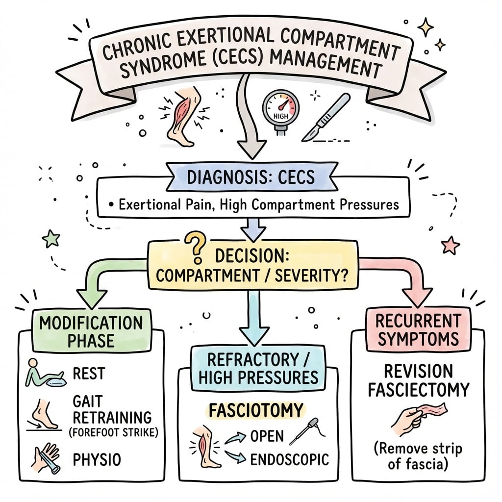
Non-Operative Management
- Activity modification (reduce intensity/duration)
- Gait retraining (forefoot vs heel strike)
- Stretching and massage
- Orthotics (theoretical benefit)
- Cross-training (swimming, cycling)
- NSAIDs (limited evidence)
- Low for return to full activity (under 50%)
- May be adequate if willing to modify sport
- Athletes usually require surgery
Conservative management should be tried first, but in dedicated athletes with confirmed CECS, fasciotomy is usually needed for return to sport.
The topic notes conservative care "usually fails," but one specific intervention is genuinely curative for many anterior CECS patients and is increasingly first-line, especially in runners and the military:
- What it is: structured forefoot-strike (or midfoot) running retraining in a habitual rearfoot (heel) striker. Heel-striking demands strong eccentric tibialis anterior activity to control foot slap, which drives anterior-compartment loading; converting to a forefoot landing markedly reduces tibialis anterior demand and anterior intracompartmental pressure.
- The evidence: a supervised gait-retraining programme can resolve symptoms and return runners/soldiers to activity without fasciotomy - in some military cohorts gait retraining has matched or exceeded surgery (the army study flagged in this topic's own systematic-review evidence). It is most applicable to anterior (not deep posterior) CECS.
- How it is delivered: a graded programme (often weeks) with cadence increase (shorter, quicker steps), forefoot/midfoot landing cues, and gradual mileage build-up - ideally with gait-lab or physiotherapy supervision to embed the new pattern and avoid metatarsal/Achilles overload during transition.
- Other non-operative adjuncts: activity modification, and (lower-grade evidence) botulinum toxin injection to the affected compartment muscles as an emerging option.
Exam point: before fasciotomy for anterior CECS, offer a supervised forefoot-strike gait-retraining programme - it lowers tibialis-anterior-driven compartment pressure and can return runners and military patients to sport without surgery.
Surgical Technique
Anterior Compartment Fasciotomy
- Supine with leg externally rotated
- Tourniquet optional (many prefer no tourniquet)
- Pad bony prominences
- Single lateral incision, 2-3cm anterior to fibula
- Length: 10-15cm for adequate release
- Identify subcutaneous fat and crural fascia
- Incise anterior compartment fascia longitudinally
- Extend proximally and distally with scissors
- Release must be complete from tibial plateau to ankle
- Visualize muscle bulging through fasciotomy
- Ensure complete release
- Identify anterior intermuscular septum
- Check lateral compartment if symptomatic
Proper technique ensures adequate decompression.
Complications
Intraoperative Complications
- Risk
- Most common nerve injury
- Prevention
- Identify and protect
- Management
- Observation if neuropraxia
- Risk
- Commonest cause of failure
- Prevention
- Full visualization
- Management
- Revision surgery
- Risk
- Rare
- Prevention
- Know anatomy
- Management
- Direct repair or ligation
- Risk
- Rare
- Prevention
- Confirm anatomy
- Management
- Release correct compartment
Superficial peroneal nerve injury during lateral release is the most common nerve complication. It causes numbness over the dorsum of the foot but does not affect motor function. Most are neuropraxias that recover.
- Incidence
- 5-10%
- Risk Factor
- Incomplete release
- Incidence
- 3-5%
- Risk Factor
- Hematoma, poor technique
- Incidence
- 1-3%
- Risk Factor
- Superficial peroneal at risk
- Incidence
- Under 1%
- Risk Factor
- Immobility
- Incidence
- 1-2%
- Risk Factor
- Standard surgical risk
Postoperative Care
Immediate Postoperative (0-2 weeks)
- Compression dressing
- Elevate leg
- Ankle ROM exercises begin same day
- Weight-bearing as tolerated
- Wound checks at 5-7 days
- Remove sutures at 10-14 days
- Active ankle dorsiflexion/plantarflexion
- Ice for swelling
- Gentle calf stretches
- Wound healing
- Maintain ankle ROM
- Control swelling
- Prevent DVT
Early mobilization is key to successful recovery.
Outcomes and Prognosis
Surgical Results
- Rate
- 90-95%
- Notes
- Most return to pre-injury level
- Rate
- 85-95%
- Notes
- High satisfaction rates
- Rate
- 5-10%
- Notes
- Usually due to inadequate release
- Rate
- Under 5%
- Notes
- Wound and nerve issues rare
Prognostic Factors
- Clear diagnosis (positive ICP)
- Anterior/lateral compartment
- Complete surgical release
- Younger patients
- Single sport athlete
- Atypical presentation
- Deep posterior involvement
- Previous failed surgery
- Military personnel (higher demands)
- Coexisting conditions
Guidelines, Registries & Global Practice
Global Epidemiology
CECS is a major cause of exercise-induced leg pain in young athletes and military recruits. In military and recreational-runner cohorts, exercise-induced lower leg pain accounts for an estimated 27 to 33 percent of all lower-leg pain presentations, with CECS a leading diagnosis after medial tibial stress syndrome (Breen et al. 2015, PMID 25709867). Pooled systematic-review data show a male-to-female ratio of roughly 7:4 and an age range spanning 12 to 70 years, with most patients in the second-to-fourth decades (Vogels et al. 2020, PMID 32526086). The anterior (and combined anterolateral) compartment is the most frequently affected subtype across populations (de Bruijn/Winkes 2020, PMID 30980096).
Guidance and Evidence Across Bodies
No single national college (AAOS, NICE, BOA, AO, EFORT) publishes a dedicated CECS clinical practice guideline; management is instead driven by sports-medicine consensus and systematic reviews. There is broad international agreement on the principles below.
- Consensus Position
- Clinical pattern plus intracompartmental pressure (Pedowitz criteria); dynamic testing physiologically preferred but underused
- Evidence Level
- Level III (Pedowitz, PMID 2301689)
- Consensus Position
- Trial of conservative care including gait/running-technique retraining, especially in military and runners
- Evidence Level
- Level IV SR (PMID 32526086)
- Consensus Position
- Fasciotomy of all symptomatic compartments when conservative care fails
- Evidence Level
- Level IV SR (PMID 37778507)
- Consensus Position
- Lower, less predictable surgical success; counsel accordingly
- Evidence Level
- Level III SR (PMID 24065078)
Registry and Population Evidence
There is no dedicated CECS registry; the evidence base is systematic reviews and cohort series rather than arthroplasty-style national registries. The largest pooled lower-leg dataset (68 studies, n=3783) reports fasciotomy satisfaction of 85 percent and return to activity of 80 percent, versus roughly 47 to 50 percent for conservative care (Vogels 2020, PMID 32526086).
Practice Variation
Most published series use static rather than dynamic intracompartmental pressure measurement, and ICP cut-offs vary widely between centres, contributing to heterogeneous reported outcomes (Dean 2024, PMID 37778507).
Civilian athletes do considerably better than military/high-demand cohorts, in whom only about half achieve complete resolution and at least 25 percent cannot return to full duty (Dunn and Waterman 2014, PMID 25280617).
MCQ Practice Points
Q: What are the Pedowitz criteria for diagnosing CECS?
A: 15-30-20: Pre-exercise over 15mmHg, 1 min post over 30mmHg, 5 min post over 20mmHg. Any ONE positive is diagnostic.
Q: Which compartment is most commonly affected in CECS?
A: Anterior compartment (45%), followed by lateral (35%), deep posterior (15%), and superficial posterior (5%). Anterior and lateral are often affected together.
Q: What percentage of CECS cases are bilateral?
A: 85% are bilateral. If truly unilateral, strongly reconsider the diagnosis and investigate other causes such as popliteal artery entrapment or stress fracture.
Q: What nerve is at risk during lateral compartment release for CECS?
A: The superficial peroneal nerve emerges through the lateral compartment and is at risk during fasciotomy. It must be identified and protected.
Q: What is the expected return to sport rate after fasciotomy for CECS?
A: 90-95% of patients return to their previous level of sport after fasciotomy. Success is highest for anterior and lateral compartment releases.
Exam Viva Scenarios
Practise clinical reasoning and management decisions out loud
“A 22-year-old long-distance runner presents with bilateral anterior leg pain that starts 20 minutes into running and forces her to stop. Pain resolves within 5-10 minutes of rest. She describes tightness and occasional numbness on the dorsum of her foot. Examination at rest is normal.”
“A 28-year-old male soccer player presents with unilateral right leg pain with exercise. He describes cramping in the calf that starts after 30 minutes of training. He has no numbness. His pain sometimes persists for hours after stopping.”
“A 25-year-old triathlete had anterior compartment fasciotomy for CECS 6 months ago. She has returned to training but symptoms have recurred. ICP measurement shows elevated pressures in the anterior compartment.”
Definition
- Exercise-induced elevated intracompartmental pressure
- Symptoms with exercise, resolve with rest
- Reversible, no tissue necrosis (unlike acute CS)
- 85% bilateral
Pedowitz Criteria (15-30-20)
- Resting (pre-exercise) over 15mmHg
- 1 minute post-exercise over 30mmHg
- 5 minutes post-exercise over 20mmHg
- Any ONE positive = diagnostic
Compartment Frequency
- Anterior: 45% (most common)
- Lateral: 35% (often with anterior)
- Deep posterior: 15%
- Superficial posterior: 5% (rare)
Treatment
- Conservative: Usually fails in athletes
- Fasciotomy: Definitive treatment
- 95% return to sport post-op
- Complete release essential
Differential (STAMPS)
- Stress fracture - point tenderness
- Tibial nerve entrapment
- Artery entrapment (popliteal)
- MTSS (shin splints)
- Peroneal nerve entrapment
Surgical Pearls
- Protect superficial peroneal nerve
- Complete release proximal to distal
- Consider releasing lateral with anterior
- Soleal bridge for deep posterior
Evidence Base
Pedowitz Modified Diagnostic Criteria (Landmark)
- Derived the still-used intramuscular pressure thresholds from slit-catheter recordings in 210 compartments: a pre-exercise pressure of 15 mmHg or more, a 1-minute post-exercise pressure of 30 mmHg or more, or a 5-minute post-exercise pressure of 20 mmHg or more is diagnostic when clinical findings fit. Muscle herniation was the only history/examination feature that differed between groups (45.9 percent vs 12.9 percent).
Treatment Outcomes in Lower-Leg CECS (Systematic Review)
- Fasciotomy significantly reduced intracompartmental pressure (mean 76 mmHg to 24 mmHg) and achieved 85 percent (plus or minus 13) satisfaction and 80 percent (plus or minus 17) return to activity. Conservative interventions (gait retraining, botulinum toxin) gave roughly 47 percent satisfaction and 50 percent return to activity. Return to activity was significantly more likely after surgery (P less than 0.01), except in one army-personnel study favouring gait retraining.
Deep Posterior CECS: Is Surgery Effective? (Systematic Review)
- Success after fasciotomy for deep posterior CECS was modest, ranging 30 to 65 percent, markedly inferior to anterior/lateral release. No single surgical technique was superior, ICP cut-offs varied widely, and prolonged high post-provocation ICP was associated with surgical success.
Diagnosis and Treatment Patterns in CECS (Systematic Review)
- Most published series (24 of 29) diagnose CECS with static rather than dynamic pressure measurement, and fasciotomy (single-incision most common) is the dominant treatment. Reported post-fasciotomy satisfaction ranged 42 to 94 percent and return to sport 26 to 100 percent; conservative return to sport was 25 to 35 percent, underscoring wide practice variation and heterogeneous outcome reporting.
CECS in Military Populations (Practice Variation)
- In military service members, surgical results are far less reliable than in civilian athletes: only about half achieve complete symptom resolution and at least 25 percent are unable to return to full duty. The five cardinal symptoms are pain, tightness, cramps, weakness and diminished sensation.
Management of Anterolateral-Leg CECS (Review)
- Anterior tibial muscle CECS is the most common subtype; dynamic intracompartmental pressure measurement is the gold-standard diagnostic test. Duplex and imaging exclude popliteal artery entrapment and nerve entrapment. Conservative measures including running-technique modification can succeed; recalcitrant cases warrant fasciotomy, and residual or recurrent disease may need partial fasciectomy.