Spinous Process Avulsion | C6-C7-T1 | Ghost Sign | Stable Injury | Conservative Treatment
- Definition: Avulsion fracture of the spinous process, typically C6, C7, or T1.
- Mechanism: Sudden flexion against resisting extensors (historically: shoveling clay). Modern: MVA, sports.
- Stability: STABLE injury. Posterior bony element only. Ligaments intact.
- Treatment: Conservative. A collar for 4-6 weeks is for SYMPTOM CONTROL, not stability - non-union of the fragment is common and asymptomatic, and union is not the treatment goal. A 10-year follow-up of recurrent golf-related fractures showed radiographic non-union with only mild intermittent discomfort (PMID 37674772).
- Key Point: Rule out associated injuries (burst, facet fractures). Isolated Clay-Shoveler's is benign.
- “STABLE injury - Treat conservatively.
- “Named after clay workers in Australia in the 1930s.
- “Must rule out other cervical injuries (especially burst, facet, ligamentous).
- “If isolated, excellent prognosis.
Overview and Epidemiology
A clay-shoveler's fracture is an avulsion fracture of the spinous process, typically at C6, C7 or T1, with C7 the most common level. It is a stable injury, because only a posterior bony element is broken, and in isolation it has an excellent prognosis with conservative treatment. The name comes from Australian workers shovelling heavy clay in the 1930s; the injury is now more commonly seen in motor vehicle accidents and sport.
Who. All ages, with no specific predilection. The modern causes are motor vehicle accidents, sports and falls.
What matters. The whole message of this page is that the injury is stable, so the failure mode is under-investigating. A spinous process fracture can accompany more significant cervical injuries (burst fractures, facet injuries, ligamentous disruption) and may be the tip of the iceberg; once those are excluded, function usually recovers fully and surgery is not indicated.
Anatomy and Pathophysiology
The spinous processes. C6 to T1 have long spinous processes, bifid at C6 and prominent at C7 and T1. C7 carries the longest, non-bifid process, the vertebra prominens, and the strongest ligamentous and muscular attachments: the nuchal ligament, trapezius, rhomboids and semispinalis.
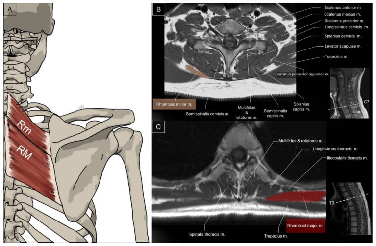
Why C7 and T1. The cervicothoracic junction is the transition from the mobile lordotic cervical spine to the rigid kyphotic thoracic spine, which concentrates flexion-tension forces there. At C7 the lever arm and the tensile load are greatest, because the process is longest and its attachments strongest.
The classic mechanism. The neck flexes suddenly and forcefully while the extensors (trapezius, semispinalis) contract to resist it. The tensile load passes through the supraspinous and nuchal ligament and the trapezius-rhomboid insertions and avulses the spinous process tip at the attachment. This is the original shovelling mechanism.
The stress mechanism. Repetitive submaximal traction from sport (paddling, weightlifting, golf, volleyball) produces a fatigue avulsion, often without a single traumatic event; the juvenile equivalent is Schmitt disease. In children and adolescents a pure soft-tissue avulsion may occur with normal radiographs and only MRI-evident marrow and soft-tissue oedema.
Stability. Only the posterior bony element is injured. The anterior and middle columns are intact, and although the interspinous and supraspinous ligaments may be partially involved, the posterior ligamentous complex is functionally intact. Avulsion of the tip is the benign entity; a fracture through the base of the spinous process with interspinous widening is a marker for posterior ligamentous complex disruption, a flexion-distraction injury.
The column concept. The stability statement borrows the Denis three-column model, defined for the thoracolumbar spine and applied conceptually to the cervical spine:
- Anterior column - anterior longitudinal ligament and the anterior two-thirds of the vertebral body and annulus
- Middle column - posterior one-third of the vertebral body and annulus and the posterior longitudinal ligament; the key load-bearing and stability column, and retropulsion implies its failure
- Posterior column - the posterior bony arch (pedicles, facets, laminae, spinous process) and the posterior ligamentous complex
A clay-shoveler's fracture injures only the bony part of the posterior column, the spinous process tip, and leaves the middle column, the critical determinant of stability, intact, which is precisely why it is stable. In the thoracolumbar Denis model, instability is suggested when two or more columns fail. Other generally stable cervical injuries, for comparison:
- Minor compression, less than 25% height loss
- Anterior listhesis less than 3.5mm
- Isolated pedicle fracture (rare)
- Minor anterior wedge compression
In the subaxial cervical spine, stability is judged not by columns alone but by the White and Panjabi checklist, a points system where a total of 5 or more = clinically unstable:
- Anterior elements destroyed (2)
- Posterior elements destroyed (2)
- Relative sagittal translation greater than 3.5 mm (2)
- Relative sagittal angulation greater than 11 degrees (2)
- Positive stretch test (2)
- Spinal cord damage (2)
- Nerve-root damage (1)
- Abnormal disc narrowing (1)
- Developmentally narrow canal (1)
- Dangerous loading anticipated (1)
An isolated clay-shoveler's fracture scores low, only part of the posterior bony element, confirming a stable injury, whereas a flexion teardrop or facet dislocation rapidly accumulates points. This complements the SLIC score for the operative-versus-conservative decision.
Classification Systems
The division that decides treatment is whether the spinous process fracture is isolated or accompanies another cervical injury; the second table records where the fracture sits.
- Description
- Spinous process fracture only, no other injury
- Stability
- Stable
- Treatment
- Conservative - collar 4-6 weeks for symptoms; union not required
- Description
- Spinous process + other cervical injury (burst, facet, ligamentous)
- Stability
- Depends on associated injury
- Treatment
- Treat the other injury, spinous process fracture incidental
- Features
- Bifid spinous process, less common
- Clinical Significance
- Still stable if isolated
- Features
- Vertebra prominens, most common
- Clinical Significance
- Most prominent, easiest to palpate
- Features
- Upper thoracic, common
- Clinical Significance
- May extend into thoracic spine
- Features
- C7+T1 or C6+C7, still stable if isolated
- Clinical Significance
- May involve 2-3 levels, still conservative
Clinical Assessment
History. A hyperflexion injury in a motor vehicle accident, a direct blow or whiplash in sport, or a fall. The pain is localised to the lower cervical spine (C6-C7-T1) and is worse with extension and rotation.
Examination. Point tenderness over the spinous process, easiest to find at C7 because the vertebra prominens is the most palpable. A step-off or gap may be felt at the process, and movement is painful, especially extension. Neurology should be intact if the fracture is isolated, and a full motor and sensory examination is what proves it.
Always assess the entire cervical spine on CT. Any of the following should stop you calling the fracture isolated:
- A high-energy mechanism
- Interspinous widening
- Facet malalignment or perched or jumped facets
- Loss of vertebral body height
- Multiple contiguous levels
- Any neurological sign
Plain films under-call all of this. Get the CT before you reassure.
- Distinguishing Features
- Isolated, oblique/avulsion fracture of C6-T1 spinous process tip; ghost sign; neurology intact; anterior/middle columns normal
- Stability / Action
- Stable - conservative
- Distinguishing Features
- Triangular antero-inferior body fragment, retrolisthesis, kyphosis, frequent cord injury (anterior cord syndrome)
- Stability / Action
- Highly UNSTABLE - urgent surgery
- Distinguishing Features
- Facet override or 'naked facet', anterolisthesis greater than 25 percent (unilateral) or 50 percent (bilateral), radiculopathy/myelopathy
- Stability / Action
- Unstable - reduction +/- fusion
- Distinguishing Features
- Comminuted vertebral body, retropulsion, loss of height, middle column involvement
- Stability / Action
- Often unstable - SLIC-guided
- Distinguishing Features
- Smooth, corticated secondary ossification centre, symmetrical, bilateral on CT; no oedema on MRI
- Stability / Action
- Normal variant - no treatment
- Distinguishing Features
- Diffuse tenderness, normal CT, no discrete bony fragment; MRI may show soft-tissue oedema without fracture
- Stability / Action
- Stable - symptomatic
The dangerous mimic. The single most important differential is the flexion teardrop fracture. Both arise from forced flexion, but the teardrop is one of the most unstable cervical injuries with a high rate of cord injury, whereas clay-shoveler's is benign. The discriminator is the column involved: clay-shoveler's affects only the posterior bony element, while the teardrop disrupts the anterior and middle columns with a displaced antero-inferior body fragment and kyphotic angulation.
Investigations
Who needs imaging. Whether the patient needed imaging at all is decided by the Canadian C-spine rule, which was more sensitive than NEXUS (99.4 vs 90.7 percent) and would have reduced imaging (55.9 vs 66.6 percent) in 8,283 patients (PMID 14695411).
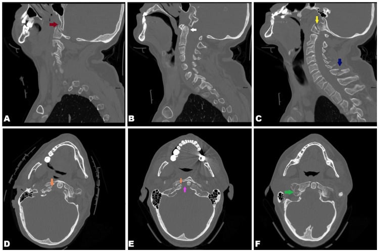
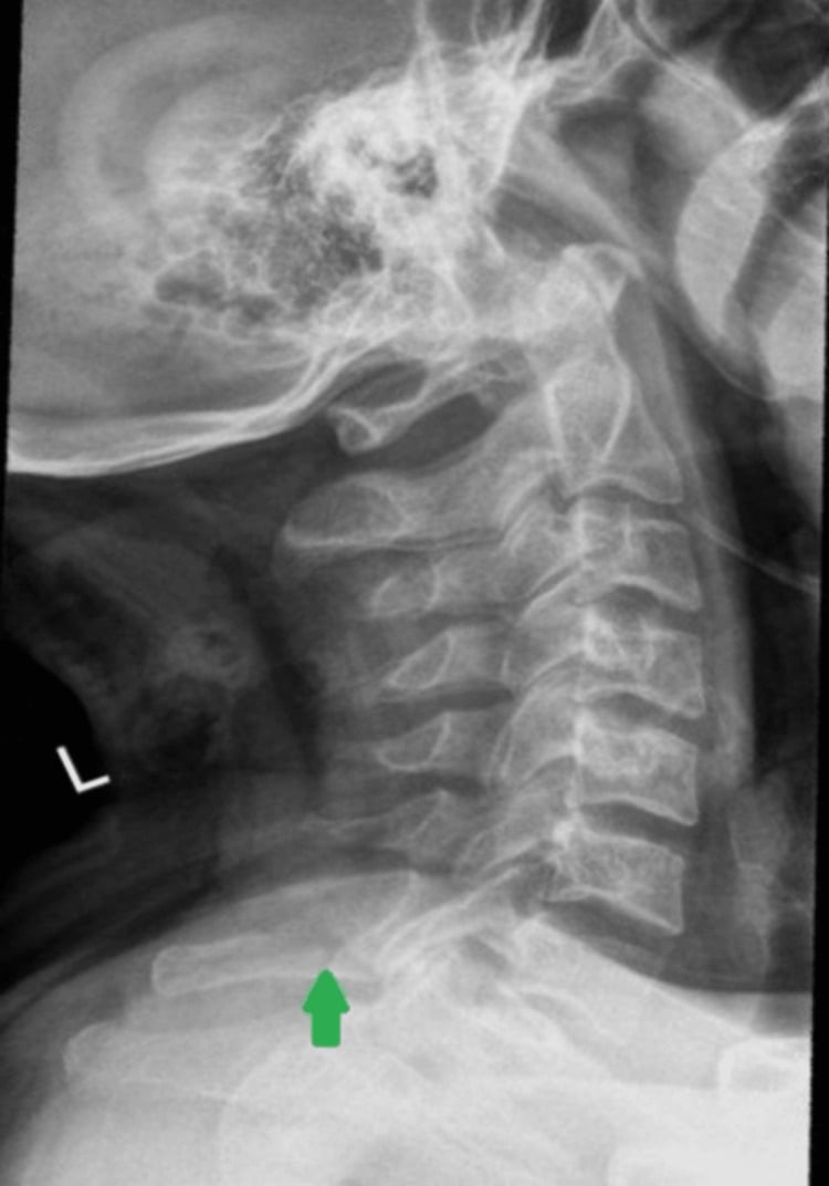
Radiographs. Start with the lateral cervical film. The ghost sign is a double shadow at the spinous process level: the avulsed fragment casts a second shadow separate from the in-situ portion, and it is diagnostic. C7 and T1 may be obscured by the shoulders, so a swimmer's view may be needed to see them.
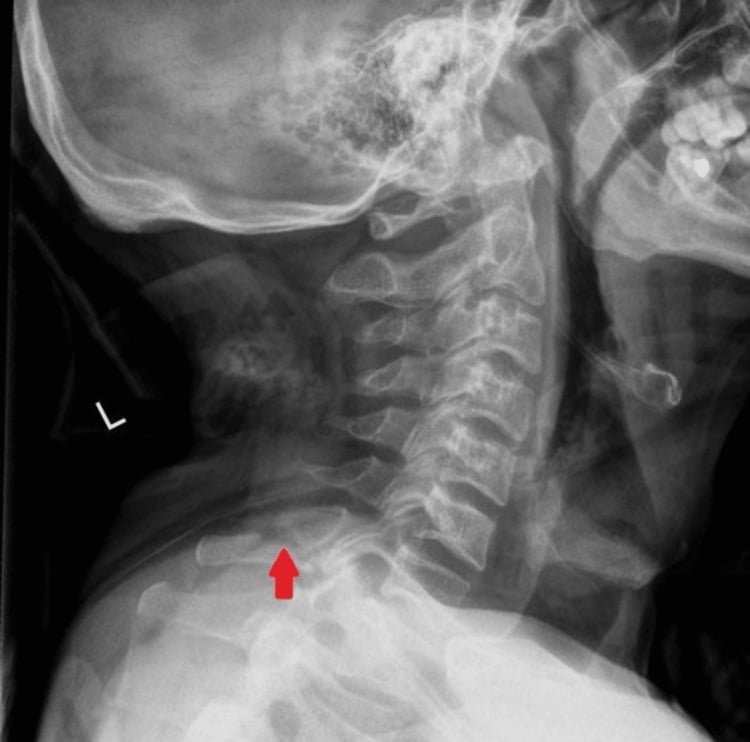
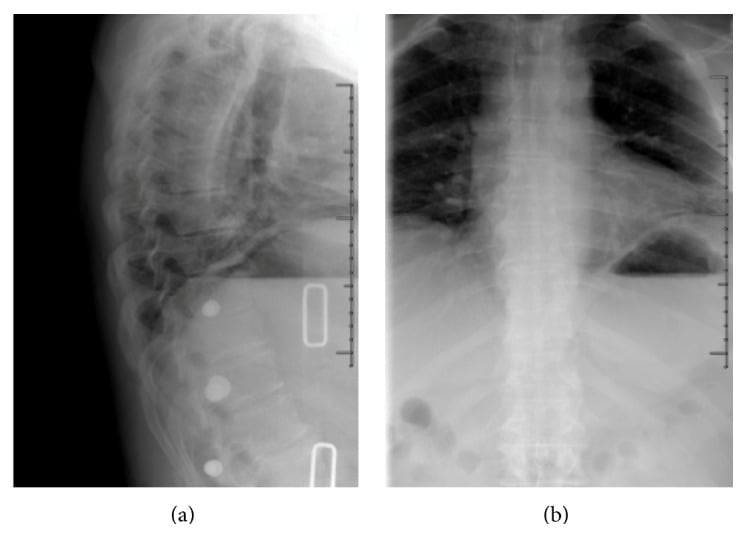
Plain films under-call multi-level and associated injury. In the multi-level case on this page, the radiographs showed only absent or deviated spinous process shadows, and CT was needed to reveal fractures from T2 to T8 plus a T11 compression fracture.
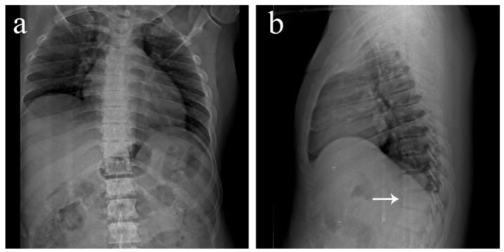
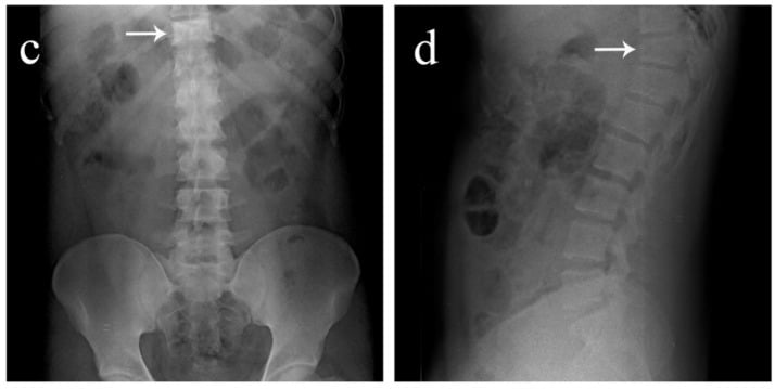
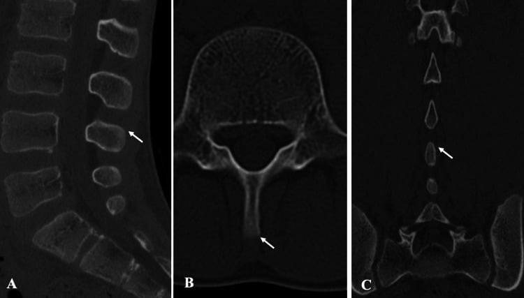
CT. The gold standard, with 1mm cuts, and essential for confirmation and assessment. It confirms the avulsion, shows fragment displacement, defines multi-level involvement (C6, C7, T1) and, most importantly, rules out the associated injuries (burst, facet, ligamentous) that determine treatment.
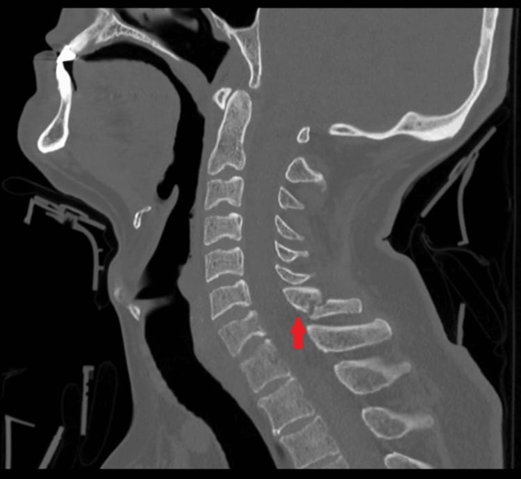
Multi-level injury. The level table says two or three levels; the cases on this page run from T2 to T8 and from C6 to T9.
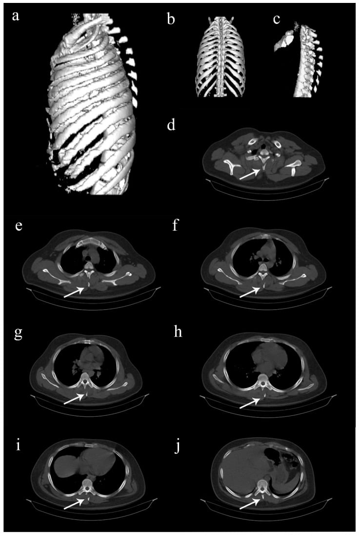
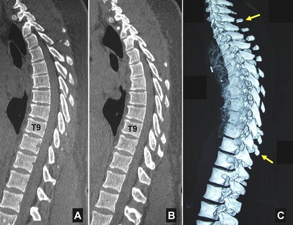
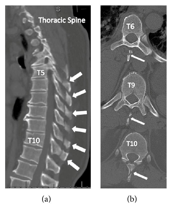
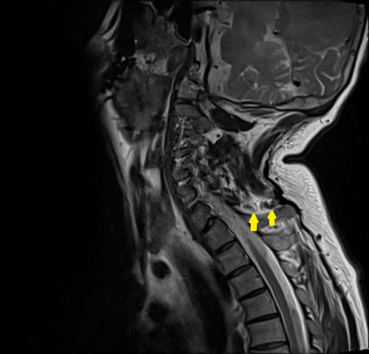
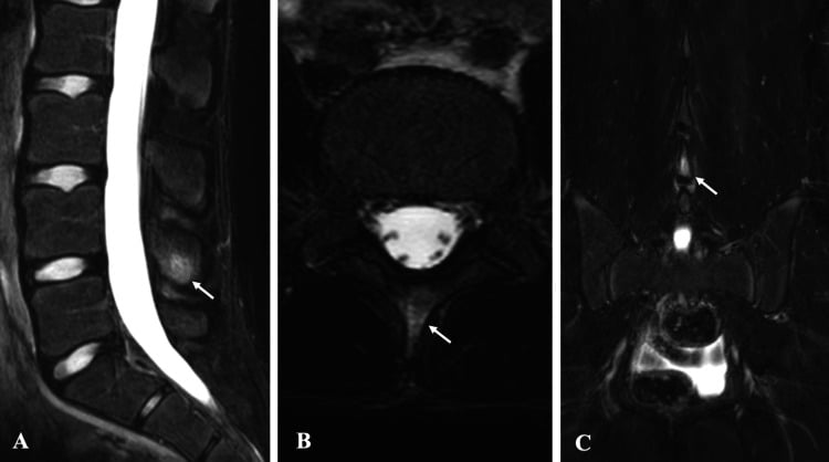
MRI. Requested when there is concern for ligamentous injury or neurological symptoms: it assesses the posterior ligamentous complex, the disc, and the spinal cord if there is neurology.
Management Algorithm
The decision. An isolated fracture is treated conservatively, and the job of the work-up is to make sure it is isolated. Any neurology, a high-energy mechanism or suspicion of an associated injury triggers CT, with MRI for ligamentous or cord concern. If a burst, facet or ligamentous injury or instability is found, the patient moves onto the spine-trauma pathway and is graded (SLIC or AO Spine); the spinous-process fragment itself is incidental and is not routinely fixed. The unstable injuries being excluded are covered in cervical spine fracture and facet dislocations, and in the thoracic segment thoracolumbar fractures.
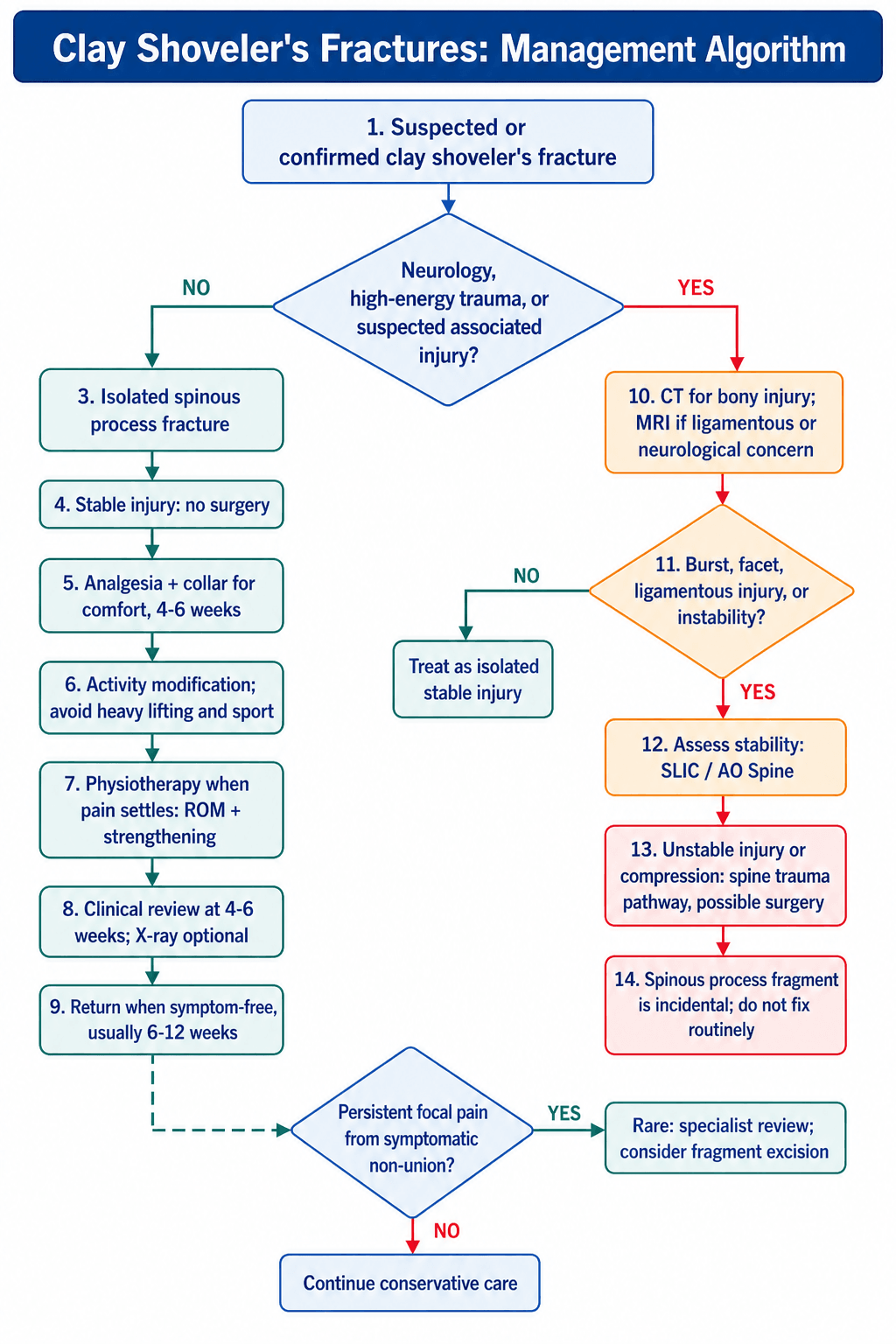
The isolated fracture. Conservative management:
- Analgesia - NSAIDs, paracetamol
- Collar - a rigid cervical collar for 4-6 weeks, for symptom control, not stability
- Activity modification - avoid heavy lifting and sport
- Physiotherapy after the pain settles - range of motion, then strengthening
- Follow-up - clinical review at 4-6 weeks; X-ray optional (union not required for symptoms)
The prognosis is excellent and full recovery is expected.
The associated injury. Treat the other injury; the spinous process fracture is incidental and the associated injury determines management:
- Burst fracture - SLIC scoring; may need surgery
- Facet injury - assess stability; surgery if unstable
- Ligamentous injury - MRI assessment; halo or surgery
Surgical Technique
Surgery is not indicated for an isolated clay-shoveler's fracture, and conservative management remains the standard of care. The rationale:
- Isolated spinous process fractures are stable
- Conservative treatment has excellent outcomes
- Non-union is common but asymptomatic
- Surgery adds risk without benefit
The rare exception. A symptomatic non-union causing persistent pain, where excision of the non-united fragment can be considered if symptomatic. It is extremely rare, and usually not needed.
Surgery for the associated injury. When an operation is required, it is for the primary injury, and the spinous process fracture does not need fixation:
- Burst fracture - anterior corpectomy and fusion (ACCF)
- Facet instability - posterior fusion with lateral mass screws
- Ligamentous injury - halo or surgical stabilisation
Complications
- Frequency
- Rare (under 5%)
- Management
- Physiotherapy, injection if needed
- Frequency
- Common (50%+), asymptomatic
- Management
- None needed if asymptomatic
- Frequency
- Risk if not imaged well
- Management
- Full C-spine CT
Non-union. Common but usually asymptomatic. The avulsed fragment may not heal back, and this is clinically irrelevant.
Postoperative Care and Rehabilitation
There is no operation to recover from; the timeline below is the conservative protocol, and the radiographs show what follow-up films look like in practice.
Rehabilitation Timeline
Collar: Rigid cervical collar
Activity: Avoid heavy lifting, contact sports
Analgesia: NSAIDs, paracetamol as needed
Follow-up: Clinical review at 4 weeks
Collar: Wean collar if pain-free
Activity: Gradual return to activities
Physiotherapy: ROM exercises, strengthening
Follow-up: X-ray optional (union not required for symptoms)
Activity: Full return to activities
Function: Usually back to normal
Non-union: Common but asymptomatic (no treatment needed)
Outcomes and Prognosis
Recovery. Isolated fractures recover fully in nearly 100% of cases, and return to activity is typically 6-12 weeks. Where an associated injury is present, it determines the outcome.
The collar duration is not the recovery timeline. Children and adolescents with the soft-tissue fracture equivalent, the avulsion visible on MRI rather than plain film, were managed non-operatively and returned to sport by 4 months (PMID 22990368), not at six weeks. The fragment often never unites, and that is not a problem: a golfer followed for 10 years after repeated clay-shoveler's fractures showed radiographic non-union of the avulsed fragments with only mild intermittent cervicothoracic discomfort (PMID 37674772).
The consent conversation. The collar is for comfort, the bone may never join, and neither of those things predicts how the patient will feel. That is an unusual and genuinely reassuring message, most fracture counselling runs the other way, and it is worth saying explicitly so that a follow-up radiograph showing non-union is not read as failure.
Guidelines, Registries & Global Practice
Clay-shoveler's fracture is rare and there is no formal registry or pooled incidence - it is reported worldwide as case reports and small series. Historically an occupational injury (1930s Australian clay shovellers, hence the eponym), it is now predominantly a sporting and high-energy-trauma entity reported across all continents: paddling, weightlifting, golf, volleyball, wrestling, rock-climbing and motor-vehicle/motorcycle crashes. The juvenile stress equivalent is termed Schmitt disease. C7 is the most common level globally, followed by T1 and C6.
- Canadian C-spine Rule (Stiell, NEJM 2003) - high-sensitivity rule deciding which alert, stable patients need cervical imaging
- NEXUS low-risk criteria - widely used in North America; slightly lower sensitivity than the Canadian rule in head-to-head data
- NICE NG41 (UK, head/spinal injury) - CT cervical spine for adults meeting trauma criteria; MRI if neurology or ligamentous concern
- EAST / trauma society guidance - CT is the standard modality for cervical clearance after significant mechanism; plain films alone are inadequate
- AO Spine subaxial classification - facet (F), and morphology grades; an isolated spinous process injury is a minor/stable pattern
- SLIC (Vaccaro/Whang) - low score for isolated posterior-element injury supports non-operative care
- No society recommends surgery for an isolated clay-shoveler's fracture
- Consensus across societies: rigid/soft collar for comfort, analgesia, activity modification 4-6 weeks, graded return
- Role for this injury
- Decides who needs imaging
- Practical recommendation
- Image if high-risk mechanism, midline tenderness or inability to rotate 45 degrees
- Role for this injury
- Modality selection
- Practical recommendation
- CT C-spine for qualifying trauma; MRI if neurology/ligamentous concern
- Role for this injury
- Stability grading
- Practical recommendation
- Isolated spinous process = minor, stable pattern; non-operative
- Role for this injury
- Clearance standard
- Practical recommendation
- CT preferred over plain films after significant mechanism
High-Resource vs Limited-Resource Practice
- CT is first-line for trauma clearance; multi-level and associated injuries reliably excluded
- MRI readily available for the soft-tissue avulsion variant (children, normal X-rays) and discoligamentous assessment
- Decision rules (Canadian C-spine / NEXUS) reduce unnecessary imaging
- Plain radiography +/- swimmer's view may be the only modality - awareness of the ghost sign is essential
- Plain films under-call associated/multi-level injuries; a low threshold to transfer for CT after high-energy trauma
- Management itself (collar, analgesia, activity modification) is low-cost and universally deliverable; the constraint is diagnosis, not treatment
Controversies and Areas of Uncertainty
There is no evidence a collar improves union or outcome. Because the injury is stable, immobilisation is purely for symptom control, and many authors favour early mobilisation as tolerated rather than rigid bracing. Practice varies from no collar to 4-6 weeks.
Fibrous non-union is frequently described and is usually asymptomatic, but the often-quoted "over 50 percent" figure derives from small case series, not robust cohorts. Rare symptomatic non-union (as in the 10-year golf follow-up) is reported.
In children/adolescents a clay-shoveler's-type injury can occur with normal radiographs and only MRI-evident avulsion. Whether this is a distinct entity (Schmitt disease) or the same process is debated, but the practical message is to image with MRI when X-rays are normal but suspicion is high.
Excision of a symptomatic non-united fragment is described only in isolated reports. There is no comparative evidence; it is a last resort after failed prolonged conservative care, not a routine option.
MCQ Practice Points
Q: What are the typical levels for clay-shoveler's fractures? A: C6, C7, T1 - C7 is most common (vertebra prominens). These levels have prominent spinous processes with strong muscle attachments.
Q: What is the mechanism of clay-shoveler's fracture? A: Sudden forceful flexion against resisting extensor muscles - the nuchal ligament and extensor muscles (trapezius, semispinalis) contract to resist flexion, causing avulsion of the spinous process at their attachment.
Q: Is clay-shoveler's fracture stable or unstable? A: STABLE - it involves the posterior bony element only (spinous process). The anterior and middle columns are intact, ligaments are functionally intact.
Q: What is the characteristic X-ray sign of clay-shoveler's fracture? A: The 'Ghost Sign' - a double shadow at the spinous process level on lateral X-ray, caused by the avulsed fragment creating a second shadow separate from the in-situ spinous process.
Q: What is the treatment for isolated clay-shoveler's fracture? A: Conservative treatment - rigid cervical collar for 4-6 weeks, analgesia, physiotherapy. Surgery is NOT indicated for isolated fractures.
Q: Why is CT essential for clay-shoveler's fractures? A: To rule out associated injuries (burst fractures, facet injuries, ligamentous disruption) which are common in high-energy mechanisms. The spinous process fracture may be incidental to a more serious injury.
Exam Viva Scenarios
Practise clinical reasoning and management decisions out loud
“A 35-year-old male presents after an MVA with neck pain. Lateral X-ray shows a 'double shadow' at the C7 spinous process level. He is neurologically intact. What is your diagnosis and management?”
“The examiner asks about the origin of the name 'Clay-Shoveler's Fracture'. Explain the history.”
“You are shown a lateral C-spine X-ray with a 'double shadow' at the C7 spinous process level. Describe this sign and its significance.”
“A 24-year-old is brought in after a high-speed motorcycle crash. He has midline cervical tenderness. The junior doctor says the lateral film shows 'just a clay-shoveler's at C6' and wants to send him home in a collar. How do you respond, and how do you decide if this is safe?”
Key Anatomy
- Spinous process = posterior bony element, muscle attachments (nuchal ligament, trapezius, semispinalis)
- C7 = vertebra prominens, most common level
- C6-C7-T1 = typical levels involved
- Posterior column only = stable injury
Classification
- Isolated = spinous process only, stable, conservative treatment
- Associated = with other injury (burst, facet), assess stability on CT
- Multi-level = C6+C7 or C7+T1, still stable if isolated to spinous process
- Classic Clay-Shoveler's = C7 or T1, historically from shoveling heavy clay
- Modern Mechanism = MVA or sports (sudden flexion against resistance)
Treatment Algorithm
- Isolated: Conservative - collar 4-6 weeks, analgesia, physiotherapy
- Associated: Treat the other injury, spinous process fracture is incidental
- Surgery: NOT indicated for isolated fractures (risk exceeds benefit)
- Non-union: Common (over 50%) but asymptomatic, no treatment needed
- Activity: Return to heavy lifting/sports at 6-12 weeks once symptom-free
Imaging Pearls
- Ghost Sign = double shadow on lateral X-ray (diagnostic)
- CT essential = confirm diagnosis, rule out associated injuries
- Swimmer's view = may be needed for C7/T1 visualization
- MRI = if concern for ligamentous injury or neurology
Complications
- Non-union: Common (50%+) but asymptomatic, no treatment needed
- Persistent pain: Rare (under 5%), physiotherapy, injection if needed
- Missed associated injury: Risk if not imaged well - full C-spine CT essential
- Delayed return to work: Temporary, typically 4-6 weeks for office work
- Excellent prognosis: Full recovery expected in isolated injuries
Evidence Base and Key Trials
There are no randomised trials for clay-shoveler's fracture - the evidence is entirely narrative reviews, case reports and case series (Level 4-5). This is itself an exam point: it is a rare, benign, self-limiting injury, so the literature is descriptive and management is by consensus. The higher-level evidence below relates to the cervical-spine clearance and classification frameworks that surround it (Canadian C-spine rule, SLIC).
Clay Shoveler's Fracture: A Pain in the Neck (contemporary review)
- Narrative review confirming clay-shoveler's is a STABLE spinous process fracture, most commonly at C7
- Origin: 1930s Australian clay workers; sudden flexion force on neck/back muscles avulses the spinous process
- Modern mechanisms include paddling, weightlifting, volleyball and other rotational sports; Schmitt disease is the juvenile (stress) equivalent
- Frequently misdiagnosed because of its rarity - clinical suspicion plus targeted imaging is needed
The Clay Shoveler's Fracture: Case Report and Review of the Literature
- Describes a clay-shoveler's avulsion sustained during occupational work, treated with rest, analgesia and graded mobilisation
- Emphasises the injury is frequently overlooked due to its rarity yet has specific clinical features that should prompt the diagnosis
- Lower cervical / upper thoracic spinous processes are the typical sites
- Summarises the historical origin and existing literature on the entity
Multiple Clay Shoveler's Fractures of the Thoracic Spine (MVA, conservative)
- 35-year-old man after a motor vehicle accident with multiple spinous process fractures T2-T8 plus a T11 compression fracture
- Plain films showed absent/deviated spinous process shadows; CT was required to delineate the multi-level fractures
- Considered stable; managed conservatively with rest, bracing and analgesia
- Returned to full functional status by six weeks
Clay-Shoveler's Fracture Equivalent in Children (MRI soft-tissue avulsion)
- Two adolescents (baseball, wrestling) with acute posterior neck pain and a clinical picture of clay-shoveler's but NORMAL radiographs
- MRI demonstrated acute soft-tissue avulsion of the spinous process (C7 and T2) - the 'fracture equivalent'
- Both treated non-operatively and returned to sport by 4 months
- First English-language report of the soft-tissue avulsion variant in children
Golf-related Isolated Spinous Process Fractures with Ten-Year Follow-up (non-union)
- 28-year-old golfer with multiple episodes of clay-shoveler's fractures of lower cervical / upper thoracic vertebrae
- At 10 years, radiographs showed NON-UNION of the avulsed fragments with mild intermittent cervicothoracic discomfort
- Authors propose non-union alters local biomechanics and predisposes to additional adjacent fractures during vigorous activity
- Patient nonetheless recovered to full activity with no limitation in activities of daily living
Subaxial Injury Classification (SLIC) - Development and Evaluation
- SLIC scores three domains: injury morphology, discoligamentous complex integrity, and neurological status
- Interobserver reliability of the primary components (ICC 0.49-0.90) is at least as good as prior schemes (ICC 0.41-0.53)
- Total score guides operative vs non-operative decision-making for subaxial cervical trauma
- An isolated spinous process (posterior bony) injury scores low - consistent with non-operative management
Canadian C-spine Rule vs NEXUS Low-Risk Criteria
- Prospective cohort of 8283 alert, stable trauma patients across nine Canadian emergency departments
- Canadian C-spine Rule more sensitive (99.4 percent vs 90.7 percent) and more specific (45.1 percent vs 36.8 percent) than NEXUS for clinically important injury
- Use of the rule would reduce imaging rates (55.9 percent vs 66.6 percent)
- The rule reliably selects which neck-pain patients need cervical imaging in the first place