Polytrauma | Temporary Stabilization | Second Hit Prevention
- DCO = temporary external fixation, delay definitive surgery until physiology normalizes
- Second hit phenomenon - surgery adds insult to already inflamed patient
- Borderline patients benefit most from DCO approach
- Start definitive surgery day 5-10 when inflammation subsides
- Lactate, pH, base deficit are key monitoring parameters
- “DCO prevents the 'second hit' of major surgery to inflammatory system
- “External fixation of femur reduces pulmonary complications in polytrauma
- “ISS greater than 20 + chest/head injury = consider DCO
- “Only borderline patients need decision-making - stable gets ETC, unstable gets DCO
Overview and Epidemiology
Definition. Damage control orthopaedics (DCO) is a staged approach to the fractures of a polytrauma patient. The skeleton is stabilised temporarily, usually with an external fixator, and definitive surgery is delayed until the patient's physiology has normalised. It runs in three phases: temporary stabilisation, a period of ICU optimisation, and conversion to definitive fixation.
How it arose. Through the 1980s and 1990s the paradigm was Early Total Care (ETC), early definitive fixation of every fracture, and femoral nailing within 24 hours became standard. Some patients deteriorated after that early surgery. The explanation, the second hit phenomenon, was that the operation itself adds an inflammatory insult, and DCO was developed for the high-risk polytrauma patient in whom that insult is not affordable.
The rationale. Major trauma produces a systemic inflammatory response, and a further surgical insult amplifies the cascade and can precipitate ARDS, multi-organ failure and death. DCO keeps the surgical trauma to a minimum until the inflammation subsides.
DCO is only for unstable or borderline patients. The majority of trauma patients (stable physiology) should receive Early Total Care (ETC) with definitive fixation. DCO in stable patients delays mobilization and increases infection risk.
Major trauma is a leading global cause of death in those under 45, with variable access to definitive care worldwide. DCO allows district and regional hospitals to stabilize fractures for safe transfer to tertiary trauma centres - a principle as relevant to remote high-income regions as to limited-resource settings. External fixation is a core skill for every orthopaedic surgeon.
Pathophysiology
The first hit is the injury. Damaged tissue releases damage-associated molecular patterns (DAMPs), which activate the innate immune system and drive the release of IL-1, IL-6 and TNF-alpha. The result is the systemic inflammatory response syndrome (SIRS), matched by a compensatory anti-inflammatory response (CARS).
The second hit is the operation. Surgery adds tissue damage and further cytokine release to a system already primed, and tips the balance toward hyperinflammation. End-organ damage follows, in the lungs (ARDS), the kidneys and the liver, and multi-organ failure (MOF).
Patient classification. Polytrauma patients fall into four categories, and the category chooses the strategy.
- Definition
- Isolated injuries, responding to resuscitation
- Physiology
- Normal lactate, pH, hemodynamics
- Management
- ETC - definitive surgery. The majority of trauma patients
- Definition
- Multiple injuries, moderate derangement, responding to resuscitation
- Physiology
- Lactate 2-4, mild acidosis, soft tissue injury
- Management
- DCO vs ETC - individualized decision. The group that benefits most from DCO
- Definition
- Ongoing hemorrhage, not responding
- Physiology
- Lactate greater than 4, pH less than 7.25, coagulopathy, persistent hypotension
- Management
- DCO mandatory - minimal intervention. Definitive surgery would be fatal
- Definition
- Moribund, cardiac arrest, perimortem
- Physiology
- Unresponsive to resuscitation
- Management
- Hemorrhage control only, no fracture fixation. Focus on survival, not fractures
Stable patients get ETC. Unstable patients get DCO. The decision-making challenge is the BORDERLINE patient. Use physiological parameters, injury pattern (ISS, chest injury), and clinical trajectory to decide. When in doubt, choose DCO.
Damage Control Resuscitation and the Lethal Triad
Damage control orthopaedics cannot be separated from damage control resuscitation (DCR); the two run in parallel. DCO controls the skeletal second hit, and DCR reverses the physiology that makes the patient unsafe to operate on.
The lethal triad. The thresholds that follow are unified by the trauma triad of death: hypothermia, acidosis and coagulopathy. Each worsens the others in a self-perpetuating cycle. Hypothermia and acidosis impair the clotting cascade and platelet function, bleeding worsens acidosis, and transfusion, cold fluids and exposure worsen hypothermia. Breaking that cycle is the whole point of damage control: do the minimum to stop bleeding and stabilise, then correct physiology before any definitive reconstruction.
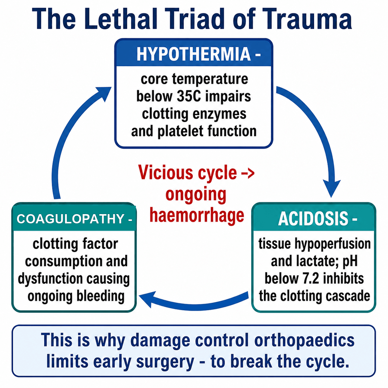
Coagulopathy in major trauma is not just dilutional — an acute traumatic coagulopathy driven by tissue hypoperfusion and inflammation is present on arrival in severely injured patients, then amplified by hypothermia, acidosis and crystalloid dilution. This is why early haemostatic resuscitation, not late correction, is key.
Permissive hypotension. Tolerate a lower blood pressure, a palpable radial pulse or a systolic of about 80-90 mmHg, until surgical haemorrhage control is achieved, to avoid "popping the clot" and dilutional coagulopathy. The exception is traumatic brain injury, where cerebral perfusion must be maintained and hypotension avoided.
Haemostatic resuscitation. Give blood products early in balanced ratios approximating whole blood, around 1:1:1 red cells to plasma to platelets, through a massive transfusion protocol, and minimise crystalloid, which dilutes clotting factors and worsens the triad. Give tranexamic acid early: CRASH-2 showed that TXA within 3 hours of injury reduces death due to bleeding in trauma haemorrhage, and that it is harmful if given late.
Goal-directed correction and endpoints. Rewarm aggressively, correct ionised calcium, and guide product use with point-of-care viscoelastic testing (ROTEM or TEG) where available. Adequate resuscitation, and readiness to escalate fracture care, is signalled by clearance of lactate and base deficit, not by blood pressure alone.
Classification
The table. The same handful of measurements decides the category on arrival. The Pro-DCO column is the set of triggers for temporary fixation; the Pro-ETC column describes the stable patient in whom definitive surgery can proceed from the outset.
- Pro-ETC
- Greater than 7.35
- Borderline
- 7.25-7.35
- Pro-DCO
- Less than 7.25
- Reasoning
- Significant acidosis indicates poor perfusion
- Pro-ETC
- Less than 2
- Borderline
- 2-4
- Pro-DCO
- Greater than 4 mmol/L
- Reasoning
- Marker of tissue hypoxia
- Pro-ETC
- Less than 4
- Borderline
- 4-6
- Pro-DCO
- Greater than 6
- Reasoning
- Reflects degree of metabolic derangement
- Pro-ETC
- Greater than 36°C
- Borderline
- 35-36°C
- Pro-DCO
- Less than 35°C
- Reasoning
- Hypothermia impairs coagulation
- Pro-ETC
- Greater than 100
- Borderline
- 50-100
- Pro-DCO
- Less than 50
- Reasoning
- Coagulopathy, ongoing haemorrhage
- Pro-ETC
- Borderline
- Pro-DCO
- Greater than 10 units in 24h
- Reasoning
- Massive transfusion = unstable
- Pro-ETC
- Borderline
- Pro-DCO
- Greater than 1.5
- Reasoning
- Coagulopathy
These are guidelines, not absolute cutoffs, and clinical judgement remains essential.
Base deficit and base excess are the same measurement with opposite signs. A base deficit of 6 is a base excess of -6. So "base deficit less than 6" and "base excess at least -5.5" describe almost the same patient, and quoting one threshold in each convention on the same page, as is common, makes them look like two separate hurdles when they are one.
Clinical Assessment
The primary survey. ATLS principles come first. Control haemorrhage with a pelvic binder or a tourniquet, resuscitate with blood products, and identify every injury; a tertiary survey is essential. Then classify the patient as stable, borderline, unstable or in extremis.
The ongoing assessment. The category is not fixed, so the physiological parameters are trended and the patient reassessed regularly; the question is whether they are improving or deteriorating. Response to resuscitation is the key decision point, status can change rapidly, and the decision is shared with the trauma team, anaesthesia and ICU.
- Adequate resuscitation
- Normalising heart rate
- Adequate resuscitation
- Decreasing blood product requirement
- Adequate resuscitation
- Falling lactate
- Adequate resuscitation
- Improving acidosis
- Adequate resuscitation
- Warm peripheries, urine output greater than 0.5 ml/kg/hr
Young patients compensate well. Blood pressure may be normal despite significant blood loss. Use lactate, base deficit, and clinical signs to assess perfusion. Hypotension is a LATE sign of decompensation.
Investigations
The investigations are the ones that feed the physiological table in the Classification section. The blood gas is the one that is repeated.
Serial lactate is the single most useful parameter. A falling lactate indicates adequate resuscitation. A rising lactate despite resuscitation indicates ongoing hemorrhage or inadequate perfusion - this patient needs DCO, not definitive surgery.
Decision Differential: ETC vs DCO vs EAC
The viva trap is treating DCO and ETC as a binary. They sit on a spectrum, with Early Appropriate Care (EAC) as the modern, resuscitation-led bridge, and the three need to be distinguished clearly.
- Early Total Care (ETC)
- Definitive fixation of all fractures early
- Early Appropriate Care (EAC)
- Resuscitate to targets, then fix definitively early
- Damage Control (DCO)
- Temporary stabilization, delay definitive surgery
- Early Total Care (ETC)
- Stable physiology
- Early Appropriate Care (EAC)
- Achieves lactate less than 4, pH at least 7.25, BE at least -5.5
- Damage Control (DCO)
- Persistent derangement / high-risk pattern
- Early Total Care (ETC)
- Definitive (nail/ORIF)
- Early Appropriate Care (EAC)
- Definitive once resuscitated (often within 36h)
- Damage Control (DCO)
- External fixation / splint
- Early Total Care (ETC)
- Bone 1989 (RCT)
- Early Appropriate Care (EAC)
- Vallier 2013/2015
- Damage Control (DCO)
- Pape 1993, EPOFF 2003
- Early Total Care (ETC)
- Second hit in the unstable patient
- Early Appropriate Care (EAC)
- Delay if resuscitation targets ignored
- Damage Control (DCO)
- Prolonged ex-fix, pin infection, delayed union
The EAC criterion. Vallier's protocol recommends definitive fixation within 36 hours if lactate is less than 4.0 mmol/L, pH is at least 7.25, or base excess is at least -5.5 mmol/L: any one of the three, not all of them. In the EAC cohort complications were 16.3% in those fixed within 36 hours versus 33.3% in those delayed.
Fixed timing or physiology-led timing. The traditional teaching, avoid days 2-4 and convert on day 5-10, is being displaced by EAC, which fixes definitively as soon as the resuscitation endpoints are met. The day-count is a guide, not a rule.
Who is truly borderline. There is no universally validated cut-off. Lactate trajectory, base deficit, chest injury severity and response to resuscitation matter more than any single number.
Reaming in the chest-injured patient. The fat embolism and second hit concern around reaming is debated, and modern data suggest that adequate resuscitation matters more than reaming per se.
Head injury and timing. Avoiding hypotension and hypoxia for the injured brain can argue for prompt fixation, which complicates a simple "delay everything" approach.
Management
The goals. Stabilise the fractures temporarily, keep the surgical insult to a minimum, control the haemorrhage that comes from the fractures, and give the patient the chance to be resuscitated and optimised. A stabilised patient can also be transferred safely to ICU.
The temporising measures are chosen by site:
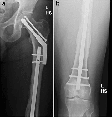
- External fixation - pelvis, femur, tibia, humerus
- Splinting - forearm, ankle, foot
- Spanning plates - an alternative for some sites
- Wound VAC - open fractures
What not to do. Definitive ORIF, intramedullary nailing with extensive reaming, prolonged surgical time, and any procedure that is not essential.
Surgical Technique
Indications. An unstable pelvic ring injury, ongoing haemorrhage, or the need for temporary stabilisation.
Technique. Position supine and identify the ASIS and the iliac crest. Make a 2 cm incision over the iliac crest, 3 cm posterior to the ASIS, and insert 5 mm Schanz pins under image guidance, aiming toward the AIIS, two pins per side. Apply the connecting bar across the anterior pelvis and tighten the frame to reduce and stabilise. The alternative for posterior injuries is the pelvic C-clamp, which carries a higher risk and requires experience.
Haemorrhage control in the bleeding pelvis. The exsanguinating pelvic ring injury is a prototypical DCO and DCR emergency, and stabilisation and haemorrhage control escalate stepwise:
- Pelvic binder — applied first, centred over the greater trochanters (not the iliac crests); reduces pelvic volume and tamponades venous and bony bleeding. A simple, immediate, life-saving step.
- Mechanical stabilisation — anterior external fixation (for anteroposterior-compression or "open-book" patterns) or a posterior C-clamp (for vertically or rotationally unstable posterior injuries) when the binder is insufficient.
- Divergent haemorrhage control by bleeding source:
- Angioembolization for arterial bleeding — suggested by a contrast blush on CT and ongoing instability despite mechanical control.
- Preperitoneal pelvic packing for venous and bony surface bleeding (the majority) — rapid, especially where angiography is unavailable or the patient is too unstable to transfer.
- REBOA (resuscitative endovascular balloon occlusion of the aorta, Zone III) — a temporising adjunct in the in extremis patient to control inflow while definitive haemorrhage control is arranged.
The binder or external fixator controls the fracture; it does not by itself control arterial bleeding, so escalate to angiography or packing if the patient remains unstable.
Complications
- DCO Risk
- Lower in unstable patients
- ETC Risk
- Higher if done in unstable patient
- DCO Risk
- 5-10%
- ETC Risk
- N/A
- DCO Risk
- Possible if prolonged external fixation
- ETC Risk
- Lower
- DCO Risk
- Reduced by avoiding second hit
- ETC Risk
- Higher in unstable patient
- DCO Risk
- Similar
- ETC Risk
- Similar
The frame has its own complications. Beyond pin site infection, pins loosen, malunion follows if pins are placed incorrectly, a pin can injure a nerve, and pins sitting in the path of the future nail make the conversion harder.
Pin site infection increases risk of deep infection after conversion to internal fixation. If significant pin site infection develops, consider a staged approach: remove external fixator, treat infection, then definitive fixation after pin sites healed.
Postoperative Care
After the frame. The patient goes to ICU for ongoing resuscitation with serial lactate monitoring, and is optimised through the inflammatory peak without further surgery. The physiological parameters are reviewed daily, because the goal is to convert to definitive fixation as soon as it is safely possible: prolonged external fixation increases infection risk and complicates the conversion.
After conversion. Once the definitive fixation is in, the patient follows standard fracture rehabilitation with early mobilisation.
Outcomes and Prognosis
What DCO is credited with. Reduced ARDS in patients with a femur and chest injury, lower mortality in borderline and unstable patients, and comparable long-term outcomes when it is used appropriately. Used in an unstable patient it improves survival; the whole difficulty is identifying the borderline patient, who may decompensate with definitive surgery.
DCO vs ETC studies suffer from selection bias. Sicker patients get DCO, so direct comparisons are challenging and observational comparisons cannot be taken at face value. The principle of minimizing surgical insult in the already-stressed patient remains sound, and the physiological rationale (EPOFF) remains the strongest support.
Guidelines, Registries & Global Practice
Global Epidemiology
- Trauma is among the leading causes of death worldwide in people aged under 45, and the leading cause of years of life lost.
- Long-bone and pelvic fractures are common in major polytrauma; the femur-plus-chest pattern is the prototypical high-risk DCO group (Pape 1993).
- Most major-trauma deaths follow a trimodal distribution; the late peak (days to weeks) from sepsis and multi-organ failure is the window DCO seeks to influence by limiting the surgical "second hit".
Side-by-Side Guidance
- Core Position
- Stable / borderline / unstable / in-extremis grading drives ETC vs DCO
- Practical Emphasis
- Physiology-led; reassess the borderline patient repeatedly
- Core Position
- Resuscitate to lactate/pH/base-excess targets, then fix definitively early (within 36h)
- Practical Emphasis
- Avoid both under-resuscitation AND unnecessary delay
- Core Position
- Major-trauma networks; definitive care at the right centre by the right team
- Practical Emphasis
- Damage control and timely transfer within a regional network
- Core Position
- Haemorrhage control and physiological correction precede skeletal reconstruction
- Practical Emphasis
- Lactate/base deficit clearance as resuscitation endpoints
- National major-trauma networks (e.g. UK TARN, German TraumaRegister DGU) link early appropriate care to lower mortality
- Registry data underpin the move from rigid "day 5-10" rules toward resuscitation-guided timing
- Damage control enables safe inter-hospital transfer to the definitive-care centre
- Well-resourced centres: ICU optimization, point-of-care coagulation (ROTEM/TEG), early conversion to definitive fixation
- Limited-resource settings: external fixation may remain the definitive construct where ICU/implant access is constrained
- External fixation is a universal core skill - low cost, rapid, life-saving for haemorrhage control
DCO is the bridge across geography and resource gaps. A patient stabilized with external fixation at a district hospital can be safely transported over long distances - relevant to remote regions of any country and to limited-resource health systems alike.
MCQ Practice Points
Q: What is the 'second hit' phenomenon in polytrauma management? A: Major surgery causes an additional inflammatory insult to an already compromised patient. This can precipitate ARDS and multi-organ failure. DCO minimizes this by delaying definitive surgery until the initial inflammatory response subsides.
Q: What are the physiological thresholds that indicate a patient needs DCO rather than ETC? A: pH less than 7.25, lactate greater than 4 mmol/L, base deficit greater than 6, temperature less than 35°C, platelets less than 50,000. Also consider DCO for ISS greater than 20, bilateral femur fractures, or femur + chest/head injury.
Q: When is the optimal time to convert external fixation to definitive fixation in DCO patients? A: Day 5-10 after injury. This is the immunological window after the initial inflammatory peak (days 2-4) has subsided. Convert when physiological parameters normalize (lactate less than 2, pH greater than 7.35).
Q: What are the four patient categories in DCO decision-making? A: Stable (ETC appropriate), Borderline (individualized decision based on parameters), Unstable (DCO mandatory), In Extremis (hemorrhage control only, no fracture fixation).
Q: Why is femoral fracture + chest injury a classic DCO indication? A: Early femoral nailing increases pulmonary complications in patients with chest injury. Pape (1993) showed early nailing with severe chest injury raised ARDS (33% vs 7.7%) and mortality (21% vs 4%). External fixation allows fracture stabilization without the second hit of intramedullary reaming.
Q: What is the single most useful parameter for monitoring resuscitation adequacy in polytrauma? A: Serial lactate. Falling lactate indicates adequate tissue perfusion. Rising or static lactate despite resuscitation suggests ongoing hemorrhage or inadequate perfusion - this patient needs DCO approach.
Clinical Decision Scenarios
Practise clinical reasoning and management decisions out loud
“A 28-year-old male is brought to ED following a high-speed motor vehicle accident. He has bilateral femoral shaft fractures and a pulmonary contusion. His lactate is 4.5, pH 7.28, and he has required 6 units of blood in the ED. How would you manage his femoral fractures?”
“Explain the concept of damage control orthopaedics and when you would apply it.”
“A polytrauma patient had a femoral external fixator applied 3 days ago for DCO. Today their lactate is 1.5, pH 7.38, and they are hemodynamically stable off vasopressors. When would you convert to definitive fixation?”
Patient Categories
- Stable: ETC - definitive fixation
- Borderline: Decision point - use parameters
- Unstable: DCO mandatory
- In Extremis: Hemorrhage control only
DCO Indications
- pH less than 7.25
- Lactate greater than 4
- ISS greater than 20
- Bilateral femur or femur + chest injury
- Ongoing hemorrhage despite resuscitation
Second Hit Phenomenon
- Surgery is inflammatory insult
- Can precipitate ARDS, MOF
- DCO minimizes surgical trauma
- Allow inflammation to subside
Timing
- Day 0-1: Window of opportunity if stable
- Day 2-4: Avoid surgery (peak inflammation)
- Day 5-10: Safe window for conversion
- Monitor lactate, pH, base deficit
Evidence Base and Key Trials
Bone (1989) - Early vs Delayed Femoral Stabilization (RCT)
- Prospective RCT of 178 adults comparing early (less than 24h) vs delayed femoral fracture stabilization
- In multiply injured patients, DELAYED fixation increased pulmonary complications (ARDS, fat embolism, pneumonia), ICU stay and hospital stay
- Hospital costs were significantly higher in the delayed-treatment group across all patients
- Established the original 'early total care' (ETC) paradigm of urgent definitive femoral fixation
Pape (1993) - Early Femoral Nailing + Chest Injury and ARDS
- Retrospective study of 106 multiply-injured patients with femoral midshaft fracture treated by intramedullary nailing
- With severe chest injury, early (less than 24h) nailing carried higher post-traumatic ARDS (33% vs 7.7%) and mortality (21% vs 4%)
- Without chest injury, early nailing REDUCED ICU and intubation time - benefit depends on the chest
- Provided the physiological rationale that femur-plus-chest is the high-risk group
Pape - EPOFF Study (Randomized, Inflammatory Burden)
- Prospective randomized multicentre trial (n=35) in clinically stable multiply-injured patients: primary IM nailing vs DCO (external fixation then secondary nailing)
- Primary IM nailing produced a sustained perioperative rise in IL-6 and IL-8; external fixation did NOT
- Secondary conversion to a nail (day 5-10 window) caused no inflammatory surge - the 'second hit' is avoided
- No difference in ARDS/sepsis/MOF rates in this small stable cohort