First Dorsal Compartment | APL and EPB | Finkelstein Test | Injection
- First dorsal compartment contains APL and EPB
- Finkelstein test is pathognomonic
- Injection success 70-80%
- Watch for aberrant EPB septum
- Common in new mothers (repetitive lifting)
- “Finkelstein: fist over thumb, ulnar deviate wrist
- “New mothers: repetitive baby lifting
- “Superficial radial nerve at risk during surgery
- “Separate septum for EPB in 30-50%
First dorsal compartment contains APL and EPB. These tendons pass over radial styloid. Stenosing tenosynovitis causes pain with thumb/wrist movement.
The tendons may have a separate septum which must be released at surgery.
Make fist over thumb, ulnar deviate wrist. Positive test reproduces radial wrist pain. Pathognomonic for de Quervain.
The test stretches the APL and EPB tendons.
Corticosteroid injection is first-line treatment. 70-80% success rate. Inject into tendon sheath, not tendon.
May repeat once if partial response.
Separate septum for EPB in 30-50% of cases. If EPB in separate compartment, both compartments must be released at surgery.
This is the most common cause of failed release.
Overview and Epidemiology
De Quervain tenosynovitis is a stenosing tenovaginitis of the first dorsal extensor compartment, where the abductor pollicis longus (APL) and extensor pollicis brevis (EPB) tendons become entrapped beneath a thickened extensor retinaculum at the radial styloid. Despite the historical name "tenosynovitis", the dominant histological finding is myxoid degeneration and fibrocartilaginous thickening of the retinaculum with little true inflammatory infiltrate - it is a degenerative/mechanical overload process rather than a primary inflammatory synovitis.
- Population prevalence roughly 0.5 to 1.3 percent
- Female predominance (around 4 to 6 : 1)
- Peak in the fourth to sixth decades
- Postpartum and lactating women, occupational repetitive users, and heavy smartphone users are over-represented
- Repetitive thumb abduction/extension with ulnar wrist deviation (lifting an infant, texting, gripping)
- Postpartum hormonal change plus repetitive lifting (often bilateral)
- Female sex, age 40 to 60
- Associations reported with diabetes and inflammatory arthropathy
New mothers are the classic patients due to repetitive lifting of babies with the thumb extended and the wrist ulnar-deviated; it is frequently bilateral in this group. The same repetitive-loading mechanism explains the rising incidence among heavy smartphone and digital-device users.
Pathophysiology and Mechanisms
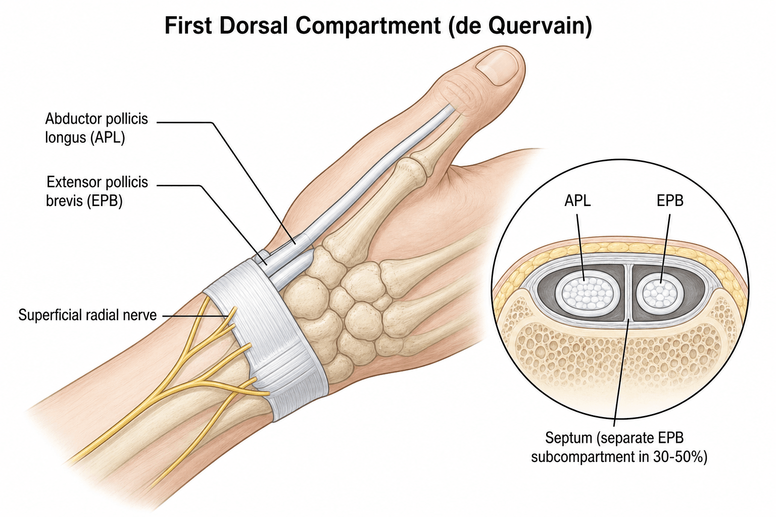
First Dorsal Compartment
- Abductor pollicis longus (APL)
- Extensor pollicis brevis (EPB)
Over radial styloid
EPB in separate compartment in 30-50%
Understanding anatomy is key to successful treatment.
Classification Systems
Clinical Staging
- Description
- Intermittent pain with activity
- Management
- Splinting, activity modification
- Description
- Constant pain, positive Finkelstein
- Management
- Corticosteroid injection
- Description
- Failed injection, chronic symptoms
- Management
- Surgical release
Clinical staging helps guide treatment intensity.
Clinical Assessment
- Radial wrist pain
- Pain with thumb use
- Worse with gripping
- Swelling over radial styloid
- New parent or repetitive work
Ask about repetitive thumb/wrist activities.
- Tenderness over first dorsal compartment
- Positive Finkelstein test
- May have visible swelling
- Crepitus with movement
Finkelstein is the key diagnostic test.
Make fist over thumb, then ulnar deviate wrist. This stretches APL and EPB over the radial styloid. Reproduction of radial wrist pain is positive.
This test is pathognomonic for de Quervain tenosynovitis.
Investigations
Usually Not Required
De Quervain is a clinical diagnosis.
Finkelstein test + radial styloid tenderness is sufficient.
Imaging rarely needed unless diagnosis uncertain.


Differential Diagnosis
- Distinguishing features
- Pain/tenderness over radial styloid, swelling of first dorsal compartment
- Discriminating test
- Finkelstein / Eichhoff positive; WHAT (wrist hyperflexion and abduction of thumb) test
- Distinguishing features
- Pain at base of thumb, more distal and volar; squaring of thumb base
- Discriminating test
- Grind test positive; radiographs show CMC joint OA
- Distinguishing features
- Pain and crepitus roughly 4 cm proximal to wrist where second compartment crosses first
- Discriminating test
- Tenderness proximal to the radial styloid, not over it
- Distinguishing features
- Dorsoradial numbness/paraesthesia, no true tendon swelling
- Discriminating test
- Positive Tinel over the nerve; sensory not tendon-stretch pain
- Distinguishing features
- History of trauma, anatomic snuffbox tenderness
- Discriminating test
- Snuffbox tenderness, axial thumb load pain; radiographs/MRI
- Distinguishing features
- Pain at the STT joint, just distal to scaphoid
- Discriminating test
- Localised STT tenderness; radiographs
The test most clinicians call "Finkelstein" is technically the Eichhoff manoeuvre: the patient makes a fist over a flexed thumb and the examiner ulnar-deviates the wrist. The true Finkelstein test has the examiner grasp the thumb and longitudinally pull while ulnar-deviating the wrist. Both stress the APL/EPB. The Eichhoff can be falsely positive; the WHAT test (resisted thumb extension/abduction with the wrist in hyperflexion) has been proposed as more specific.
Clinically Predicting a Separate EPB Compartment
Because a separate EPB sub-compartment (the septum) is the dominant cause of injection and surgical failure, it is worth trying to detect it before treatment rather than only on ultrasound or at operation. Selective provocative testing helps.
- What it isolates
- Stresses APL and EPB together (passive ulnar deviation, thumb in palm)
- Interpretation
- Confirms first-compartment disease but does not localise to one tendon
- What it isolates
- Resisted thumb MCP extension with the wrist slightly flexed - isolates EPB
- Interpretation
- Pain reproduced suggests EPB involvement in a separate sub-compartment
- What it isolates
- Isolates APL
- Interpretation
- Pain mainly here points to APL-dominant disease
A positive EPB entrapment test raises the probability of a septated compartment, which predicts failure of a single blind injection - so it is a reason to either inject under ultrasound guidance into both sub-compartments or counsel the patient earlier about surgical release.
Finkelstein/Eichhoff confirms first-compartment disease but cannot tell APL from EPB. The EPB entrapment test (pain on resisted thumb MCP extension, wrist slightly flexed) isolates the EPB and, if positive, suggests a separate EPB sub-compartment - the variant that defeats a blind injection and an incomplete release. Use it to decide on ultrasound-guided injection of both sub-compartments or earlier surgery.
Management Algorithm
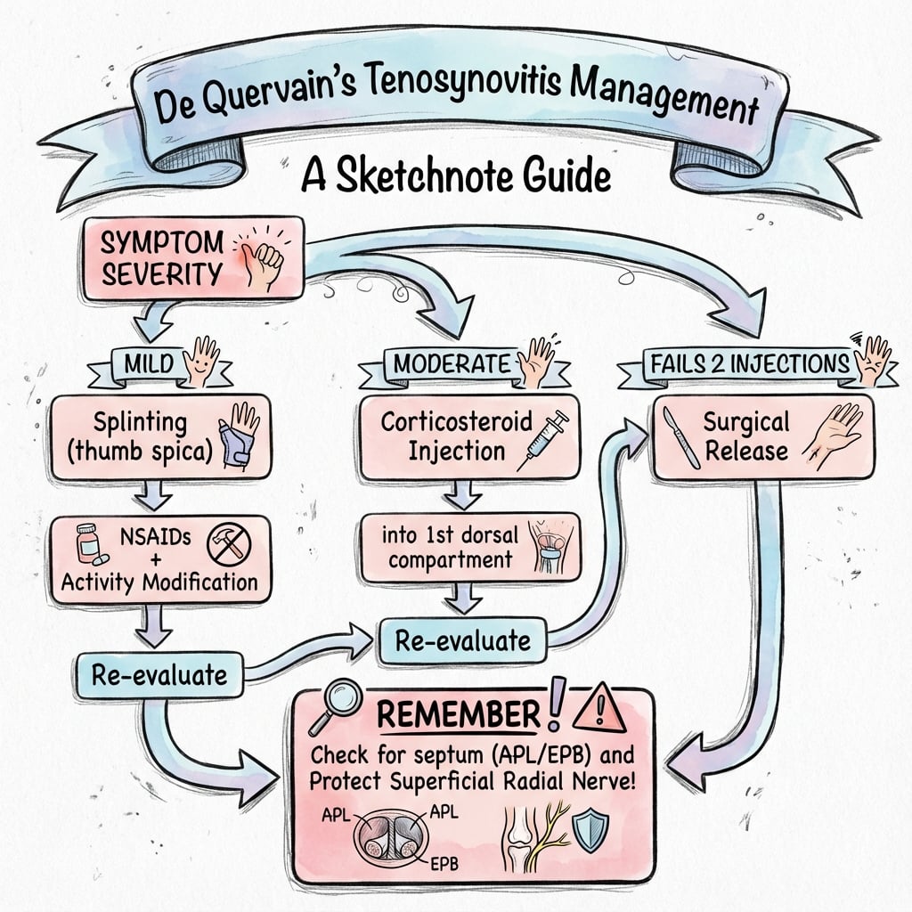
First-Line Treatment
- Thumb spica splint
- Rest the first dorsal compartment
- Corticosteroid into tendon sheath
- Success rate 70-80%
- May repeat once
- Avoid aggravating activities
- Ergonomic advice
Conservative treatment is effective in majority of cases.
Surgical Technique
First Dorsal Compartment Release
Transverse or longitudinal over first DC
- Protect superficial radial nerve branches
- Identify first dorsal compartment
- Release retinaculum longitudinally
- CHECK for separate EPB septum (30-50%)
- Release EPB compartment if present
- Ensure both tendons glide freely
Always look for separate EPB compartment.
Always look for separate EPB compartment. Present in 30-50% of patients. If only APL compartment released, EPB remains stenosed and symptoms persist. This is the most common cause of failed surgery.
Preventing Tendon Subluxation: Where to Incise the Retinaculum
A specific technical pitfall of first-compartment release is volar (palmar) subluxation of the tendons - if the retinaculum is divided in the wrong place, the APL and EPB can bowstring across the radial styloid when the wrist flexes, causing painful snapping.
The preventive principle is where you incise the retinaculum:
- Divide the retinaculum along its dorsal (dorso-ulnar) margin, not its volar edge. This leaves the bulk of the retinaculum as a volar leaf against which the tendons rest, so they cannot fall palmar-ward.
- Avoid a complete circumferential release. Some surgeons additionally preserve or reconstruct a retinacular sling (for example a step-cut or Z-lengthening) in patients judged at higher risk of subluxation.
- After release, take the wrist through flexion-extension and radial-ulnar deviation to confirm both tendons stay seated and glide freely.
Release the first-compartment retinaculum along its dorsal margin so a volar leaf remains to buttress the tendons - dividing it volarly or circumferentially lets the APL/EPB sublux palmarly and bowstring on wrist flexion. Always check intra-operatively that the tendons stay seated through a full range before closing, and protect the superficial radial nerve throughout.
Complications
- Incidence
- 5-10%
- Prevention/Management
- Careful dissection and protection
- Incidence
- 5-10%
- Prevention/Management
- Check for EPB septum
- Incidence
- Rare
- Prevention/Management
- Preserve some retinaculum dorsally
- Incidence
- Variable
- Prevention/Management
- Proper incision placement
Postoperative Care
Recovery Timeline
Soft dressing. Gentle ROM immediately. Avoid forceful gripping.
Remove sutures. Progressive thumb use. Return to light duties.
Full activity as tolerated. Complete recovery expected.
Outcomes and Prognosis
Prognostic Factors
Shorter symptom duration, successful injection response, complete surgical release.
Missed EPB septum at surgery, nerve injury, delayed treatment.
Guidelines, Registries & Global Practice
- Overall population prevalence is roughly 0.5 to 1.3 percent, with a female-to-male ratio of about 4 to 6 to 1 and a peak in the fourth to sixth decades.
- High-risk groups consistently identified across studies: postpartum and lactating women, occupational repetitive thumb/wrist users, and a newer cohort of high-intensity smartphone and digital-device users.
- Reported prevalence in infant caregivers reached 26.8 percent in one cross-sectional study, and a positive Finkelstein test was found in 36.8 percent of heavy device users among university students - far higher than the general population, underlining the role of repetitive loading.
No single orthopaedic society publishes a dedicated, named de Quervain guideline, so practice is anchored by high-level synthesis rather than a formal guideline document.
- Position
- Corticosteroid injection (ideally ultrasound-guided) plus 3-4 weeks thumb spica
- Notes
- Strongest current evidence base; 30 trials, 1663 patients
- Position
- Stepwise: splint and activity modification, then injection, then release
- Notes
- Emphasis on counselling re repeat injection and surgical anatomy
- Position
- Conservative first; injection in primary or secondary care; release if refractory
- Notes
- Pragmatic, primary-care-led injection pathways common in the NHS
- Position
- Conservative-first, surgery reserved for failed injection
- Notes
- Consistent with above; mini-open release widely taught
De Quervain release is a soft-tissue decompression and is not captured by joint-replacement registries (NJR, AJRR, AOANJRR). Outcome data therefore come from RCTs, systematic reviews and institutional series rather than national implant registries.
- In well-resourced settings ultrasound-guided injection is increasingly standard, improving accuracy and allowing both subcompartments to be targeted when a septum is seen.
- In limited-resource settings landmark-guided (blind) injection remains the norm and is effective; the diagnosis is clinical (Finkelstein test) so no imaging is required to start treatment.
- Surgical release is a low-cost, high-yield day-case procedure under local or regional anaesthesia, making it feasible across resource levels.
Know the Finkelstein test technique, the injection approach (sheath not tendon), and the importance of the separate EPB septum. Be able to describe surgical release with superficial radial nerve protection, and cite that injection plus short thumb-spica immobilisation is the best-supported first-line strategy.
Controversies and Areas of Uncertainty
The 2023 network meta-analysis ranked ultrasound-guided injection highest for pain, and a septum predicts failure of a single blind injection. However, blind injection remains effective and far cheaper, and no adequately powered head-to-head RCT has shown a clinically meaningful functional difference. Reasonable to reserve guidance for failed blind injection or a known septum.
Adding 3-4 weeks of thumb spica to injection improves function statistically but the difference may not reach a clinically important threshold, and splints are poorly tolerated by caregivers. The optimal duration and whether splinting alone has any durable role remain unsettled.
Mini-open, percutaneous and ultrasound-guided releases report fewer wound issues, but evidence is largely low-level (case series). Open release with direct visualisation of any EPB septum and protection of the superficial radial nerve remains the reference standard.
Commonly quoted single-injection cure rates of 70 to 80 percent derive from observational and mixed cohorts; the only included trial in the Cochrane review was tiny and confined to pregnant/lactating women. True durable single-injection success is likely lower when a septum is present (around 30 percent recurrence at six weeks in one ultrasound cohort).
MCQ Practice Points
Q: What tendons are in the first dorsal compartment? A: APL (abductor pollicis longus) and EPB (extensor pollicis brevis).
Q: What is the Finkelstein test? A: Fist over thumb, ulnar deviate wrist. Positive when this reproduces radial wrist pain.
Q: What is the most common cause of failed surgical release? A: Missed separate EPB septum. Present in 30-50%; both compartments must be released.
Q: What is the success rate of corticosteroid injection for de Quervain? A: 70-80% success rate. It is first-line treatment. May be repeated once if partial response.
Q: What nerve is at risk during de Quervain release? A: Superficial radial nerve. Branches cross the surgical field. Injury causes numbness over dorsal thumb web or painful neuroma.
Additional Quiz Questions
At a Glance
- Key Points
- Stenosing tenosynovitis of 1st dorsal compartment (APL, EPB)
- Key Points
- New mother with radial wrist pain, worse with lifting
- Key Points
- Finkelstein test - fist over thumb, ulnar deviate - reproduces pain
- Key Points
- Corticosteroid injection (70-80% success)
- Key Points
- Failed conservative treatment (2 injection trials)
- Key Points
- Check for separate EPB septum (30-50%) - release both compartments
APL EPBFirst Dorsal Compartment
Hook:APL and EPB are in first dorsal compartment!
SQUEEZEDEQUERVAIN - Key Facts
Hook:SQUEEZE thumbs up for de Quervain!
1-2-3-4-5-6Dorsal Compartments
Hook:Six dorsal compartments - know them all!
Exam Viva Scenarios
Practise clinical reasoning and management decisions out loud
“A 32-year-old new mother has 2 months of radial wrist pain. It is worse when lifting her baby. Finkelstein test is positive. There is tenderness over the radial styloid. What is your diagnosis and management?”
“A 45-year-old woman had surgical release for de Quervain 6 weeks ago but her symptoms have not improved. The wound healed well. Finkelstein test remains positive. What is the most likely cause and how would you manage?”
“You are about to perform a corticosteroid injection for de Quervain tenosynovitis. Describe your technique and what complications you would warn the patient about.”
Anatomy
- First dorsal compartment
- APL and EPB
- Over radial styloid
- Separate EPB septum 30-50%
Clinical
- Radial wrist pain
- New mothers classic
- Finkelstein test positive
- Tender over radial styloid
Conservative Treatment
- Injection: 70-80% success
- Thumb spica splint
- Activity modification
- May repeat injection once
Surgery
- Release first dorsal compartment
- Check for EPB septum
- Protect superficial radial nerve
- Confirm both tendons glide
Complications
- Superficial radial nerve injury
- Missed EPB septum (failure)
- Tendon subluxation (rare)
- Scar sensitivity
- Recurrence: usually missed septum
Key Points
- Finkelstein is pathognomonic
- Injection first-line
- EPB septum is key at surgery
- 90%+ success with proper release
- New mothers: postpartum hormones
Evidence Base and Key Studies
- 30 randomised trials, 1663 patients (mean age 46 years, 80% female)
- Adding 3-4 weeks of thumb spica immobilisation to corticosteroid injection (CSI) gave statistically (not clinically) significant short- and mid-term functional benefit (Q-DASH mid-term mean difference 9.4 points)
- Ultrasound-guided CSI ranked highest for pain relief in the network analysis
- CSI plus thumb spica had the highest probability of being the most effective option for short- and mid-term function
- Only one small controlled trial met inclusion criteria (18 pregnant or lactating women)
- All patients given methylprednisolone plus bupivacaine injection achieved complete pain relief (9/9) versus none with thumb spica splinting alone (0/9), number needed to treat = 1
- No injection side effects or local complications observed
- Authors stressed limited applicability owing to the single small, methodologically weak trial
- Reviewed 15 randomised controlled trials in de Quervain disease (plus trigger finger and Dupuytren)
- Moderate evidence for very-short-term benefit of corticosteroid injection
- A thumb splint added to corticosteroid injection was effective at short and mid term (moderate evidence)
- Called for more high-quality RCTs to support evidence-based practice
- 87 embalmed cadaveric wrists dissected (Thai population)
- Complete intracompartmental septum in 17.2% and incomplete septum in 42.5% (roughly 60% had some septation)
- Superficial radial nerve crossed over the first extensor compartment in 59.5% of specimens
- Two APL tendon slips were most common (54%); a single EPB slip in 97.7%, with EPB absent bilaterally in one cadaver
- 50 consecutive patients given a single ultrasound-assessed corticosteroid injection
- Symptom recurrence in 15 patients (30%) within six weeks
- An intracompartmental septum (adjusted OR 18.4) and a greater number of tendon slips (adjusted OR 24.7 per slip) independently predicted recurrence
- Mean DASH improved from 74.1 to 19.3 and VAS pain from 8.5 to 2.0
- Cross-sectional study of 508 university students
- 36.8% had a positive Finkelstein test, 95.6% in the dominant hand
- Positivity rose with screen time, from 12.5% at under 2 hours/day to 46.2% at 8 hours/day or more
- Wrist ulnar deviation during device use was strongly associated with a positive test
- Cross-sectional study of 190 infant caregivers screened with Finkelstein test
- 26.8% prevalence of de Quervain disease
- Infant age, lifting frequency and hand dominance were significant risk factors
- Caregiver age, infant weight and relationship to the infant were not significant