Idiopathic Inflammatory Myopathies
- The idiopathic inflammatory myopathies (dermatomyositis, polymyositis, and overlapping subtypes) are autoimmune, immune-mediated muscle diseases characterised by SYMMETRIC PROXIMAL muscle WEAKNESS (trouble rising from a chair, climbing stairs, lifting overhead) with a raised CREATINE KINASE; two-thirds of adults have a CHRONIC course and require MULTIDISCIPLINARY management.
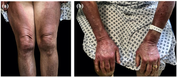
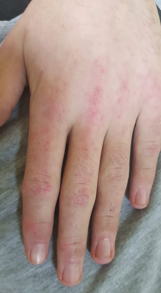
- DERMATOMYOSITIS adds characteristic SKIN signs - the violaceous periorbital HELIOTROPE rash and GOTTRON'S PAPULES over the knuckles (also the shawl/V sign and mechanic's hands) - and is a complement-mediated, interferon-driven microangiopathy; POLYMYOSITIS has the myopathy WITHOUT the skin signs and is a diagnosis of exclusion (inclusion-body myositis and dystrophies must be excluded).
- Two SYSTEMIC ASSOCIATIONS are critical: in adults, dermatomyositis is strongly PARANEOPLASTIC, so a new diagnosis mandates a SEARCH for occult MALIGNANCY; and INTERSTITIAL LUNG DISEASE is a major, sometimes severe, complication (especially with the anti-synthetase syndrome and anti-MDA5 antibodies) - autoantibodies help identify these high-risk subtypes.
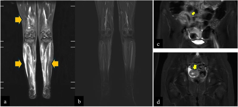
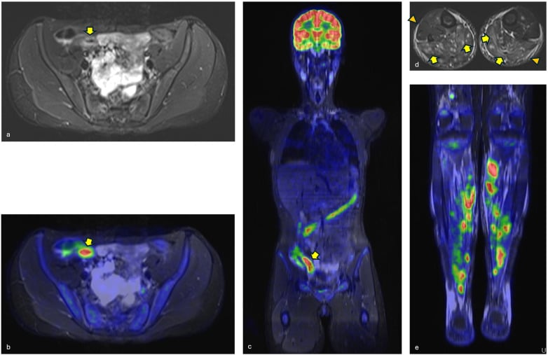
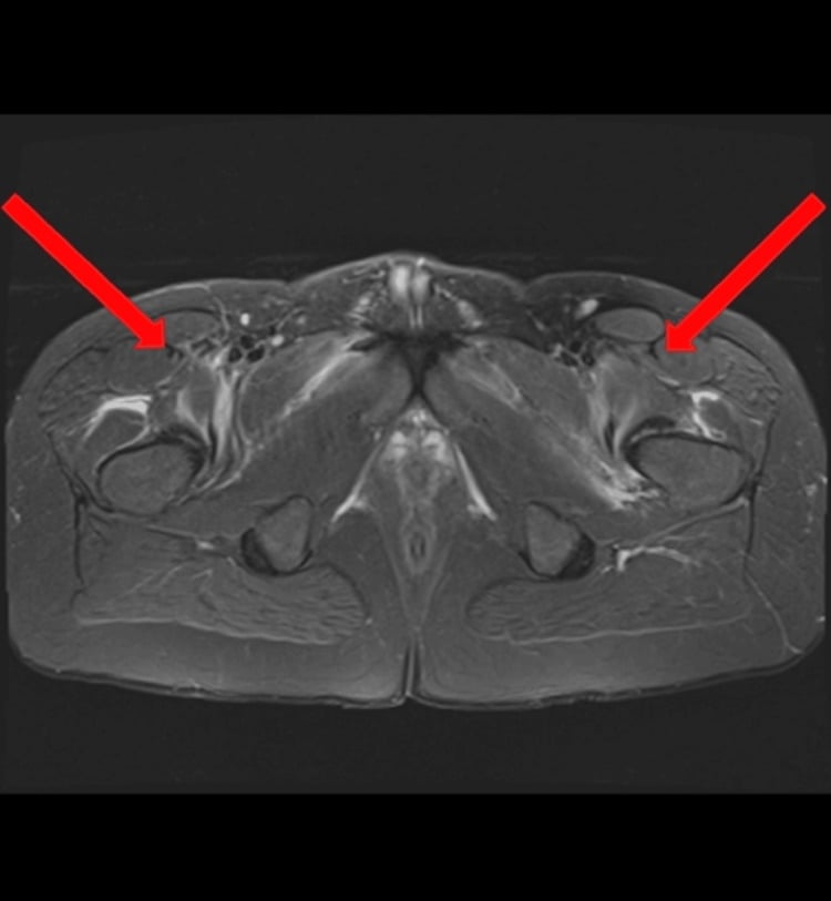
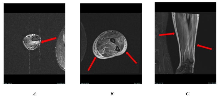
- DIAGNOSIS integrates the clinical picture with MYOSITIS-SPECIFIC and -associated ANTIBODIES (which define subtypes and predict complications such as cancer or ILD), ELECTROMYOGRAPHY, MRI (muscle oedema, and a guide to biopsy site), MUSCLE BIOPSY, and elevated muscle enzymes - the antibody profile increasingly guides classification and risk stratification.
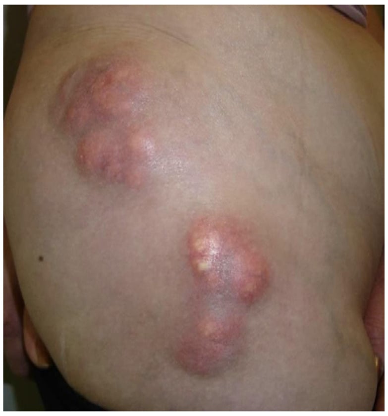
- JUVENILE DERMATOMYOSITIS is distinguished by a prominent VASCULOPATHY and the development of CALCINOSIS (dystrophic calcium deposits in skin/soft tissue) which can ulcerate, become infected, restrict joints and cause significant morbidity - and, with chronic weakness, can lead to joint CONTRACTURES.
- PUT NUMBERS ON 'SCREEN FOR CANCER'. The risk is not uniform across adult dermatomyositis - it is concentrated in an antibody-defined subset. In a meta-analysis of 18 studies and 1,962 patients, 41 percent of adults with ANTI-TIF-1-GAMMA had cancer-associated dermatomyositis (95% CI 36-45), with a diagnostic odds ratio for cancer of 9.37 (95% CI 5.37-16.34) and no heterogeneity between studies. So the antibody panel is not merely a classification exercise: it tells you WHOM to investigate hard and whom to reassure.
- IVIG IS NOT JUST 'EFFICACIOUS' - IT HAS A PLACEBO-CONTROLLED TRIAL, AND A SAFETY SIGNAL THAT MATTERS BEFORE SURGERY. In ProDERM, 95 adults with active dermatomyositis were randomised to IVIG 2.0 g/kg or placebo every 4 weeks: 79 percent (37 of 47) versus 44 percent (21 of 48) reached the primary endpoint at 16 weeks, a 35-percentage-point difference (95% CI 17-53). But 6 of the 9 IVIG-related serious adverse events were THROMBOEMBOLIC - relevant to any patient coming to an orthopaedic operation. Note too that CREATINE KINASE did not differ meaningfully between the groups, so CK is a poor marker of treatment response even though it is used to make the diagnosis.
- MANAGEMENT is medical and multidisciplinary: CORTICOSTEROIDS with steroid-sparing IMMUNOSUPPRESSION (methotrexate, azathioprine, mycophenolate), INTRAVENOUS IMMUNOGLOBULIN (efficacious, particularly in refractory dermatomyositis), and newer agents targeting the interferon/JAK pathway or B cells; the ORTHOPAEDIC relevance is the management of weakness-related disability, joint CONTRACTURES, and the complications of CALCINOSIS (surgical excision of problematic deposits), always alongside the treating rheumatology/neurology team.
- “Inflammatory myopathies = symmetric PROXIMAL weakness + raised CK. DERMATOMYOSITIS = heliotrope rash + Gottron's papules (skin signs); POLYMYOSITIS = no skin signs (diagnosis of exclusion).
- “Adult DM = PARANEOPLASTIC → SCREEN for occult malignancy. INTERSTITIAL LUNG DISEASE (anti-synthetase, anti-MDA5) is a major complication. Myositis-specific antibodies define subtypes/risk.
- “Juvenile DM = CALCINOSIS + vasculopathy. Treat with corticosteroids + immunosuppression + IVIG; orthopaedic role = weakness/contractures + excising problematic calcinosis.
- “Quote the numbers, not the adjectives: anti-TIF-1-gamma DM is cancer-associated in 41% (OR 9.37); IVIG beat placebo 79% vs 44% in ProDERM (n=95) - but 6 of 9 IVIG serious adverse events were THROMBOEMBOLIC, and CK did NOT track the response.
Symmetric proximal weakness + raised CK; dermatomyositis adds heliotrope rash + Gottron's papules. Confirm with myositis antibodies, EMG, MRI, biopsy.
Adult dermatomyositis is paraneoplastic - screen for occult malignancy. Interstitial lung disease (anti-synthetase, anti-MDA5) can be severe. Juvenile DM → calcinosis.
Recognition, Diagnosis & Associations
The inflammatory myopathies present with symmetric proximal muscle weakness and a raised creatine kinase. Dermatomyositis adds the heliotrope rash and Gottron's papules (and shawl/V sign, mechanic's hands); polymyositis has the myopathy without the skin signs and is a diagnosis of exclusion. Two associations are critical: adult DM is paraneoplastic (search for occult malignancy), and interstitial lung disease (especially anti-synthetase syndrome and anti-MDA5) can be severe. Diagnosis uses myositis-specific antibodies (which define subtypes and predict cancer/ILD risk), EMG, MRI (oedema; biopsy guide) and muscle biopsy. Juvenile DM features a vasculopathy and calcinosis.
- Dermatomyositis
- Yes - heliotrope rash, Gottron's papules
- Polymyositis
- No characteristic skin signs
- Dermatomyositis
- Complement-mediated microangiopathy (perifascicular)
- Polymyositis
- T-cell-mediated endomysial
- Dermatomyositis
- Strong paraneoplastic association - screen
- Polymyositis
- Weaker association
- Dermatomyositis
- Yes (calcinosis, vasculopathy)
- Polymyositis
- Rare
- Dermatomyositis
- Amyopathic form (skin only)
- Polymyositis
- Diagnosis of exclusion (exclude IBM, dystrophy)
Management & Orthopaedic Relevance
- Medical (mainstay): corticosteroids with steroid-sparing immunosuppression (methotrexate, azathioprine, mycophenolate); IVIG (efficacious, particularly in refractory dermatomyositis); newer interferon/JAK-pathway and B-cell-targeted therapies in refractory disease.
- Screen and treat the associations: malignancy work-up in adult DM; assess/treat interstitial lung disease.
- Orthopaedic relevance: rehabilitation for weakness; prevent/manage joint contractures; excise problematic CALCINOSIS (ulcerating/infected/joint-restricting deposits, especially in juvenile DM); coordinate perioperative immunosuppression.
- Multidisciplinary: managed with rheumatology/neurology and (where relevant) oncology/respiratory.
The single most important systemic point in adult dermatomyositis is its strong paraneoplastic association: a new diagnosis should prompt an age- and risk-appropriate search for an occult malignancy, because the myositis can be the presenting sign of an underlying cancer (and the myositis-specific antibody profile helps identify the highest-risk patients). The second critical association is interstitial lung disease, which can be rapidly progressive and life-threatening, particularly with the anti-synthetase syndrome and anti-MDA5 antibody, so lung involvement must be actively assessed. For the orthopaedic surgeon, the relevance is mostly the consequences of the disease and its treatment - proximal weakness and disability, joint contractures from chronic weakness, and the calcinosis of juvenile dermatomyositis, whose deposits may ulcerate, become infected or restrict joints and sometimes need surgical excision - all managed alongside the rheumatology/neurology team, with attention to perioperative steroids and immunosuppression.
The Myositis-Specific Antibody Map
- anti-Jo-1 (and the other anti-tRNA-synthetases - anti-PL-7, anti-PL-12) → the antisynthetase syndrome: myositis + interstitial lung disease + mechanic's hands + Raynaud + non-erosive arthritis + fever.
- anti-Mi-2 → classic dermatomyositis (shawl/V sign, Gottron papules) with a good prognosis and low cancer risk.
- anti-MDA5 → clinically-amyopathic dermatomyositis with rapidly-progressive ILD, skin ulcers and palmar papules (high mortality).
- anti-TIF1-gamma (and anti-NXP2) → cancer-associated dermatomyositis (the highest adult malignancy risk).
- anti-SRP and anti-HMGCR → immune-mediated necrotizing myopathy (very high CK, necrosis with sparse inflammation); anti-HMGCR is the statin-associated one.
Q: Match the key myositis-specific antibodies to their subtype.
A: anti-Jo-1 (+ PL-7/PL-12) = antisynthetase syndrome (myositis + ILD + mechanic's hands + Raynaud + arthritis); anti-Mi-2 = classic DM, good prognosis; anti-MDA5 = amyopathic DM + rapidly-progressive ILD (+ skin ulcers); anti-TIF1-gamma / anti-NXP2 = cancer-associated DM; anti-SRP / anti-HMGCR = immune-mediated necrotizing myopathy (anti-HMGCR = statin-associated).
The Muscle Biopsy: DM vs PM vs the IBM Mimic
Three biopsy patterns separate the myopathies that look alike at the bedside, and the third is the one that changes the answer when a "polymyositis" fails to respond.
- Dermatomyositis biopsy. The hallmark is perifascicular atrophy (atrophic fibres at the fascicle periphery from the microangiopathy), with perivascular/perimysial inflammation, complement (MAC, C5b-9) on capillaries with capillary dropout, and MxA (an interferon marker) in perifascicular fibres.
- Polymyositis biopsy. Endomysial CD8+ T cells surround and invade non-necrotic fibres that aberrantly express MHC-I (the CD8/MHC-I complex) - with no perifascicular atrophy.
- The IBM mimic. Inclusion-body myositis shares the endomysial CD8/MHC-I picture but adds rimmed vacuoles, protein aggregates (p62/TDP-43) and COX-negative fibres; clinically it is distal and asymmetric (finger flexors, quadriceps) in older patients and responds poorly to steroids - so a 'polymyositis' that fails steroids is often IBM.
Two neighbouring pages carry the parts of this picture that reach an orthopaedic clinic. The calcium deposits of juvenile dermatomyositis are dystrophic and differ in mechanism from the metastatic deposits of tumoral calcinosis, which is worth reading for the contrast. A granulomatous myopathy with weakness and raised muscle enzymes can mimic these diseases closely, so musculoskeletal sarcoidosis belongs in the differential. The thromboembolic risk that IVIG adds to an already immobile patient is set against the general framework in venous thromboembolism.
Q: How do the DM, PM and IBM muscle biopsies differ?
A: DM = perifascicular atrophy + perivascular inflammation + complement (MAC/C5b-9) on capillaries + MxA in perifascicular fibres. PM = endomysial CD8 T cells invading non-necrotic MHC-I-expressing fibres (no perifascicular atrophy). IBM = the same CD8/MHC-I plus rimmed vacuoles + p62/TDP-43 aggregates + COX-negative fibres (distal/asymmetric, older, steroid-unresponsive - a 'PM' failing steroids is often IBM).
Two Numbers That Change What You Actually Do
"Adult dermatomyositis is paraneoplastic, so screen for cancer" is true but blunt, and a candidate who can stratify it is a better one. A meta-analysis of 18 studies and 1,962 patients with dermatomyositis found that in the presence of anti-TIF-1-gamma, 41 percent had cancer-associated disease (95% CI 36 to 45), with a diagnostic odds ratio for cancer of 9.37 (95% CI 5.37 to 16.34) and no heterogeneity between studies (I-squared zero). The mirror image is just as useful: anti-Mi-2 marks classic dermatomyositis with a good prognosis and low cancer risk, and anti-MDA5 points not at cancer but at rapidly progressive lung disease. So the antibody result should change the intensity of the malignancy work-up, not merely label the subtype. What no study establishes is how long to keep screening or at what interval, so no surveillance schedule is given here.
ProDERM randomised 95 adults with active dermatomyositis to IVIG 2.0 g/kg or placebo every four weeks. At 16 weeks 79 percent (37 of 47) met the primary endpoint against 44 percent (21 of 48) on placebo - a 35-percentage-point difference (95% CI 17 to 53). This is the evidence behind the word "efficacious", and it is worth quoting rather than the adjective.
Of nine IVIG-related serious adverse events, six were thromboembolic - which matters when the same patient is being consented for an operation and considered for thromboprophylaxis. And creatine kinase did not differ meaningfully between the IVIG and placebo groups despite the clinical improvement, so a CK that has not fallen is not evidence that treatment has failed.
Mnemonics & Memory Aids
MYOSITIS
Hook:MYOSITIS: Malignancy, Young (juvenile/calcinosis), Occult ILD, Symmetric proximal weakness, Immunosuppression+IVIG, Tests, Inflammatory skin (DM), Skin-sparing (PM).
Clinical Decision Scenarios
Practise clinical reasoning and management decisions out loud
“An adult presents with symmetric proximal weakness, a violaceous rash around the eyes and scaly papules over the knuckles. What is the diagnosis and what must you not miss?”
Recognise
- Symmetric proximal muscle weakness + raised creatine kinase
- Dermatomyositis: heliotrope rash, Gottron's papules (shawl/V sign, mechanic's hands)
- Polymyositis: no skin signs (diagnosis of exclusion - exclude IBM/dystrophy)
Critical associations
- Adult dermatomyositis = paraneoplastic → screen, stratified by antibody (anti-TIF-1-gamma: 41% cancer, OR 9.37)
- Interstitial lung disease (anti-synthetase syndrome, anti-MDA5) - can be severe
- Juvenile DM: vasculopathy + calcinosis
Diagnosis
- Myositis-specific antibodies (subtype/risk stratification)
- EMG, MRI (oedema, biopsy guide), muscle biopsy
- Elevated muscle enzymes (CK)
Management
- Corticosteroids + steroid-sparing immunosuppression (methotrexate/azathioprine/MMF)
- IVIG 2 g/kg: 79% vs 44% response at 16 weeks (ProDERM, n=95) - but 6 of 9 serious adverse events thromboembolic, and CK did not track response
- Interferon/JAK and B-cell therapies in refractory disease
- Orthopaedic: weakness/contractures; excise problematic calcinosis
Evidence & Key Studies
Advances in the classification and management of idiopathic inflammatory myopathies
- The idiopathic inflammatory myopathies are immune-mediated disorders with multisystem involvement and a chronic course in two-thirds of adults; autoantibodies aid identification of disease subtypes and their associated severe complications such as cancer or interstitial lung disease.
- Patients should be managed in a multidisciplinary setting; intravenous immunoglobulin is efficacious in refractory dermatomyositis, improving skin and muscle disease activity.
- Advances include interferon-pathway biomarkers and therapies (anti-interferon monoclonals, JAK inhibitors) and B-cell (CD19 CAR-T) approaches for refractory disease.
Paraneoplastic dermatomyositis secondary to an underlying plasma-cell dyscrasia
- Dermatomyositis is often associated with malignancies; this case was paraneoplastic, secondary to an underlying (smouldering multiple myeloma) plasma-cell dyscrasia, reinforcing the malignancy association.
- Recognition of the dermatomyositis presentation (proximal weakness, skin changes, raised creatine kinase) led to investigation for secondary causes.
- Routine screening for secondary causes/malignancy is recommended as part of the dermatomyositis work-up, to allow early diagnosis before organ damage.
Trial of Intravenous Immune Globulin in Dermatomyositis (ProDERM)
- 95 adults with active dermatomyositis randomised 1 to 1 to IVIG 2.0 g/kg or placebo every 4 weeks for 16 weeks, with an open-label extension to 40 weeks. The primary endpoint was a Total Improvement Score of at least 20 with no confirmed deterioration.
- 79 percent of the IVIG group (37 of 47) reached the endpoint versus 44 percent on placebo (21 of 48) - a difference of 35 percentage points (95% CI 17 to 53, p less than 0.001). Secondary endpoints ran in the same direction, with one exception: change in creatine kinase did not differ meaningfully between groups.
- 282 treatment-related adverse events occurred over 40 weeks in the IVIG group - headache in 42 percent, pyrexia in 19 percent, nausea in 16 percent - and 6 of the 9 IVIG-related serious adverse events were thromboembolic. The trial was industry-funded and ran only 16 weeks to the primary endpoint.
Use of anti-transcriptional intermediary factor-1 gamma autoantibody in identifying adult dermatomyositis patients with cancer: a systematic review and meta-analysis
- 18 studies and 1,962 patients with dermatomyositis pooled from EMBASE, MEDLINE and the Cochrane Library to May 2018.
- Pooled prevalence of cancer-associated dermatomyositis among anti-TIF-1-gamma-positive adults was 0.41 (95% CI 0.36 to 0.45), with an overall diagnostic odds ratio for cancer of 9.37 (95% CI 5.37 to 16.34) and no heterogeneity (I-squared zero).
- The antibody therefore identifies a subset of adults warranting the most intensive malignancy work-up; the meta-analysis does not establish how long screening should continue or at what interval.
The multisystem nature, antibody-based subtyping (identifying cancer/ILD risk), chronic course, multidisciplinary management and the efficacy of IVIG in refractory dermatomyositis come from the cited Raaphorst review; the paraneoplastic malignancy association and the recommendation to screen for secondary causes from the cited Meel case. The randomised evidence for IVIG, including the thromboembolic signal and the failure of creatine kinase to track the clinical response, comes from ProDERM; the anti-TIF-1-gamma cancer figures from the Best meta-analysis. The dermatomyositis skin signs (heliotrope, Gottron's), the polymyositis diagnosis of exclusion, the three biopsy patterns, juvenile DM calcinosis, and the orthopaedic relevance (weakness, contracture, calcinosis excision) are standard, well-established teaching. No trial establishes an interval or a duration for malignancy surveillance after a dermatomyositis diagnosis, and none compares surgical excision of calcinosis against leaving it alone, so neither is quoted as though settled.