Dermatomes, Myotomes & Spinal Nerve Root Anatomy
Weak great-toe extension (EHL), dorsiflexion and hip abduction (gluteus medius, superior gluteal nerve); sensory change over the dorsum of the foot; reflexes preserved, because L5 has no deep tendon reflex of its own. Usually a paracentral L4/5 disc compressing the traversing L5 root.
Weak ankle plantarflexion (test by single-leg heel raises, not by pushing against your hand — gastrocnemius is far too strong to fatigue manually), sensory change over the lateral border and heel, and a reduced or absent Achilles reflex. Usually a paracentral L5/S1 disc.
C5-T1Upper-Limb Myotomes (count down the arm)
Hook:Down the arm C5→T1: flex elbow, extend wrist, extend elbow, flex fingers, spread fingers. Pair with the lower-limb L2→S1 card.
Overview
Dermatome and myotome knowledge is the backbone of the neurological examination in orthopaedics and spine practice — it is how you turn "leg weakness and numbness" into "an L5 root lesion" or "a T6 sensory level". The two facts that make it clinically usable are that segments overlap (sensory loss is a fuzzy band, weakness is partial) and that the body has been standardised by the ISNCSCI into reproducible key sensory points and key muscles. Approach the topic in three layers: the maps (which level supplies which skin, muscle and reflex), the localisation logic (combine three findings to pinpoint a root), and the two big applications — distinguishing the common lumbar radiculopathies and performing the ASIA examination that grades and prognosticates a spinal cord injury.
Definitions & Principles
Dermatome vs Myotome
- Dermatome: the area of skin supplied by a single spinal (dorsal root) nerve.
- Myotome: the group of muscles supplied by a single spinal nerve root (most muscles are supplied by more than one root).
- Sclerotome / viscerotome: bone and viscera supplied by a segment (less examined).
The Key Maps
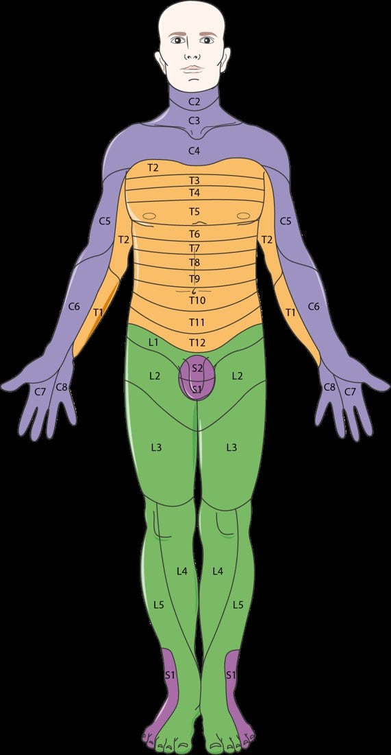
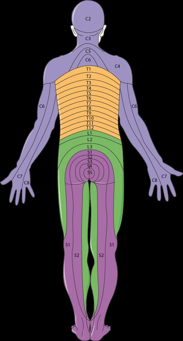
Landmark Dermatomes (ISNCSCI key points)
- C2 occiput - C3 posterior neck/supraclavicular fossa - C4 acromioclavicular joint
- C5 lateral antecubital fossa (lateral arm) - C6 thumb - C7 middle finger - C8 little finger - T1 medial antecubital fossa (medial forearm)
- T4 nipple - T6 xiphisternum - T10 umbilicus - L1 inguinal region/groin
- L2 anterior mid-thigh - L3 medial femoral condyle (medial knee) - L4 medial malleolus - L5 dorsum of the foot at the 3rd MTP joint / great toe - S1 lateral heel
- S2 popliteal fossa - S3 ischial tuberosity - S4-5 perianal
L2-S2Lower-Limb Myotomes (count up the leg)
Hook:Up the back of the leg: L2 flex hip, L3 straighten knee, L4 lift foot, L5 lift toe, S1 push off.
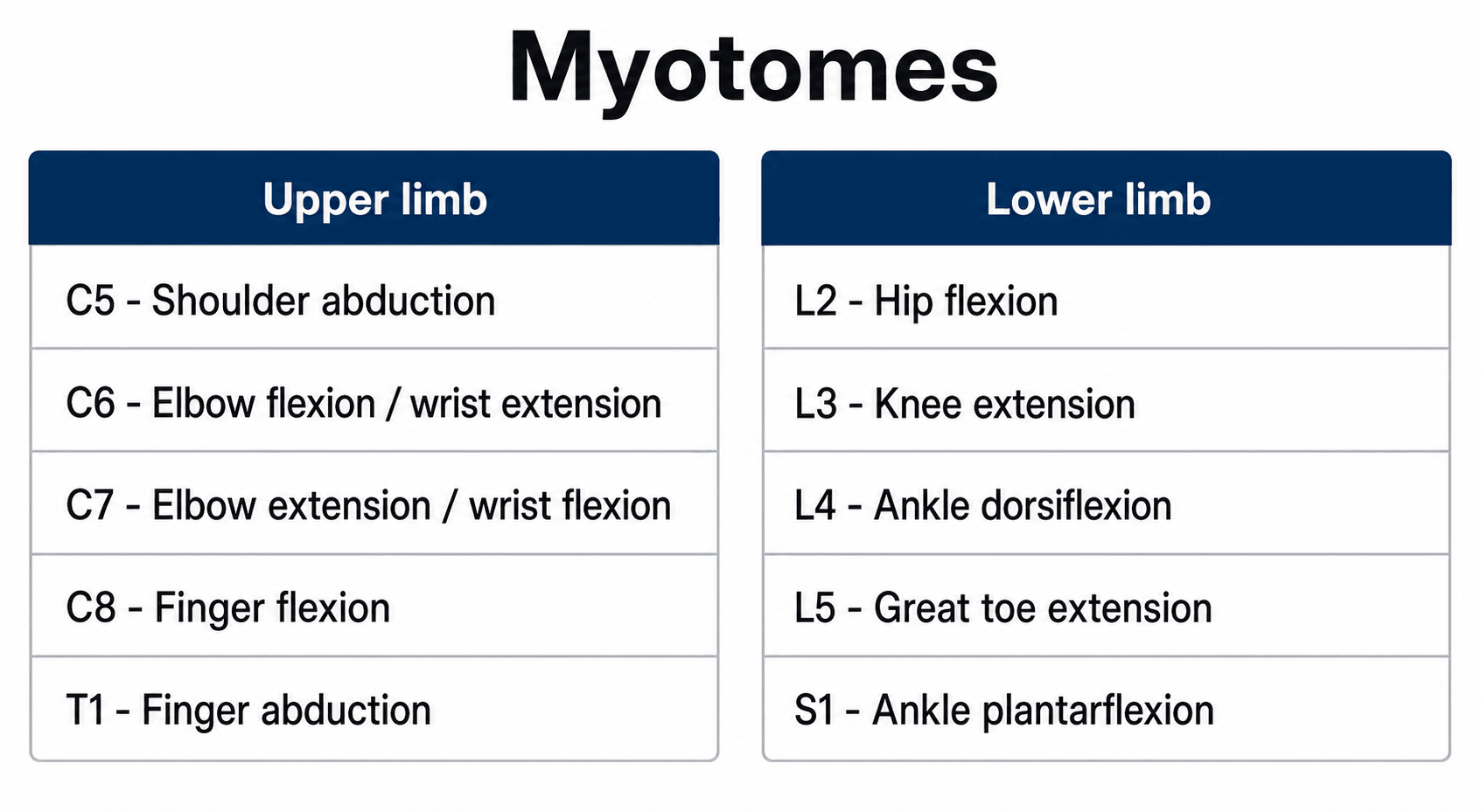
Candidates lose marks here because two different, both-correct lists are in circulation:
- The traditional clinical scheme (the chart above) starts the upper limb at C5 = shoulder abduction, then C6 elbow flexion, C7 elbow extension. It follows the order in which movements are lost as you descend the plexus, and it is what most bedside teaching uses.
- The ISNCSCI key-muscle list starts at C5 = elbow flexion (biceps) and C6 = wrist extension. It deliberately omits shoulder abduction because deltoid is hard to grade reproducibly across the 0-5 range in a supine, injured patient, and ISNCSCI needs one testable muscle per level that any examiner can score identically.
Say which scheme you are using. In a spinal-cord-injury or ASIA question, use the ISNCSCI list. In a general radiculopathy or peripheral-nerve question, the clinical scheme is expected. The lower limb is the same in both (L2 hip flexion → S1 plantarflexion), which is why only the upper limb causes trouble.
Which Root Does a Disc Hit? Exiting versus Traversing
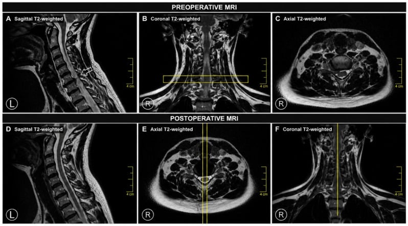
Knowing the dermatomes is useless at the MRI viewer unless you can convert a disc level into a root level. That conversion is one of the most reliably examined ideas in spinal anatomy, and it depends on two facts.
Fact 1 — the numbering shifts between the cervical and thoracolumbar spine. There are 8 cervical nerve roots but only 7 cervical vertebrae. C1 to C7 roots exit above the pedicle of the correspondingly numbered vertebra; C8 exits below C7 (there is no C8 vertebra); and from T1 downwards every root exits below the pedicle of its own vertebra.
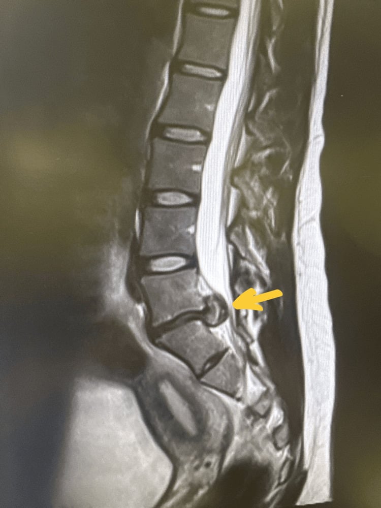
Fact 2 — a root's course determines which disc can reach it. At any level two roots are in play: the exiting root, already turning out into its foramen, and the traversing root, still descending to leave one level lower.
- Root compressed
- The TRAVERSING root - i.e. the root numbered for the level BELOW
- Worked example
- L4/5 paracentral disc → L5 root → weak EHL, dorsum-of-foot numbness, reflexes intact
- Root compressed
- The EXITING root - i.e. the root numbered for the level ABOVE
- Worked example
- L4/5 far-lateral disc → L4 root → weak dorsiflexion, medial malleolus numbness, reduced knee jerk
- Root compressed
- Multiple traversing roots - the cauda equina
- Worked example
- L4/5 large central disc → cauda equina syndrome, not a single radiculopathy
- Root compressed
- The EXITING root, because cervical roots exit above their pedicle and the disc sits at that foramen
- Worked example
- C5/6 disc → C6 root → weak wrist extension, thumb numbness, reduced brachioradialis reflex
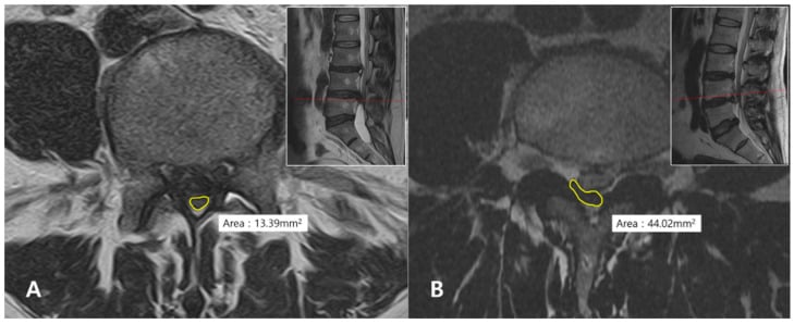
In the cervical spine the disc hits the root of the LOWER number of the two vertebrae — a C5/6 disc gives C6, a C6/7 disc gives C7. In the lumbar spine a paracentral disc also hits the lower-numbered root (L4/5 gives L5, L5/S1 gives S1) — but for the opposite anatomical reason, because it catches the traversing root rather than the exiting one. The exception that examiners use to separate candidates is the far-lateral disc, which skips past the traversing root and catches the exiting root instead: a far-lateral L4/5 disc produces an L4, not an L5, syndrome.
A related trap is the "far-out" syndrome (Wiltse): extraforaminal entrapment of the L5 root between the L5 transverse process and the sacral ala, typically with a collapsed disc or an isthmic spondylolisthesis at L5/S1. It produces an L5 radiculopathy from a lumbosacral junction lesion rather than from an L4/5 disc, and it is easily missed because the MRI must be reviewed lateral to the foramen.
Is It the Root or the Peripheral Nerve?
A root lesion and a peripheral nerve lesion can produce almost the same map. The discriminator is always a muscle or sensory area supplied by the same root through a different nerve.
- Peripheral nerve lesion
- Axillary nerve (the 'regimental badge' patch)
- Root lesion
- C5 root
- The discriminating test
- Axillary palsy weakens deltoid alone; C5 also weakens biceps and depresses the biceps reflex
- Peripheral nerve lesion
- Radial or posterior interosseous nerve
- Root lesion
- C6 root
- The discriminating test
- Radial palsy spares elbow flexion and the biceps; C6 weakens brachioradialis and depresses its reflex
- Peripheral nerve lesion
- Ulnar nerve at the elbow
- Root lesion
- C8 or T1 root
- The discriminating test
- Ulnar palsy spares the C8-innervated FDP to the index and thumb (median), and spares APB; a T1 root lesion may add a Horner sign
- Peripheral nerve lesion
- Common peroneal nerve at the fibular neck
- Root lesion
- L5 root
- The discriminating test
- Peroneal palsy SPARES inversion (tibialis posterior is L5 but travels in the TIBIAL nerve) and spares hip abduction; both are weak in an L5 root lesion
- Peripheral nerve lesion
- Lateral femoral cutaneous nerve (meralgia paraesthetica)
- Root lesion
- L2/L3 root
- The discriminating test
- Meralgia is purely sensory - any quadriceps weakness or knee-jerk change means a root
The dorsal root ganglion sits in the intervertebral foramen, distal to the point where most root avulsions occur. So in a preganglionic lesion (root avulsion) the sensory cell body stays connected to its peripheral axon, which therefore does not degenerate: the limb is anaesthetic but the sensory nerve action potentials (SNAPs) are PRESERVED. In a postganglionic lesion (rupture distal to the ganglion) the axon is separated from its cell body and the SNAPs are lost.
This is the electrodiagnostic basis for separating an avulsion, which is not repairable and needs a nerve transfer, from a rupture, which can be grafted. Supporting signs of a preganglionic injury include Horner's syndrome (T1 sympathetic outflow), winging from serratus anterior (long thoracic, C5-7 off the roots), rhomboid weakness (dorsal scapular, C5) and an elevated hemidiaphragm (phrenic, C3-5) — all branches taken directly off the roots and therefore only lost when the lesion is proximal.
Nerve Root Reflex Levels and Grading
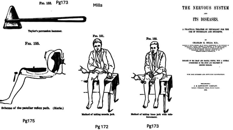
The deep tendon reflexes pin a few roots precisely and are an independent third data point alongside the myotome and dermatome:
- Root level
- C5–C6
- Note
- Predominantly C5
- Root level
- C6
- Note
- Inversion of this reflex localises a C5–6 cord level
- Root level
- C7
- Note
- Root level
- C8
- Note
- Rarely graded; a positive Hoffmann is an UMN sign, not a C8 root sign
- Root level
- L3–L4
- Note
- Predominantly L4
- Root level
- S1
- Note
- L5 has NO deep tendon reflex — the single most useful negative in the leg
The superficial (cutaneous) reflexes are separately examinable and are the only way to test some segments:
- Root level
- T8–T12
- How and why
- Stroke each quadrant; the umbilicus moves toward the stimulus. Lost below a cord lesion, and asymmetry can localise a thoracic level
- Root level
- L1–L2
- How and why
- Stroke the inner thigh; the testis elevates. Absent in cord lesions above L1 and in testicular torsion
- Root level
- S2–S4
- How and why
- Perianal scratch produces external sphincter contraction — part of the mandatory sacral examination
- Root level
- S2–S4
- How and why
- Squeeze the glans or clitoris (or tug a urinary catheter); the anal sphincter contracts. Its return marks the END OF SPINAL SHOCK
Spinal shock is the transient loss of all reflex activity below an acute cord injury, producing flaccid areflexia that mimics a complete lesion. A complete (AIS A) injury cannot be declared while the patient is in spinal shock, because the picture may improve; the conventional marker of its resolution is the return of the bulbocavernosus reflex, usually within 24–72 hours. Note the exception: the bulbocavernosus reflex is normally absent in a conus medullaris or cauda equina lesion, because the reflex arc itself is destroyed — so its absence there does not mean spinal shock.
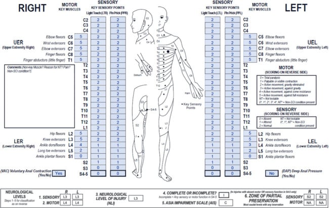
In the ISNCSCI/ASIA examination each key muscle is graded 0–5 on the MRC scale (0 no contraction, 1 flicker, 2 movement with gravity eliminated, 3 movement against gravity, 4 movement against resistance, 5 normal) and each key sensory point 0–2 (0 absent, 1 impaired/altered, 2 normal) for both light touch and pin-prick, tested against the face as the normal reference. Summing these yields the motor and sensory scores, the sensory and motor levels, and the single neurological level of injury (the most caudal segment with normal sensation and at least antigravity power, provided everything above it is intact).
The zone of partial preservation (ZPP) is the term for the dermatomes and myotomes below the neurological level that retain some function in an otherwise complete (AIS A) injury. It is recorded for all four (right/left, sensory/motor), it applies only to complete injuries, and a larger motor ZPP carries a better prognosis.
The same reflex that localises a root also flags a cord lesion — by whether it is reduced or exaggerated, the single most important interpretive step examiners test:
- Radiculopathy = a LOWER motor neuron pattern at the level: the affected reflex is diminished or absent, with hypotonia, segmental wasting and fasciculations and weakness in that myotome (e.g. an absent Achilles in S1, an absent biceps jerk in C5-6).
- Myelopathy / cord compression = an UPPER motor neuron pattern BELOW the level: reflexes are brisk/exaggerated, with clonus, spasticity, an up-going plantar (Babinski), a positive Hoffmann sign (flicking the middle-finger nail flexes the thumb/index), and Lhermitte's phenomenon — while a single root at the level of the cord lesion may still be LMN (a wasted, areflexic segment with brisk reflexes below it).
- The inverted supinator (radial) reflex is the classic crossover sign: tapping brachioradialis gives little brachioradialis contraction but brisk finger flexion, localising a C5-C6 cord/myelopathic level (the C5-6 LMN arc is interrupted while C8 below is disinhibited) — a peripheral musculocutaneous/C6 lesion does NOT do this.
So a "depressed reflex" is a root sign, a "brisk reflex with clonus/Babinski/Hoffmann" is a cord (UMN) sign, and the two can coexist at the compressed cervical level — distinguishing radiculopathy, myelopathy and myeloradiculopathy from the same examination.
Clinical Application
Localising a Radiculopathy
- Combine the key muscle (myotome) + dermatome + reflex to localise the level.
- C6: weak wrist extension/elbow flexion, thumb sensation, reduced brachioradialis reflex.
- C7: weak elbow extension (triceps), middle-finger sensation, reduced triceps reflex.
- L4: weak dorsiflexion, medial-malleolus sensation, reduced knee reflex.
- L5: weak great-toe extension, dorsum-of-foot sensation, reflexes preserved.
- S1: weak plantarflexion, lateral-foot sensation, reduced Achilles reflex.
ASIA Impairment Scale and Cauda Equina
The ASIA Impairment Scale (AIS)
The completeness and severity of a spinal cord injury are graded A–E, anchored on sacral sparing:
- Definition
- No motor or sensory function preserved in the sacral segments S4–S5 (no sacral sparing)
- Definition
- Sensory but not motor function preserved below the neurological level, including the sacral segments S4–S5
- Definition
- Motor incomplete status (voluntary anal contraction, OR sacral sensory sparing plus motor sparing more than three levels below the motor level), AND fewer than half the key muscles below the neurological level grade 3 or more
- Definition
- Motor incomplete status as for C, but at least half the key muscles below the neurological level grade 3 or more
- Definition
- Sensory and motor function normal in all segments (in a patient who had prior deficits)
The crucial conceptual point is that sacral sparing distinguishes complete (A) from incomplete (B–D) injuries — which is why the perianal examination (S4–5 light touch/pin-prick, deep anal pressure and voluntary anal contraction) is mandatory and never omitted. Spinal shock (transient areflexia) must resolve — confirmed by return of the bulbocavernosus reflex — before a complete injury is declared.
"Incomplete" (AIS B-D) is not one thing: the dermatome/myotome map plus the three long tracts (corticospinal = motor, dorsal columns = proprioception/vibration/light touch, spinothalamic = pain/temperature) define recognisable syndromes the exam expects you to name:
- Central cord syndrome — the commonest incomplete injury; a hyperextension injury in an older patient with cervical spondylosis (often without fracture). Upper limbs (especially the hands) are weaker than the lower limbs (medial-to-lateral somatotopy of the corticospinal tract), with variable "cape" sensory loss and usual sacral sparing. Generally managed without emergent surgery; most regain ambulation, hand recovery is least.
- Brown-Séquard (cord hemisection) — penetrating/lateral mass injury: ipsilateral loss of motor (corticospinal) and dorsal-column (proprioception/vibration) function, with contralateral loss of pain and temperature (spinothalamic) beginning a few levels below (fibres decussate within 1-2 segments). Best prognosis of the incomplete syndromes.
- Anterior cord syndrome — anterior spinal artery territory / flexion-compression: loss of motor and pain/temperature with PRESERVED proprioception, vibration and light touch (dorsal columns spared). Worst prognosis.
- Posterior cord syndrome — rare: isolated dorsal-column loss (proprioception/vibration → sensory ataxia and a positive Romberg) with motor and pain/temperature preserved.
Conus medullaris vs cauda equina (both involve the lumbosacral roots/segments and saddle anaesthesia, but localise differently): the conus (cord segments around L1-L2) gives an early, symmetrical, severe bladder/bowel and saddle deficit with mixed UMN + LMN signs (an up-going plantar can persist), relatively symmetric and less radicular pain; the cauda equina (roots below the conus) gives an asymmetric, LMN picture with prominent radicular pain, areflexia and a later but progressive bladder dysfunction. Both demand urgent MRI; cauda equina compression is the classic decompression emergency.
Cauda equina syndrome
The same lumbosacral roots that the dermatome/myotome map describes form the cauda equina below the conus (which ends at about L1–L2 in adults). Compression — classically a large central lumbar disc — produces cauda equina syndrome, a surgical emergency: bilateral sciatica, saddle (S2–4) anaesthesia, bladder dysfunction (retention with overflow), bowel/faecal incontinence, and reduced anal tone. It is the lower-limb counterpart of the sacral-sparing concept and demands urgent MRI and decompression (the evidence favours early surgery to optimise recovery of continence). Always check perianal sensation, anal tone and post-void residual in any patient with bilateral leg symptoms or new urinary dysfunction.
Guidelines, Registries & Global Practice
Global Practice Picture
Dermatome and myotome mapping is universal clinical knowledge, standardised internationally by the ISNCSCI (ASIA) for spinal cord injury and used everywhere to localise radiculopathy. The key sensory points and key muscles provide a common, reproducible language across clinicians and registries.
Side-by-Side Synthesis
- Key muscle (myotome)
- Elbow flexion (biceps)
- Key sensory point (dermatome)
- Lateral arm
- Reflex
- Biceps (C5-6)
- Key muscle (myotome)
- Wrist extension
- Key sensory point (dermatome)
- Thumb
- Reflex
- Brachioradialis
- Key muscle (myotome)
- Elbow extension (triceps)
- Key sensory point (dermatome)
- Middle finger
- Reflex
- Triceps
- Key muscle (myotome)
- Finger flexion
- Key sensory point (dermatome)
- Little finger
- Reflex
- -
- Key muscle (myotome)
- Ankle dorsiflexion
- Key sensory point (dermatome)
- Medial malleolus
- Reflex
- Knee (L3-4)
- Key muscle (myotome)
- Great toe extension (EHL)
- Key sensory point (dermatome)
- Dorsum great toe
- Reflex
- -
- Key muscle (myotome)
- Ankle plantarflexion
- Key sensory point (dermatome)
- Lateral foot/heel
- Reflex
- Achilles
Viva Scenarios
Practise clinical reasoning and management decisions out loud
“A patient has weak great-toe and ankle dorsiflexion, numbness over the dorsum of the foot, and normal ankle and knee reflexes. Which root is affected and how would you confirm it?”
“A patient with acute severe low back pain now has bilateral leg pain, numbness around the perineum and difficulty passing urine. Using your knowledge of the sacral roots, how do you assess and manage this?”
Definitions
- Dermatome = skin of one spinal nerve
- Myotome = muscles of one root
- Dermatomes overlap (band, not anaesthesia)
- ISNCSCI = standardised key points/muscles
Landmarks
- C6 thumb, C7 middle, C8 little finger
- T4 nipple, T10 umbilicus
- L4 medial malleolus, L5 great toe, S1 lateral foot
- Reflexes: biceps C5-6, triceps C7, knee L3-4, Achilles S1
Clinical
- L5: EHL/dorsiflexion, dorsum foot, reflexes spared
- S1: plantarflexion, lateral foot, absent Achilles
- Localise with myotome + dermatome + reflex
- ASIA A-E: sacral sparing = incomplete (B-D); A = complete
- Cauda equina: bilateral sciatica, saddle anaesthesia, retention, low anal tone → urgent MRI + decompression
Evidence Base
The maps are not as settled as they look, and that is itself examinable. Lee, McPhee and Stringer's systematic review found that the dermatome maps in standard anatomy texts differ significantly from one another and that "current dermatome maps are inaccurate and based on flawed studies" — the classical Foerster and Keegan-and-Garrett maps were derived by very different methods (residual sensibility after sectioning multiple roots, versus the pattern of hypoaesthesia in single-root compression) and disagree most in the limbs. They built an evidence-based composite and argued that overlap and inter-individual variability deserve far more emphasis than the tidy textbook diagram implies. Use the ISNCSCI key points precisely because they are a convention chosen for reproducibility, not a claim about where one root's skin truly ends. The rest of the modern evidence concerns the ISNCSCI examination built on those points: Kirshblum confirms it is the predominant tool for classifying and prognosticating spinal cord injury (most AIS conversion and motor recovery within the first 6-9 months, fastest in the first 3 months), and Walden shows it is reproducible enough to be computerised and used worldwide. The practical message: the value is in performing the exam consistently.
Characterizing Natural Recovery after Traumatic Spinal Cord Injury
- The ISNCSCI (with the ASIA Impairment Scale) is the predominant tool to classify and predict outcomes after traumatic spinal cord injury
- Completeness is defined by the sacral sparing definition
- Most AIS conversion and motor recovery occurs within the first 6-9 months, fastest in the first 3 months
- Initial AIS grade and zone of partial preservation influence prognosis
Computer International Standards for Neurological Classification of Spinal Cord Injury (ISNCSCI) algorithms: a review
- Reviews validated computerised ISNCSCI algorithms (EMSCI and Praxis Spinal Cord Institute)
- Algorithms support education, clinical documentation, and consistent classification worldwide
- Over half of surveyed users apply the algorithm regularly in their workflow
- They support, not replace, trained clinicians and allow reclassification with updated ISNCSCI versions
An Evidence-Based Approach to Human Dermatomes
- Systematic review of the primary evidence for the distribution of human dermatomes, appraising the method of ascertainment, the extent of each dermatome and the number of subjects in each source study
- Found significant variation between the dermatome maps printed in standard anatomy texts
- Concluded that current dermatome maps are inaccurate and based on flawed studies, and constructed a novel evidence-based map of the most consistent tactile dermatomal area for each dorsal root
- Argued that the overlap and inter-individual variability of dermatomes deserve much greater emphasis than textbook diagrams convey