Spinal Nerve Root Mapping
- A dermatome is the skin area supplied by a single spinal nerve; a myotome is the muscle group supplied by a single nerve root.
- Adjacent dermatomes OVERLAP, so a single root lesion rarely gives complete sensory loss in its dermatome.
- Key upper-limb dermatomes: C6 thumb, C7 middle finger, C8 little finger.
- Key lower-limb dermatomes: L4 medial malleolus, L5 dorsum of the great toe, S1 lateral border/heel of the foot.
- Key myotomes: L4 ankle dorsiflexion, L5 great toe extension (EHL), S1 ankle plantarflexion; C5 elbow flexion, C7 elbow extension, T1 finger abduction.
- Reflex levels: biceps C5-6, brachioradialis C6, triceps C7, patellar L3-4, Achilles S1.
- “The ISNCSCI defines standard key sensory points (each dermatome) and key muscles (each myotome) for reproducible spinal-cord-injury assessment.
- “L5 vs S1 radiculopathy: L5 weakens great-toe extension and dorsiflexion (dorsum great toe sensation); S1 weakens plantarflexion (lateral foot sensation, absent Achilles reflex).
- “Sacral sparing (perianal sensation/voluntary anal contraction, S4-5) defines an incomplete spinal cord injury.
Weak great-toe extension (EHL) and dorsiflexion/foot inversion-eversion; sensory change over the dorsum of the foot / great toe; weak hip abduction; reflexes usually preserved. (Often a far-out or paracentral L4/5 disc.)
Weak ankle plantarflexion (calf), sensory change over the lateral border and sole/heel of the foot, and a reduced/absent Achilles (ankle) reflex. (Often an L5/S1 disc.)
Definitions & Principles
Dermatome vs Myotome
- Dermatome: the area of skin supplied by a single spinal (dorsal root) nerve.
- Myotome: the group of muscles supplied by a single spinal nerve root (most muscles are supplied by more than one root).
- Sclerotome / viscerotome: bone and viscera supplied by a segment (less examined).
The Key Maps
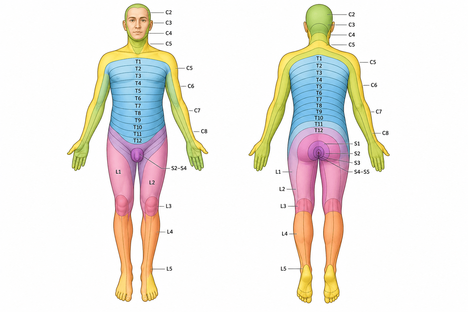
Landmark Dermatomes (ISNCSCI key points)
- C2 occiput - C3 posterior neck/supraclavicular fossa - C4 acromioclavicular joint
- C5 lateral antecubital fossa (lateral arm) - C6 thumb - C7 middle finger - C8 little finger - T1 medial antecubital fossa (medial forearm)
- T4 nipple - T6 xiphisternum - T10 umbilicus - L1 inguinal region/groin
- L2 anterior mid-thigh - L3 medial femoral condyle (medial knee) - L4 medial malleolus - L5 dorsum of the foot at the 3rd MTP joint / great toe - S1 lateral heel
- S2 popliteal fossa - S3 ischial tuberosity - S4-5 perianal
L2-S2Lower-Limb Myotomes (count up the leg)
Hook:Up the back of the leg: L2 flex hip, L3 straighten knee, L4 lift foot, L5 lift toe, S1 push off.
Clinical Application
Localising a Radiculopathy
- Combine the key muscle (myotome) + dermatome + reflex to localise the level.
- C6: weak wrist extension/elbow flexion, thumb sensation, reduced brachioradialis reflex.
- C7: weak elbow extension (triceps), middle-finger sensation, reduced triceps reflex.
- L4: weak dorsiflexion, medial-malleolus sensation, reduced knee reflex.
- L5: weak great-toe extension, dorsum-of-foot sensation, reflexes preserved.
- S1: weak plantarflexion, lateral-foot sensation, reduced Achilles reflex.
Evidence Base
ISNCSCI / ASIA and Recovery after SCI
- The ISNCSCI (with the ASIA Impairment Scale) is the predominant tool to classify and predict outcomes after traumatic spinal cord injury
- Completeness is defined by the sacral sparing definition
- Most AIS conversion and motor recovery occurs within the first 6-9 months, fastest in the first 3 months
- Initial AIS grade and zone of partial preservation influence prognosis
Computerised ISNCSCI Classification Algorithms
- Reviews validated computerised ISNCSCI algorithms (EMSCI and Praxis Spinal Cord Institute)
- Algorithms support education, clinical documentation, and consistent classification worldwide
- Over half of surveyed users apply the algorithm regularly in their workflow
- They support, not replace, trained clinicians and allow reclassification with updated ISNCSCI versions
Viva Scenarios
Practise clinical reasoning and management decisions out loud
“A patient has weak great-toe and ankle dorsiflexion, numbness over the dorsum of the foot, and normal ankle and knee reflexes. Which root is affected and how would you confirm it?”
Guidelines, Registries & Global Practice
Global Practice Picture
Dermatome and myotome mapping is universal clinical knowledge, standardised internationally by the ISNCSCI (ASIA) for spinal cord injury and used everywhere to localise radiculopathy. The key sensory points and key muscles provide a common, reproducible language across clinicians and registries.
Side-by-Side Synthesis
- Key muscle (myotome)
- Elbow flexion (biceps)
- Key sensory point (dermatome)
- Lateral arm
- Reflex
- Biceps (C5-6)
- Key muscle (myotome)
- Wrist extension
- Key sensory point (dermatome)
- Thumb
- Reflex
- Brachioradialis
- Key muscle (myotome)
- Elbow extension (triceps)
- Key sensory point (dermatome)
- Middle finger
- Reflex
- Triceps
- Key muscle (myotome)
- Finger flexion
- Key sensory point (dermatome)
- Little finger
- Reflex
- -
- Key muscle (myotome)
- Ankle dorsiflexion
- Key sensory point (dermatome)
- Medial malleolus
- Reflex
- Knee (L3-4)
- Key muscle (myotome)
- Great toe extension (EHL)
- Key sensory point (dermatome)
- Dorsum great toe
- Reflex
- -
- Key muscle (myotome)
- Ankle plantarflexion
- Key sensory point (dermatome)
- Lateral foot/heel
- Reflex
- Achilles
Definitions
- Dermatome = skin of one spinal nerve
- Myotome = muscles of one root
- Dermatomes overlap (band, not anaesthesia)
- ISNCSCI = standardised key points/muscles
Landmarks
- C6 thumb, C7 middle, C8 little finger
- T4 nipple, T10 umbilicus
- L4 medial malleolus, L5 great toe, S1 lateral foot
- Reflexes: biceps C5-6, triceps C7, knee L3-4, Achilles S1
Clinical
- L5: EHL/dorsiflexion, dorsum foot, reflexes spared
- S1: plantarflexion, lateral foot, absent Achilles
- Localise with myotome + dermatome + reflex
- Sacral sparing = incomplete SCI