SRS-Schwab Classification | PI-LL Mismatch | Osteotomies | Global Balance
- PI-LL mismatch is the strongest predictor of disability
- LL should match PI within 10 degrees (LL = PI ± 9)
- SVA over 50mm strongly correlates with pain and disability
- Pelvic retroversion is a compensatory mechanism (PT increases)
- Age-adjusted goals may be appropriate for elderly patients
- “PI is fixed - cannot be changed surgically
- “PT increases as compensation for sagittal imbalance
- “Global Alignment and Proportion (GAP) score predicts complications
- “Three-column osteotomies (PSO, VCR) carry highest complication risk
Overview and Epidemiology
Adult spinal deformity (ASD) encompasses a spectrum of conditions characterised by abnormal spinal curvature in adults, with increasing recognition of the importance of sagittal plane alignment. Unlike adolescent idiopathic scoliosis, ASD is predominantly a sagittal plane problem.
The types. Deformity in the adult arises in one of the following ways:
- De novo degenerative scoliosis - develops in adulthood from asymmetric disc degeneration and facet arthropathy
- Progressive idiopathic scoliosis - adolescent scoliosis that progresses in adulthood
- Iatrogenic deformity - after prior spinal surgery (flatback syndrome, adjacent segment disease)
- Secondary deformity - metabolic bone disease, trauma or infection
Why the sagittal plane matters. Sagittal imbalance correlates strongly with pain and disability, and the health-related quality of life (HRQOL) measures (ODI, SF-36) correlate with the sagittal parameters. An SVA over 50mm is associated with significant disability, and PI-LL mismatch is the strongest predictor of poor HRQOL.
In a volunteer cohort of just 75 healthy people over 60 (mean age 70.5 years, no prior spine history), Schwab and colleagues found that 68% met the radiographic definition of scoliosis (Cobb greater than 10 degrees), against prevalence figures of up to 32% in earlier general-population studies (Schwab et al. Spine 2005, PMID 15864163). Read the rest of that paper before using the number as a burden-of-disease argument: these volunteers were recruited because they were healthy, the authors attributed the high figure to that targeted selection, and they found no significant correlation between the radiographic deformity and visual-analogue pain scores. Their own conclusion is that a large group with adult scoliosis has "no marked physical or social impairment". That is the lesson the rest of this page teaches — coronal Cobb angle is a poor guide to disability; sagittal malalignment is what tracks with symptoms.
Pathophysiology
The chain. The spine functions as a chain of segments that must keep the centre of gravity over the pelvis and lower extremities. Loss of lumbar lordosis is the primary driver of sagittal imbalance.
Normal sagittal alignment. The values against which a deformity is judged:
- Cervical lordosis 20-40 degrees
- Thoracic kyphosis 20-50 degrees (T4-T12)
- Lumbar lordosis 40-60 degrees (L1-S1)
- Sacral slope 30-50 degrees
- Pelvic tilt low, under 20 degrees, which is the SRS-Schwab grade boundary; the ideal rises with age (Lafage, PMID 26689395; figures in Controversies)
Pelvic incidence (PI). The angle between the perpendicular to the sacral endplate and the line to the femoral head centre, normal range 40-65 degrees. It is a fixed anatomical parameter that does not change after skeletal maturity, and it determines the amount of lumbar lordosis required for balance. The fundamental equation is PI = PT + SS.
Pelvic tilt (PT). The angle between the vertical and the line from the S1 midpoint to the femoral head centre. It is positional, changing with posture, and it increases with pelvic retroversion as the pelvis compensates; a PT over 30 degrees indicates exhausted compensation.
Sacral slope (SS). The angle between the sacral endplate and the horizontal. It decreases with pelvic retroversion, and it follows from the equation: SS = PI - PT.
The compensation cascade. When lumbar lordosis is insufficient for a given PI, a predictable cascade of compensation occurs:
- Mechanism
- Thoracic hypokyphosis
- Clinical Effect
- Reduces thoracic curve
- Mechanism
- Pelvic retroversion
- Clinical Effect
- Increases PT, decreases SS
- Mechanism
- Hip extension
- Clinical Effect
- Extends hip joint
- Mechanism
- Knee flexion
- Clinical Effect
- Bent-knee gait
- Mechanism
- Decompensation
- Clinical Effect
- Positive SVA, disability
When compensation is exhausted, the C7 plumb line falls anterior to the sacrum (positive SVA) and the patient becomes symptomatic.
Classification Systems
The SRS-Schwab classification is the most validated and widely used system for ASD. It combines a coronal curve type with three sagittal modifiers, and the modifier grades were derived by cutting a multicentre database against HRQOL scores, so they are validated to correlate with HRQOL outcomes.
- Description
- Thoracic only
- Apex Location
- Apex at T9 or higher
- Description
- Thoracolumbar or lumbar only
- Apex Location
- Apex T10-L2 or L2-L4
- Description
- Double curve (thoracic and lumbar)
- Apex Location
- Both regions affected
- Description
- No major coronal deformity
- Apex Location
- Sagittal plane only
The sagittal modifiers. PI-LL mismatch, pelvic tilt and SVA are each graded 0 (non-pathological, within the normal range), + (moderate: pathological but not severe) or ++ (marked: severely abnormal). Note that the SVA grade boundaries, 4cm and 9.5cm, are not the 50mm figure that marks significant disability; the two sets of thresholds sit side by side.
- Non-pathological (0)
- Under 10°
- Moderate (+)
- 10-20°
- Marked (++)
- Over 20°
- Non-pathological (0)
- Under 20°
- Moderate (+)
- 20-30°
- Marked (++)
- Over 30°
- Non-pathological (0)
- Under 4cm
- Moderate (+)
- 4-9.5cm
- Marked (++)
- Over 9.5cm
SRS-Schwab and GAP tell you the magnitude of correction; the Roussouly classification describes the normal SHAPE of sagittal alignment and is increasingly examinable because restoring the patient's native type (not just the PI-LL number) reduces mechanical failure and PJK:
- Type 1: low sacral slope (under 35 degrees), short lower-lumbar lordosis with a low apex and a thoracolumbar kyphosis - lordosis concentrated in the lowest segments.
- Type 2: low sacral slope (under 35 degrees) with a flat, hypolordotic lumbar spine and a flat back - low-PI "flat" spines.
- Type 3: average sacral slope (35-45 degrees) with a well-balanced, harmonious lordosis (apex around L4) - the "average" spine.
- Type 4: high sacral slope (over 45 degrees) with a long, deeply curved hyperlordosis and many vertebrae in the lordosis - high-PI spines.
- The principle: lordosis is governed by pelvic incidence (a low PI gives Type 1 or 2; a high PI gives Type 4), and the apex and distribution of lordosis matter, not just total degrees. Restoring the patient to their appropriate Roussouly type - including a correctly distributed, lower-apex lordosis - lowers the risk of junctional failure compared with creating a "flat" PI-LL-correct but malshaped spine.
Exam point: use Roussouly to set the shape (apex and distribution) of the reconstructed lordosis to the patient's pelvic morphology, complementing the PI-LL magnitude target.
Clinical Assessment
History. The symptoms that bring the patient in:
- Back pain (axial, often positional)
- Radiculopathy or neurogenic claudication
- Difficulty standing upright
- Need to support the trunk with the hands on the thighs
- Decreased walking tolerance
- Progressive postural change
The elements of the history that shape the plan are the duration and progression of symptoms, prior spinal surgery (fusion levels, approach), walking capacity in blocks or time, pain location and character, neurological symptoms (weakness, numbness, bowel or bladder), and medical comorbidities and bone health.
Observation. Standing posture in the sagittal and coronal planes, forward trunk lean, a hip and knee flexion posture, and shoulder balance.
The spine. Flexibility on the forward bend test, sagittal balance by plumb line, coronal balance, and the chin-brow vertical angle in a fixed deformity.
Neurology. Motor strength through the L2-S1 myotomes, sensation, reflexes, straight leg raise and gait.
Hips, knees and function. Hip range of motion for a flexion contracture, examination of the knees, and overall mobility and function.
Assess the patient's compensatory mechanisms: Can they stand with hips and knees extended? If they require hip and knee flexion to stand upright, their pelvic compensation is exhausted and they likely have significant sagittal imbalance.
Patient-reported outcomes. The measures used to assess surgical outcome:
- Oswestry Disability Index (ODI)
- SF-36 (physical and mental component scores)
- Scoliosis Research Society-22 (SRS-22)
- Visual Analogue Scale (VAS) for pain
- EQ-5D
Investigations
Standing full-length radiographs. The gold standard for alignment assessment: standing AP and lateral on a 36-inch cassette, including C2 to the femoral heads, with the arms positioned (hands on the clavicles or held in front).
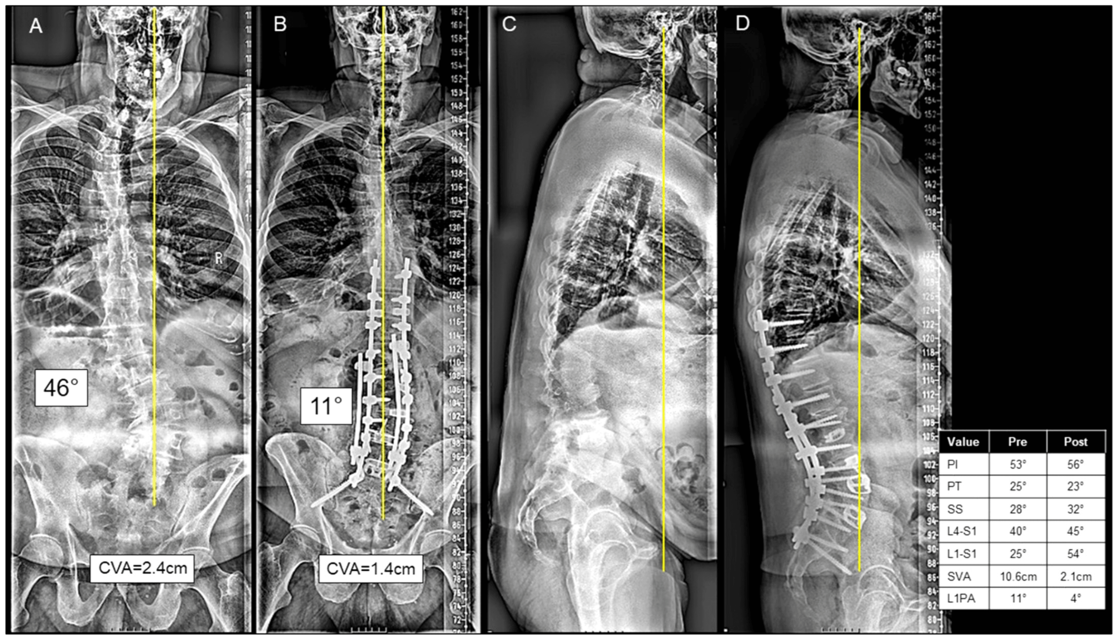
Supine and bending radiographs. These assess the flexibility of the curves and help plan the fusion levels and the need for an osteotomy. A supine lateral over a bolster assesses sagittal flexibility.
MRI of the whole spine. For neural compression, disc degeneration, the cord and the cauda equina, and to rule out intraspinal pathology.
CT, when indicated. For bone quality, a previous fusion mass, existing hardware, and osteotomy planning.
The measurements. Each parameter against its normal range:
- Measurement
- C7 plumb to S1 posterior corner
- Normal Range
- Under 50mm
- Significance
- Positive = C7 anterior to S1
- Measurement
- Sacral endplate perpendicular to femoral head
- Normal Range
- 40-65°
- Significance
- Fixed, determines LL target
- Measurement
- Vertical to femoral head-S1 line
- Normal Range
- Under 20°
- Significance
- Rises with pelvic retroversion
- Measurement
- Sacral endplate to horizontal
- Normal Range
- 30-50°
- Significance
- SS = PI - PT
- Measurement
- L1 superior to S1 superior endplate
- Normal Range
- 40-60°
- Significance
- Should match PI
- Measurement
- T4-T12 (or T5-T12)
- Normal Range
- 20-50°
- Significance
- Should balance LL
- Measurement
- Difference between PI and LL
- Normal Range
- Under 10°
- Significance
- Key outcome predictor
- Measurement
- T1 to femoral head angle
- Normal Range
- Under 14°
- Significance
- Global alignment measure
Bone density. DEXA of the hip and spine, and consider CT-based bone density (Hounsfield units). Bone quality shapes surgical planning and the fixation strategy, and osteoporosis significantly increases complication risk.
Additional studies. Ordered when the question demands them:
- CT myelogram if MRI is contraindicated
- Flexion-extension radiographs for instability
- Standing hip-to-ankle films for limb length and hip assessment
- Pulmonary function tests for severe deformity
ASD is dominated by sagittal teaching, but coronal malalignment is a distinct, examinable problem and a feared iatrogenic complication:
- How to measure it: the coronal balance is the horizontal offset of the C7 plumb line from the central sacral vertical line (CSVL); greater than 3 cm (often quoted as 4 cm) of coronal trunk shift is clinically significant coronal malalignment.
- Classification (Obeid / Bao): coronal malalignment is grouped by the direction of the C7 shift relative to the major curve concavity - broadly, the trunk shifted towards the concavity (often correctable) versus away from the curve (more rigid, harder to correct) - which guides whether and how to address it.
- The iatrogenic trap: a powerful asymmetric three-column osteotomy (e.g. PSO) corrected for sagittal balance can throw the patient into NEW coronal decompensation ("coronal malalignment after sagittal correction"). This is a recognised cause of revision and dissatisfaction despite a "perfect" PI-LL.
- Practical message: plan and check both planes - assess the CSVL/C7 coronal offset and the fractional lumbosacral curve before and after correction, and be especially vigilant for coronal decompensation after an asymmetric osteotomy.
Exam point: define coronal balance by the C7-CSVL offset (significant over ~3-4 cm), know that asymmetric PSO can cause iatrogenic coronal malalignment, and always reconstruct to a balanced spine in BOTH the sagittal and coronal planes.
Management
Who. Non-operative care suits:
- Mild deformity without significant symptoms
- Patient preference or surgical contraindications
- Adequate compensation with acceptable function
- High surgical risk patients
Physiotherapy. Core strengthening, flexibility exercises, postural training and aerobic conditioning.
Pain management. Analgesics (paracetamol, NSAIDs) and neuropathic agents (gabapentin, pregabalin). Epidural steroid injections give temporary relief; facet injections are both diagnostic and therapeutic.
Bracing. A limited role in adults. It may provide temporary symptom relief, does not prevent progression, and is considered for the high surgical risk patient.
Lifestyle modification. Weight optimisation, smoking cessation, activity modification and assistive devices.
Untreated sagittal imbalance tends to progress over time due to continued disc degeneration and muscle fatigue. Curves with PI-LL mismatch over 20 degrees or SVA over 50mm are more likely to progress.
Complications
ASD surgery has significant complication rates. Understanding and communicating these risks is essential, but quote figures that come from a named population, because the ranges in circulation ("25-80%") span populations that are not comparable.
The gradient. Across 578 thoracolumbar fixed sagittal-deformity cases in the SRS Morbidity and Mortality database the overall complication rate was 29.4%, and it rose stepwise with the osteotomy performed (the figures are in the table below). Performing any osteotomy roughly doubled the adjusted odds of a complication (OR 2.07). A VCR buys 45-70 degrees at roughly three and a half times the complication rate of operating without an osteotomy, so the osteotomy grade should be dictated by the correction the deformity actually requires, never by what is technically available. This is a large multicentre registry (Level III) of a fixed-deformity population, so these are the rates for the harder end of the spectrum.
A frequently repeated table gives roughly equal major-complication rates for SPO, PSO and VCR (around 38-40% each). That reading comes from Auerbach's 105 three-column osteotomies (PMID 22366971), which contained no SPOs at all and only 18 VCRs; the PSO and VCR figures it reported are in the table below, and P = 0.28 means no detectable difference in a sample far too small to find one. Read alongside the stepwise SRS gradient, the flat table is an artefact of a small underpowered series, not a finding. The gradient is the answer.
- SPO
- 28.1%
- PSO
- 39.1%
- VCR
- 61.1%
- Source
- SRS M&M, 578 FSPD cases (21192289); no osteotomy 17.0%
- SPO
- not studied
- PSO
- 38%
- VCR
- 22% (n=18, P=0.28)
- Source
- Auerbach 105 cases (22366971) - underpowered for VCR
- SPO
- no procedure-specific series
- PSO
- 11.1% (2.8% permanent)
- VCR
- 8% pooled (95% CI 2-16); cord injury 2%
- Source
- Buchowski 108 PSOs (17873818); Yang PVCR review, 390 patients (25599850)
- SPO
- least
- PSO
- 1.9L (Auerbach); 1.5-3L typical
- VCR
- 2.6L pooled (25599850)
- Source
- 22366971 / 25599850
These figures are not a head-to-head comparison and no randomised comparison exists. Frames of reference differ (a whole-cohort registry, a single-institution three-column series, a pooled PVCR review), and the patients differ from the constructs: a VCR is chosen for the more rigid, more severe, more often revision deformity, so part of every gradient is the deformity and not the operation.
Early complications. The incidence and the response to each:
- Incidence
- 3.8% new deficit across 578 FSPD cases (21192289); 11.1% after PSO in a dedicated series (17873818)
- Management
- Monitoring, urgent MRI, revision if progressive
- Incidence
- 5.9% (21192289)
- Management
- Primary repair, fibrin sealant
- Incidence
- 3.8% (21192289); higher in revision and long constructs
- Management
- Antibiotics, debridement
- Incidence
- 21192289
- Management
- Evacuation if symptomatic or neurology
- Incidence
- 15-30%
- Management
- Appropriate specialty management
- Incidence
- 1.0% (21192289)
- Management
- Prophylaxis, anticoagulation
Proximal junctional kyphosis (PJK). The most common mechanical complication. It is measured as the sagittal Cobb angle from the inferior endplate of the UIV to the superior endplate of UIV+2, two levels above, not one, and the Glattes criterion requires a proximal junctional angle of 10° or more that is also at least 10° greater than the preoperative value (PMID 16025035). No universal definition exists: some groups measure to UIV+1 and some compare against the early postoperative rather than the preoperative film, which is a large part of why reported incidence ranges from 10% to 48% (Glattes' own incidence was 26%).
Risk factors for PJK. Later series report older age, over-correction, osteoporosis, an upper-thoracic UIV and posterior-only surgery. Glattes himself identified none, and found SRS-24 scores were not worse in PJK patients, so radiographic PJK is not automatically a clinical problem. It may require extension of the fusion.
Proximal junctional failure (PJF) is the structural form: fracture of the UIV or UIV+1, instrumentation pull-out, sagittal subluxation, or myelopathy, not simply "PJK that gets revised".
Rod fracture. Occurs in 5-20% of cases, with the higher risk at the osteotomy site. It may be asymptomatic if the fusion is solid; revise if painful or progressing.
Pseudarthrosis. Failure of fusion. The risk factors are smoking, diabetes, multilevel surgery and a previous failed fusion; treatment is revision with bone grafting and possibly an osteotomy.
Adjacent segment disease. Degeneration above or below the fusion, more common with long rigid constructs. It may require extension of the fusion.
Risk factors for complications. The patient and operative factors that raise the rate:
- Advanced age (over 70)
- Higher ASA grade
- Obesity
- Osteoporosis
- Smoking
- Three-column osteotomies
- Revision surgery
- Long operative time
All patients must be counselled about the significant complication risk. The harms worth quoting at consent are the SRS Morbidity and Mortality figures above (the overall rate, the osteotomy gradient, and the new neurological deficit and durotomy rates in the early-complications table) together with the mortality in that database, 0.5% (PMID 21192289). Realistic expectations and shared decision-making are essential.
Postoperative Care
The first fortnight. The sequence from theatre to discharge:
Immediate postoperative sequence
ICU or high-dependency monitoring for major cases. Hourly neurological observations initially - a new deficit is an emergency and warrants urgent MRI to exclude epidural haematoma. Drain management, VTE prophylaxis, multimodal analgesia, and early mobilisation as soon as the patient is stable.
Progressive mobilisation with formal physiotherapy assessment, wound surveillance, DVT screening if clinically indicated, and medical optimisation (haemoglobin, nutrition, bone health, glycaemic control) before discharge.
Rehabilitation. Activity advances in phases:
- Timeframe
- Weeks 0-6
- Goals
- Protected mobilisation, wound healing
- Timeframe
- Weeks 6-12
- Goals
- Increase activity, core activation
- Timeframe
- Months 3-6
- Goals
- Strengthening, return to light activities
- Timeframe
- Beyond 6 months
- Goals
- Full activity as tolerated
Bracing. Practice varies with surgeon preference. Some protocols use a TLSO for 6-12 weeks; it may provide comfort and remind the patient of precautions.
Follow-up. Clinical and radiographic review at fixed points:
- Assessment
- Wound check, early mobilisation
- Assessment
- Clinical review, AP and lateral radiographs
- Assessment
- Clinical and radiographic assessment; full-length standing films at 3-6 months
- Assessment
- Full-length films, HRQOL measures
- Assessment
- Fusion assessment (CT if concern), outcome measures
- Assessment
- Long-term surveillance, imaging as clinically indicated
Outcomes and Prognosis
Radiographic outcomes. Correction of the SVA and the PI-LL mismatch to the alignment targets, restoration of coronal balance, and the fusion rate.
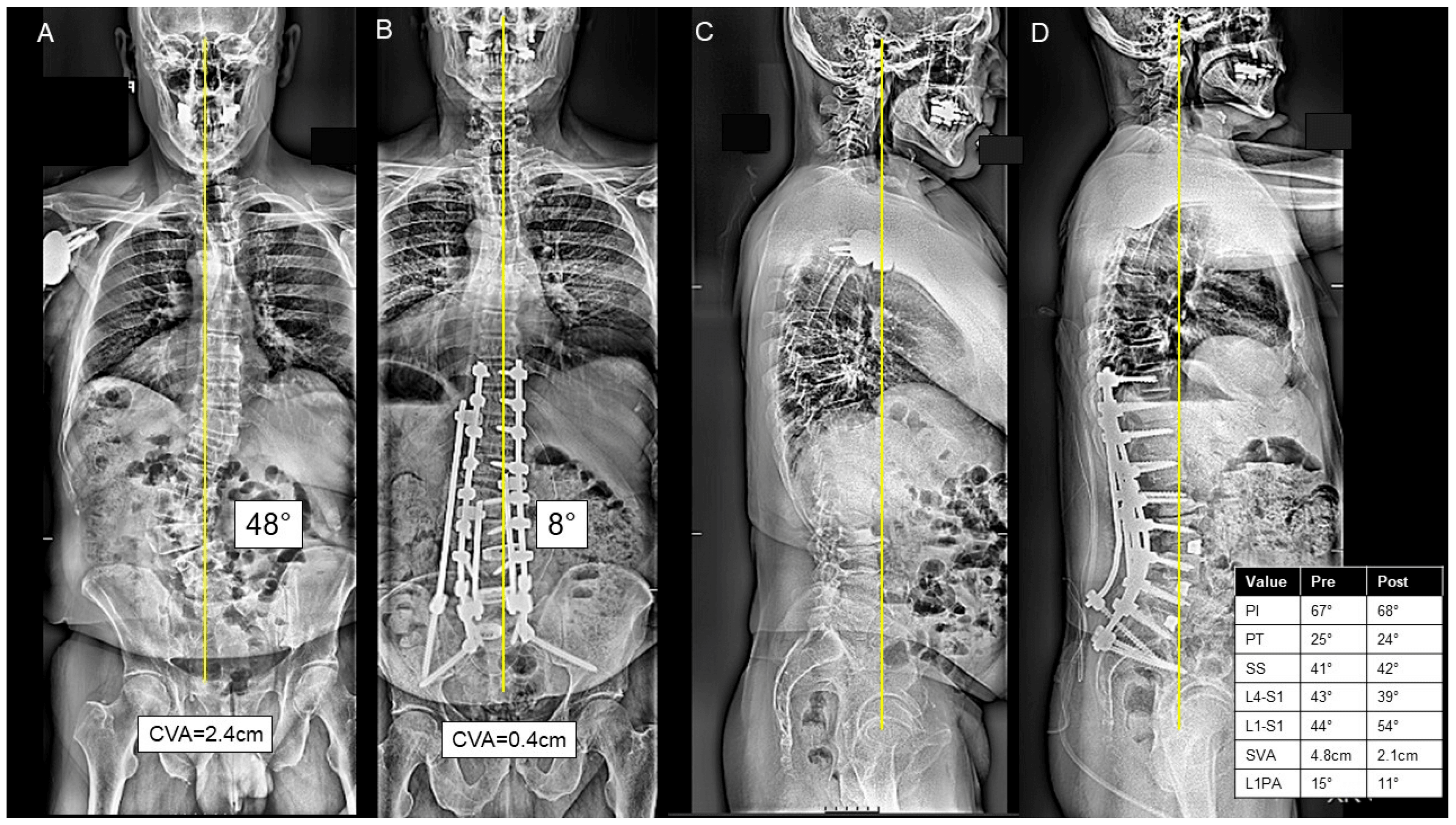
Clinical outcomes. The measures and their MCIDs:
- ODI improvement (MCID: 12-15 points)
- VAS pain reduction (MCID: 2 points)
- SRS-22 improvement
- SF-36 improvement
What to expect. Approximate rates after successful surgery:
- SVA correction achieved: 70-85%
- Fusion rate: 85-95%
- Significant pain improvement: 60-75%
- Patient satisfaction: 70-80%
What shapes the result. The most consistent predictor of a good outcome is achieving the appropriate PI-LL match (under 10 degrees mismatch): under-correction is associated with persistent symptoms, and over-correction increases the risk of PJK. Beyond alignment, the result depends on the baseline severity of the deformity, whether a major complication occurs, the patient's age, comorbidities and bone quality, whether they keep smoking, and whether the surgery is a revision rather than a primary procedure.
Guidelines, Registries & Global Practice
Global Epidemiology
Adult spinal deformity is common and strongly age-related. In a healthy elderly volunteer cohort, 68% met the radiographic definition of scoliosis (Cobb greater than 10 degrees), against earlier general-population estimates of up to 32% (Schwab et al. Spine 2005, PMID 15864163). Disability tracks with sagittal malalignment rather than coronal Cobb angle: positive sagittal balance is the radiographic parameter most strongly correlated with poor health status, and severity worsens linearly with C7 plumb-line deviation (Glassman et al. Spine 2005, PMID 16166889). As populations age worldwide, demand for both non-operative and operative ASD care is rising in every region.
Classification and Alignment Frameworks (side by side)
- What it adds
- Coronal curve type + 3 sagittal modifiers (PI-LL, PT, SVA)
- Evidence
- Validated, excellent reliability (PMID 22045006)
- Practice role
- Standard descriptive classification worldwide
- What it adds
- Age-specific ideal PT, PI-LL, SVA
- Evidence
- Multicentre, n=773 (PMID 26689395)
- Practice role
- Avoids over-correcting elderly
- What it adds
- PI-based proportional score predicting mechanical failure
- Evidence
- AUC 0.92 (PMID 28976431)
- Practice role
- Individualised planning / risk stratification
- What it adds
- Anatomic resection grade
- Evidence
- Descriptive consensus
- Practice role
- Communicates surgical aggressiveness
The SRS-Schwab system and its sagittal modifiers are accepted across SRS (international), AOSpine / AO Foundation educational frameworks, EuroSpine / European deformity societies, and North American (AANS/CNS, NASS) practice. There is broad international consensus that sagittal realignment goals (PI-LL within roughly 10 degrees, SVA under 50mm, PT under 20-25 degrees) drive outcomes, with a clear modern shift toward individualised (age-adjusted and GAP-proportioned) rather than fixed population targets.
Registry and Database Evidence
Unlike arthroplasty, ASD has no single dominant implant registry; the strongest comparative evidence comes from large prospective multicentre deformity databases (the International Spine Study Group in North America and the European Spine Study Group). These have generated the landmark complication and alignment data summarised above: the stepwise osteotomy complication gradient (SRS Morbidity and Mortality database; Smith et al. Spine 2011, PMID 21192289) and the persistent mechanical-failure risk even in well-aligned patients (Haddad/Yilgor et al. Spine J 2024, PMID 39332683).
Global Practice Variation
In high-resource settings, ASD correction is concentrated in tertiary deformity units with full-length EOS/stereoradiography, intraoperative neuromonitoring, cell salvage and high-dependency/ICU support; staged or multidisciplinary "deformity MDT" pathways and bone-health optimisation (DEXA, anti-resorptive/anabolic therapy for osteoporosis) are increasingly standard. In limited-resource settings, access to long-cassette imaging, neuromonitoring and revision capacity is constrained, so thresholds for major three-column osteotomy are often higher and non-operative management or shorter constructs may predominate. Across all settings, the high complication burden mandates careful patient selection, shared decision-making and referral to experienced deformity surgeons.
Differential Diagnosis
- Key distinguishing feature
- Stiff fixed sagittal imbalance; high PI-LL, raised PT
- Confirmatory finding
- Full-length films: positive SVA, structural LL loss
- Key distinguishing feature
- Marked forward flexion that corrects when supine/lying
- Confirmatory finding
- Posture resolves recumbent; paraspinal myopathy on EMG/MRI
- Key distinguishing feature
- Tremor, rigidity, bradykinesia; postural instability
- Confirmatory finding
- Neurological exam; levodopa response
- Key distinguishing feature
- Acute focal angular kyphosis, fragility history
- Confirmatory finding
- Vertebral wedge/collapse on plain film, low DEXA
- Key distinguishing feature
- Inflammatory back pain, fused 'bamboo' spine, young onset
- Confirmatory finding
- Sacroiliitis, syndesmophytes, HLA-B27
- Key distinguishing feature
- Prior lumbar fusion in kyphosis
- Confirmatory finding
- Loss of segmental lordosis at fused levels
- Key distinguishing feature
- Underlying neuromuscular disorder, often C-shaped collapsing curve
- Confirmatory finding
- Primary neurological diagnosis; pelvic obliquity
Controversies and Areas of Uncertainty
The single PI-LL target under 10 degrees is the classic teaching, and the age-adjusted evidence says it is wrong for older patients. Across 773 patients, the ideal spinopelvic values shifted markedly with age: under 35 years the targets are approximately PT 11 degrees, PI-LL minus 10 degrees, SVA 4mm, while over 75 years they are approximately PT 28 degrees, PI-LL plus 17 degrees, SVA 78mm. Applying a fixed population target to a 78-year-old therefore does not merely miss the mark — it over-corrects them, and over-correction is exactly what raises PJK risk, which the Outcomes section above already identifies as the failure mode.
Both positions are defensible and a good answer holds them together: PI-LL under 10 degrees remains the right instinct in the younger patient and the right thing to quote as the classic Schwab modifier, while realignment goals in the elderly should be relaxed deliberately rather than by accident. State the classic target, then state that it is age-dependent.
Proportional alignment refines the target further, and its gradient is the argument for it. The GAP score grades the postoperative spinopelvic state as proportioned, moderately disproportioned or severely disproportioned, with mechanical complication rates of 6%, 47% and 95% respectively — a spread wide enough that the score is about planning, not description.
But alignment alone does not abolish mechanical failure, and this is the honest limit. In patients over 55 who achieved a proportioned GAP state, the mechanical complication rate was still 40% at four years, with 18% requiring revision specifically for mechanical failure. Independent risk factors were the residual coronal lumbosacral curve, the number of instrumented levels and relative spinopelvic alignment, alongside higher body weight and frailty.
And "proportioned" is not one thing. Within that supposedly well-aligned group the failure rate still climbed with every GAP point: 31% at GAP 0, 54% at GAP 1, 75% at GAP 2 — so a score of 2 is not the same reassurance as a score of 0, and the useful operative thresholds derived from the same data are a residual lumbosacral curve of 4 degrees or less and a relative spinopelvic alignment of 3 degrees or less. The lesson is that radiographic perfection is necessary but not sufficient — the coronal plane and the host both matter, and a patient should not be promised that a well-planned correction removes the risk of reoperation. Note also what this cohort did not find: three-column osteotomy, postoperative bracing and an added anterior approach made no difference to mechanical failure here, which with only 83 patients is far more likely to be absent power than absent effect.
MCQ Practice Points
Q: What is the target PI-LL relationship in adult spinal deformity surgery?
A: PI-LL mismatch should be less than 10 degrees (LL = PI ± 9). PI is fixed and determines the lumbar lordosis required for sagittal balance. Every degree of mismatch beyond 10 degrees correlates with worsened quality of life scores.
Q: What SVA value correlates with significant disability in adult spinal deformity?
A: SVA over 50mm (or 5cm) is strongly associated with pain and disability. The SRS-Schwab classification uses 4cm and 9.5cm as thresholds for moderate and marked sagittal imbalance respectively.
Q: What correction is expected from each type of osteotomy?
A: SPO (Ponte): 5-10 degrees per level (posterior only, requires mobile disc). PSO: 30-40 degrees (three-column, through vertebral body). VCR: 45-70 degrees (complete vertebral resection, highest risk).
Q: What indicates exhausted pelvic compensation in sagittal imbalance?
A: Pelvic tilt above 30 degrees is the SRS-Schwab "++" grade and the point at which retroversion should be regarded as maximal; 20-30 degrees is the "+" grade, i.e. already pathological compensation. When PT is high and SVA remains positive, the patient has exhausted compensation. Beware of treating a raised PT in an older patient as an automatic surgical target: the age-adjusted ideal PT is about 28 degrees over 75 years (PMID 26689395), so correcting an 80-year-old to a PT of 15 degrees is over-correction and raises PJK risk.
Q: How is proximal junctional kyphosis (PJK) defined?
A: PJK is the proximal junctional angle measured from the inferior endplate of the UIV to the superior endplate of UIV+2 (two levels above), and Glattes' criterion requires both limbs: an angle of 10 degrees or more AND at least 10 degrees greater than the preoperative value (PMID 16025035). It is the most common mechanical complication, with a reported incidence of 10-48% — the spread is largely definitional, because some groups measure to UIV+1 and some compare against the early postoperative rather than the preoperative film. PJF is the structural form (UIV/UIV+1 fracture, implant pull-out, subluxation, myelopathy), not simply PJK that was revised.
Clinical Decision Scenarios
Practise clinical reasoning and management decisions out loud
“A 65-year-old woman presents with progressive difficulty standing upright and lower back pain. She reports needing to lean on a shopping trolley to walk. Examination shows forward trunk lean with hip and knee flexion. Full-length radiographs show SVA of 12cm, PI of 55 degrees, LL of 20 degrees, and PT of 35 degrees.”
“A 70-year-old man underwent T10-pelvis fusion for adult spinal deformity 6 months ago. He presents with new thoracic pain and difficulty standing. The proximal junctional angle - measured from the inferior endplate of T10 (the UIV) to the superior endplate of T8 (UIV+2) - has increased to 25 degrees, some 20 degrees more than on the preoperative and early postoperative films.”
“A 55-year-old woman has flatback syndrome following L4-S1 fusion performed 10 years ago. Her current LL is 10 degrees and PI is 60 degrees. SVA is 8cm positive. She has no radicular symptoms.”
Key Parameters
- PI-LL mismatch: Target under 10 degrees (LL = PI ± 9)
- SVA: Under 50mm (positive = anterior to S1)
- PT: grade 0 under 20 deg, + 20-30 deg, ++ over 30 deg - but the age-adjusted ideal rises to ~28 deg over 75 years
- PI = PT + SS (fundamental equation)
Osteotomy Selection
- SPO (Ponte): 5-10 degrees per level, requires mobile disc
- PSO: 30-40 degrees, three-column, fixed deformity
- VCR: 45-70 degrees, maximum correction, highest risk
- Choose based on correction needed and flexibility
Complications
- PJK: most common mechanical complication - angle 10 deg or more AND 10 deg or more change, measured UIV to UIV+2 (incidence 10-48%, definition-dependent)
- Overall complications rise with the ladder: no osteotomy 17%, SPO 28%, PSO 39%, VCR 61% (SRS M&M, 578 cases)
- Neurological: 3.8% new deficit overall; 11.1% after PSO (2.8% permanent); 8% pooled after PVCR - NOT a head-to-head comparison
- Risk factors: age, osteoporosis, three-column osteotomy
Surgical Goals
- Restore SVA to under 50mm
- Achieve PI-LL under 10 degrees
- Reduce PT toward under 20 degrees - but use the age-adjusted target in the elderly, not the population number
- Consider age-adjusted targets for elderly
Exam Triggers
- Cannot stand upright = sagittal imbalance
- High PT with positive SVA = exhausted compensation
- Prior fusion + flatback = consider PSO
- New kyphosis above fusion = PJK
Evidence Base
SRS-Schwab Classification: Validation (defining classification)
- Scoliosis Research Society effort revising the earlier Schwab classification to add pelvic parameters
- Adds three sagittal modifiers (PI-LL, PT, SVA) to coronal curve type
- Excellent inter-rater reliability (kappa 0.80-0.87 for curve type; 0.75-0.98 for modifiers)
- Modifier cut-offs derived from HRQOL analysis of a multicentre database
Positive Sagittal Balance Drives Disability (landmark)
- Multicentre series; positive sagittal balance was the radiographic parameter most highly correlated with adverse health status
- All health-status measures (SRS, SF-12, ODI) worsened as C7 plumb-line deviation increased
- Symptom severity increased linearly with progressive sagittal imbalance
- Lumbar kyphosis was very poorly tolerated; upper-thoracic kyphosis better tolerated
Age-Adjusted Alignment Targets
- Ideal spinopelvic values increase with age across 773 patients
- Under 35 years: PT approximately 11 degrees, PI-LL approximately -10 degrees, SVA approximately 4mm
- Over 75 years: PT approximately 28 degrees, PI-LL approximately 17 degrees, SVA approximately 78mm
- Younger patients require more rigorous realignment objectives