Cervical Extensor Weakness | Chin-on-Chest Deformity | Multifactorial Etiology
- Chin-on-chest deformity with inability to extend neck against gravity
- INEM (isolated neck extensor myopathy) is non-inflammatory myopathy of elderly
- MRI shows fatty infiltration of paraspinal muscles, especially semispinalis cervicis
- Rule out MG and ALS before diagnosing INEM - these require different treatment
- Surgery indicated when conservative care fails and fixed deformity develops
- “Check for ptosis, diplopia, limb weakness to exclude MG/ALS
- “EMG shows myopathic changes in neck extensors with normal limbs in INEM
- “Muscle biopsy shows non-inflammatory myopathy with fiber size variation
- “Posterior-only fusion may fail - often need combined anterior-posterior approach
Always exclude systemic causes: Myasthenia gravis (check AChR antibodies, ptosis, fatigability), ALS (check for fasciculations, hyperreflexia, tongue atrophy), inflammatory myopathy (check CK, muscle biopsy). INEM is a diagnosis of exclusion.
Key findings: Patient uses hand to support chin ("chin-on-hand sign"), horizontal gaze impaired, cannot extend neck against gravity, dysphagia common. Symptoms worse at end of day suggests MG.
MRI shows fatty infiltration of paraspinal muscles, especially semispinalis cervicis and capitis. T1 hyperintensity indicates fat replacement. May see secondary cervical kyphosis and cord compression.
Consider 360-degree approach: Posterior-only fusion has high failure rate due to poor muscle quality. Combined anterior release (if fixed) plus posterior instrumented fusion provides best correction and stability.
- Key Features
- Elderly, isolated to neck, non-inflammatory
- Investigations
- EMG myopathic in neck, normal limbs, muscle biopsy
- Key Features
- Ptosis, diplopia, fatigability, fluctuating
- Investigations
- AChR antibodies, repetitive nerve stimulation, Tensilon test
- Key Features
- Fasciculations, hyperreflexia, tongue atrophy
- Investigations
- EMG widespread denervation, normal sensory
- Key Features
- Proximal weakness, elevated CK, rash (dermatomyositis)
- Investigations
- CK, ANA, muscle biopsy with inflammation
- Key Features
- Myelopathy signs, radiculopathy, mechanical
- Investigations
- MRI cord compression, CT osteophytes
- Key Features
- Rigidity, tremor, bradykinesia, anterocollis
- Investigations
- Clinical diagnosis, response to L-dopa
Overview and Epidemiology
Dropped Head Syndrome (DHS) refers to severe weakness of the neck extensor muscles resulting in an inability to hold the head up against gravity. The characteristic presentation is a "chin-on-chest" deformity with impaired horizontal gaze.

Terminology:
- Definition
- Clinical syndrome of neck extensor weakness
- Definition
- Isolated Neck Extensor Myopathy - the primary/idiopathic form
- Definition
- Alternative term for the same condition
- Definition
- Similar deformity seen in Parkinson disease
Epidemiology:
DHS is uncommon but increasingly recognized in the aging population. INEM specifically affects:
- Age typically over 65-70 years
- Female predominance (approximately 2:1)
- No racial predilection identified
Clinical Significance:
DHS significantly impacts quality of life due to impaired horizontal gaze, difficulty with eating and swallowing, respiratory compromise, and social embarrassment. Progressive cervical kyphosis can lead to myelopathy from cord compression.
Isolated Neck Extensor Myopathy (INEM) was defined by Katz et al. (1996) as a non-inflammatory myopathy restricted to the paraspinal muscles of the neck. It is a diagnosis of exclusion after ruling out systemic neuromuscular disease.
Anatomy of Cervical Extensors
Primary Neck Extensor Muscles
Semispinalis Group (Most Important):
- Origin
- T1-T6 transverse processes
- Insertion
- C2-C5 spinous processes
- Action
- Extends cervical spine
- Origin
- C7-T7 transverse processes
- Insertion
- Occipital bone
- Action
- Extends head
The semispinalis cervicis is the PRIMARY muscle affected in INEM and is the most important for head extension.
Secondary Extensors:
- Function
- Extends and rotates head
- Function
- Extends and rotates cervical spine
- Function
- Extends head, lateral flexion
- Function
- Extends head, elevates scapula
- Function
- Assists extension when scapula fixed
Muscle Layers
- Trapezius
- Splenius capitis and cervicis
- Erector spinae (longissimus, iliocostalis)
- Semispinalis cervicis and capitis
- Multifidus
- Rotatores
Innervation
Posterior Rami of Cervical Nerves:
- Suboccipital nerve (C1) - suboccipital muscles
- Greater occipital nerve (C2) - semispinalis capitis
- Posterior rami C2-C7 - deep extensors
Biomechanics
The head weighs approximately 4-5 kg. The neck extensors must generate sufficient force to counteract this weight and maintain horizontal gaze. In DHS, extensor weakness shifts the center of gravity forward, creating a progressive kyphotic moment.
- Use of hand to support chin (pathognomonic)
- Hyperextension of lumbar spine
- Shoulder elevation to provide passive support
Pathophysiology
Mechanisms of Muscle Weakness
INEM (Primary/Idiopathic):
The pathophysiology of isolated neck extensor myopathy involves:
- Selective muscle involvement - Semispinalis cervicis is preferentially affected
- Non-inflammatory myopathy - No immune infiltrate on biopsy
- Fiber size variation - Type 2 fiber atrophy predominates
- Fat replacement - Progressive fatty infiltration visible on MRI
Proposed Mechanisms:
- Evidence
- Prolonged forward head posture (computer use)
- Evidence
- Watershed zone between vertebral and occipital arteries
- Evidence
- Ragged red fibers on biopsy in some cases
- Evidence
- Accelerated in neck extensors
Neuromuscular Causes
- Antibodies against acetylcholine receptors
- Fatigability of neuromuscular junction
- Neck extensors may be selectively vulnerable
- Responds to cholinesterase inhibitors
- Motor neuron degeneration
- Combined upper and lower motor neuron signs
- Neck weakness may be early presentation
- Progressive, no specific treatment
- Immune-mediated muscle destruction
- Dermatomyositis, polymyositis, inclusion body myositis
- Elevated CK, responds to immunosuppression
- May overlap with INEM
Secondary/Mechanical Causes
- Extensive cervical laminectomy
- Loss of posterior tension band
- Extensor muscle denervation
- Fibrosis of paraspinal muscles
- Usually delayed onset (months to years)
- May be progressive
Cascade of Deformity
- Initial weakness - Difficulty holding head up at end of day
- Compensatory postures - Hand support, chin on chest
- Fixed kyphosis - Soft tissue contracture develops
- Secondary myelopathy - Cord compression from kyphosis
- Functional decline - Dysphagia, respiratory compromise
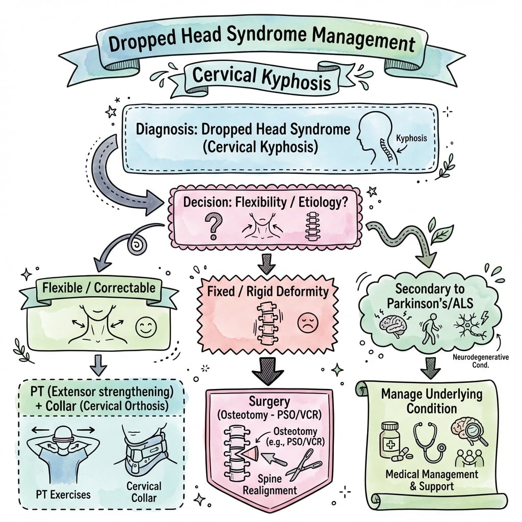
DHS typically progresses from "flexible" to "fixed" deformity over 3-6 months. Early intervention during the flexible phase may prevent fixed deformity. Once fixed, anterior release may be required for surgical correction.
When an inflammatory myopathy causes dropped head in the elderly, the single most important one to recognise is inclusion body myositis (IBM) - and it behaves completely differently from polymyositis and dermatomyositis. IBM is the commonest acquired myopathy over the age of 50 and a leading myopathic cause of both head drop and dysphagia, so the blanket statement that "inflammatory myopathy responds to immunosuppression" is a trap.
- Pattern: insidious, often asymmetric weakness with characteristic early involvement of the deep finger/wrist flexors (weak grip) and the quadriceps (knee buckling, falls); dysphagia is common.
- CK: only mildly raised (unlike the markedly elevated CK of polymyositis/dermatomyositis).
- Biopsy: endomysial inflammation with rimmed vacuoles and protein aggregates (plus mitochondrial changes); anti-cN1A (anti-NT5C1A) antibodies may be positive.
- The crucial point: IBM is largely refractory to corticosteroids and immunosuppression - misdiagnosing it as steroid-responsive polymyositis leads to futile, harmful treatment.
facioscapulohumeral dystrophy (FSHD), myotonic dystrophy, mitochondrial myopathy, and late-onset nemaline myopathy (sometimes with a monoclonal gammopathy). The exam point: recognise the finger-flexor-plus-quadriceps pattern and rimmed vacuoles, and do not assume immunosuppression will work.
Classification
Etiological Classification
Isolated Neck Extensor Myopathy
- Weakness limited to neck extensors
- No evidence of systemic neuromuscular disease
- EMG shows myopathic changes in neck muscles only
- Muscle biopsy: non-inflammatory myopathy
- Normal serum CK
- Negative autoantibodies
Age over 65 typically, female predominance, gradual onset over weeks to months, may stabilize or slowly progress.
Generally better than secondary causes. Some spontaneous improvement reported. Conservative management effective in early/mild cases.
Flexibility Classification
- Definition
- Passively correctable
- Clinical Implication
- May respond to conservative care
- Definition
- Some passive correction possible
- Clinical Implication
- Soft tissue release may help
- Definition
- No passive correction
- Clinical Implication
- Anterior release required
Clinical Presentation
History
Presenting Symptoms:
- Difficulty holding head up, especially end of day
- Need to use hand to support chin
- Impaired horizontal gaze
- Difficulty with eating and drinking
- Social embarrassment
- Progressive over weeks to months
Important History Points:
- Significance
- Morning improvement suggests MG
- Significance
- Suggests systemic myopathy
- Significance
- Suggests MG
- Significance
- Common in DHS, also suggests MG
- Significance
- Post-surgical DHS
- Significance
- Post-radiation myopathy
- Significance
- Statins, steroids
Physical Examination
- Chin-on-chest deformity
- Use of hand to support chin (pathognomonic)
- Compensatory lumbar hyperlordosis
- Shoulder elevation
- Assess flexibility of deformity
- Gently extend neck to assess passive correction
- Note any fixed contracture
- Ask patient to extend neck actively
- Prone head lift test (extend neck against gravity)
- Sustained gaze test (maintain horizontal gaze for 60 seconds)
- Assessment
- Ptosis, diplopia, facial weakness (MG)
- Assessment
- Atrophy, fasciculations (ALS)
- Assessment
- Proximal weakness (myopathy), fasciculations (ALS)
- Assessment
- Weakness, hyperreflexia (ALS)
- Assessment
- Hyperreflexia, Hoffman sign, gait
Specific Tests:
- Ice pack test (improvement in ptosis suggests MG)
- Fatigability test (repeated arm abduction)
Red Flags
Rapidly progressive weakness, limb involvement, respiratory difficulty, bulbar symptoms (dysarthria, aspiration), or hyperreflexia require urgent neurology referral to exclude ALS or MG crisis. These conditions require different management than INEM.
Investigations
Laboratory Investigations
First-Line:
- Purpose
- Elevated in inflammatory myopathy
- Purpose
- Inflammatory markers
- Purpose
- Hypothyroidism can cause myopathy
- Purpose
- Myasthenia gravis
- Purpose
- Seronegative MG
Second-Line:
- Indication
- Inflammatory myopathy
- Indication
- Suspected muscular dystrophy
- Indication
- Suspected paraneoplastic syndrome
Electrodiagnostic Studies
EMG (Electromyography):
- Interpretation
- Small, short, polyphasic
- Interpretation
- Supports INEM
- Interpretation
- Suggests systemic disease
- Interpretation
- Denervation (ALS, radiculopathy)
Nerve Conduction Studies:
- Normal in INEM
- Decremental response in MG
- Helpful to exclude neuropathy
Imaging
- Assess degree of kyphosis
- Measure chin-brow vertical angle (CBVA)
- Evaluate for fracture or spondylosis
- T1-weighted: Fatty infiltration appears hyperintense
- T2-weighted: Edema in acute phase
- Key muscles: Semispinalis cervicis, capitis
- Assess for cord compression
- Evaluate cord signal (myelomalacia)

CT:
- Bone detail for surgical planning
- Fusion assessment if prior surgery
- Fracture detection
Muscle Biopsy
- Diagnosis uncertain after non-invasive workup
- Suspected inflammatory myopathy
- Atypical presentation
- Fiber size variation
- Type 2 fiber atrophy
- No inflammation
- No necrosis
- Possible fatty replacement

Technique:
- Open biopsy of paraspinal muscle
- Usually semispinalis cervicis
- Sufficient sample for histology and special stains
Management
Treatment Algorithm
Initial Assessment:
- Complete history and examination
- Exclude systemic neuromuscular disease (MG, ALS)
- Imaging (X-ray, MRI)
- EMG and laboratory studies
- Flexible deformity
- Early presentation (less than 3-6 months)
- Mild symptoms
- High surgical risk
- Neck extensor strengthening exercises
- Postural retraining
- Stretching of anterior structures
- Aquatic therapy
- Cervical collar (soft or rigid)
- Custom orthosis for chin support
- May not halt progression but provides comfort
- Treat underlying condition (e.g., steroids for inflammatory myopathy)
- Pyridostigmine for MG
- No specific medication for INEM
Trial of 3-6 months of conservative management. If deformity progresses or becomes fixed, surgery is indicated.
Posterior-only fusion has a high failure rate in DHS due to poor muscle quality and high mechanical demand. Extending fusion to T2-T4 and considering combined anterior-posterior surgery for fixed deformity improves outcomes. Always assess flexibility preoperatively.
Surgical Technique
Posterior Cervical Fusion for Dropped Head Syndrome
- Restore horizontal gaze
- Stabilize cervical kyphotic deformity
- Decompress neural elements if myelopathy present
- Prevent progression of deformity
- Failed conservative management (3-6 months)
- Fixed or progressive deformity
- Functional goals: horizontal gaze, eating, ambulation
- Adequate bone quality for fixation
- Acceptable surgical risk profile
- Full-length standing spine radiographs
- CT cervical spine (bone quality, anatomy)
- MRI if myelopathy suspected
- Neurological assessment
- Cardiopulmonary optimization
- Patient may not tolerate prone positioning initially
- May require staged correction
- Awake fiberoptic intubation often needed
- Careful head positioning to avoid further flexion
Surgical Approach
- Prone on Jackson table or Mayfield head holder
- Position head in as much correction as safely tolerated
- Neuromonitoring (SSEP, MEP) essential
- Fluoroscopy to confirm alignment
- Posterior midline approach
- Subperiosteal dissection to expose lateral masses
- Identify occiput if occipitocervical fusion planned
- Extend to upper thoracic if long construct needed
- Lateral mass screws (C3-C6)
- Pedicle screws (C2, C7, thoracic)
- Occipital plate if occipitocervical fusion
- Rods contoured to restore lordosis
Exam Viva Point: "What are the key surgical considerations in dropped head syndrome?" Answer: Positioning is critical - patient may not tolerate prone initially. Require neuromonitoring. Long posterior fusion (often occiput to T2) to correct kyphosis. Staged correction if severe. High complication rates in elderly/comorbid patients.
Surgery aims to restore horizontal gaze and stabilize the cervical spine in corrected alignment.
A core peri-operative examinable point: a fixed chin-on-chest deformity is a PREDICTED DIFFICULT AIRWAY, and the plan must be made before the patient reaches the table.
- Why laryngoscopy fails: the fixed cervical flexion prevents the neck extension and oral-pharyngeal-laryngeal axis alignment needed for direct or video laryngoscopy, and restricts access to the mouth - standard laryngoscopy may be impossible and forced neck manipulation is dangerous.
- Airway plan: awake fibreoptic intubation is the technique of choice, maintaining spontaneous ventilation and the patient's own protective tone; have a difficult-airway trolley and a surgical-airway backup ready. Pre-existing dysphagia raises aspiration risk, which must be balanced against the difficult airway when planning induction.
- Positioning: the same fixed flexion makes safe prone positioning difficult - support and pad the head in its tolerated (often still kyphotic) position with Mayfield/Gardner-Wells fixation, do NOT force correction before instrumentation, and accept that some patients need staged or traction-assisted correction because they cannot lie flat/prone.
- Around surgery: optimise this elderly, comorbid (cardiopulmonary) population pre-operatively, use neuromonitoring during correction, and watch post-operatively for airway oedema and worsening dysphagia.
Exam point: anticipate the difficult airway - awake fibreoptic intubation and an explicit positioning/airway plan are essential before correcting a fixed chin-on-chest deformity.
Complications
Conservative Management Complications
- Fixed deformity development
- Secondary myelopathy
- Dysphagia and aspiration
- Skin breakdown under chin
- Skin pressure sores
- Discomfort and poor compliance
- May not prevent progression
Surgical Complications
Intraoperative:
- Prevention/Management
- Careful positioning, neuromonitoring
- Prevention/Management
- Anatomic knowledge, image guidance
- Prevention/Management
- Primary repair, dural sealant
- Prevention/Management
- Image guidance, careful technique
Early Postoperative:
- Management
- Speech therapy, swallow evaluation
- Management
- Antibiotics, debridement if deep
- Management
- Revision if symptomatic
- Management
- Evacuation if neurological compromise
Late Complications:
- Management
- Revision fusion, extend levels
- Management
- Monitor, extend fusion if symptomatic
- Management
- Revision, address pseudarthrosis
- Management
- More common if stopped at C7, extend to thoracic
- Management
- Expected due to underlying myopathy
Specific Risks in DHS
- Elderly population
- Consider cement augmentation
- Longer constructs for load sharing
- Reduces posterior tension band
- Higher construct demands
- May need anterior column support
- Up to 30% complication rate in some series
- Related to patient age and comorbidities
- Careful patient selection important
Postoperative Care
Immediate Postoperative Care
- Hourly neurological observations
- Upper and lower limb motor assessment
- Document any new deficits immediately
- Urgent imaging if deterioration
- New weakness or numbness
- Respiratory compromise
- Swallowing difficulty (airway edema)
- Wound hematoma
- Rigid collar (Miami-J or Aspen type)
- Worn at all times except wound care
- Duration: 6-12 weeks until radiographic fusion
- May need custom collar if alignment specific
- Elevate head of bed 30 degrees
- Log-roll precautions
- Avoid excessive neck flexion or extension
- Soft collar for sleeping comfort (over rigid collar)
Early Postoperative Management
- Check wound daily for hematoma, drainage
- Drain removal at 24-48 hours when output minimal
- Suture/staple removal at 2-3 weeks
- Multimodal analgesia
- Avoid excessive opioids (respiratory depression)
- Muscle relaxants may help spasm
- Sit upright day 1 if stable
- Walk with assistance day 1-2
- Physical therapy for gait and balance
- Avoid lifting greater than 2 kg for 6 weeks
Exam Viva Point: "What is postoperative care after cervical fusion for dropped head syndrome?" Answer: Rigid cervical collar for 6-12 weeks. Close neurological monitoring. Early mobilization. Watch for wound hematoma and dysphagia. Serial radiographs to confirm fusion.
Close monitoring in the immediate postoperative period is essential given the high-risk nature of this surgery.
Outcomes
Clinical Outcomes
Outcome Measures
- Improvement Expected
- 75-85% achieve improved forward gaze
- Factors Affecting Outcome
- Severity and flexibility of deformity preoperatively
- Improvement Expected
- Variable - 60-70% improvement
- Factors Affecting Outcome
- Underlying cause (INEM vs secondary DHS)
- Improvement Expected
- Often improves if due to chin-on-chest
- Factors Affecting Outcome
- May persist if unrelated cause
- Improvement Expected
- Improved eating, ambulation, social interaction
- Factors Affecting Outcome
- Patient goals and expectations
- Improvement Expected
- Significant improvement in most
- Factors Affecting Outcome
- Preoperative functional status
- Restores ability to look forward and see horizon
- Improves eating and swallowing (if due to chin-on-chest position)
- Enhances social interaction (eye contact)
- Reduces neck pain from chronic flexion
- Does not cure underlying neuromuscular disease
- Does not restore normal neck motion
- May not prevent progression of underlying condition
- Does not guarantee pain-free outcome
Exam Viva Point: "What outcomes can a patient expect from surgery for dropped head syndrome?" Answer: 75-85% achieve improved horizontal gaze. Quality of life improves. However, neck motion is lost, pseudarthrosis occurs in 10-25%, and complications are common (20-40%). Surgery treats the deformity but not the underlying cause.
Patient expectations must be realistic - surgery improves alignment and function but does not cure the underlying condition.
Guidelines, Registries & Global Practice
Global Epidemiology
Dropped head syndrome (DHS) is rare and predominantly affects older adults. The largest pooled cohort to date (systematic review of 129 patients across 74 studies) reports a mean age of 63.6 years and a 63% female predominance, with four diagnoses accounting for most cases.
- Pooled estimate
- 63.6 years
- Source
- Drain 2019 (PMID 30844858)
- Pooled estimate
- ~63%
- Source
- Drain 2019 (PMID 30844858)
- Pooled estimate
- 31.8% of cases
- Source
- Drain 2019 (PMID 30844858)
- Pooled estimate
- 20.2% of cases
- Source
- Drain 2019 (PMID 30844858)
- Pooled estimate
- 12.4% of cases
- Source
- Drain 2019 (PMID 30844858)
- Pooled estimate
- 7.0% of cases
- Source
- Drain 2019 (PMID 30844858)
Guidance Landscape (No Disease-Specific CPG)
There is no dedicated DHS clinical practice guideline from AAOS, NICE, BOA, AO Spine or EFORT. Practice is therefore derived from narrative reviews, consensus on cervical sagittal alignment and the underlying-disease guidelines that drive treatment.
- Relevant guidance
- CBVA (horizontal gaze), C2-7 SVA, TS-CL as planning/correction targets
- Evidence basis
- Expert Delphi, moderate reliability (PMID 26273762)
- Relevant guidance
- Restore horizontal gaze and sagittal balance; instrument to a stable caudal foundation (cervicothoracic junction)
- Evidence basis
- Expert consensus / review (PMID 23203936)
- Relevant guidance
- Treat the causative neuromuscular disorder first; DHS often improves with disease control
- Evidence basis
- Disease-specific guidelines
- Relevant guidance
- Medical/immunomodulatory therapy first; surgery for non-responders
- Evidence basis
- Systematic review, Level V (PMID 30844858)
Registry Evidence
No national joint or spine registry captures DHS as a discrete diagnosis; arthroplasty registries (AOANJRR, NJR, AJRR) are not applicable to this cervical extensor disorder. The evidence base is limited to single-centre series, the pooled systematic review above and narrative updates (PMID 32105239), all Level IV-V. This evidentiary weakness should temper any strong treatment claims.
Practice Variation
- First-line emphasis varies by referral pathway: neurology-led services prioritise antibody/EMG-guided medical therapy, whereas spine-led referrals more readily consider deformity surgery.
- Surgical extent differs between centres, from subaxial constructs to occiput/C2-to-upper-thoracic fusions; extension across the cervicothoracic junction is increasingly favoured to limit junctional failure.
- Anterior release / circumferential surgery is reserved for fixed or biplanar deformity and is concentrated in high-volume deformity units (PMID 36106864).
- Decision-making and rehabilitation for complex correction is typically multidisciplinary (spine surgeon, neurologist, radiologist); postoperative care routinely includes physiotherapy together with swallow/speech-pathology assessment for dysphagia.
MCQ Practice Points
Q: What is INEM and why is it a diagnosis of exclusion?
A: Isolated Neck Extensor Myopathy (INEM) is a non-inflammatory myopathy affecting only cervical extensors, predominantly in elderly patients. It is a diagnosis of exclusion because systemic causes (myasthenia gravis, ALS, inflammatory myopathy) must be ruled out first. EMG shows myopathic changes in neck extensors but normal limb muscles.
Q: What is the characteristic MRI finding in dropped head syndrome due to INEM?
A: T1 hyperintensity (fatty infiltration) of the paraspinal muscles, especially semispinalis cervicis and capitis. This indicates chronic muscle degeneration with fat replacement. The splenius, multifidus, and deep extensors may also be involved.
Q: What clinical features help differentiate myasthenia gravis from INEM as a cause of dropped head?
A: Myasthenia gravis:
- Fatigability (symptoms worsen with activity/end of day)
- Ptosis and diplopia (ocular involvement)
- Fluctuating weakness
- Positive AChR antibodies
INEM: Isolated to neck extensors, non-fatiguing, no ocular symptoms, negative antibodies.
Q: Why is a combined anterior-posterior approach often needed for surgical correction of dropped head syndrome?
A: Posterior-only fusion has high failure rates (up to 30-50%) due to poor extensor muscle quality and high mechanical demands. A 360-degree approach provides:
- Anterior release if fixed kyphosis
- Anterior structural support
- Posterior instrumented fusion for correction
- Better biomechanical stability
At a Glance
Dropped head syndrome (chin-on-chest deformity) results from weakness of cervical extensor muscles, most commonly due to isolated neck extensor myopathy (INEM)—a non-inflammatory myopathy of the elderly. Systemic causes must be excluded first: myasthenia gravis (ptosis, diplopia, fatigability, AChR antibodies), ALS (fasciculations, hyperreflexia, tongue atrophy), and inflammatory myopathy (elevated CK, proximal weakness). MRI demonstrates fatty infiltration of paraspinal muscles, particularly semispinalis cervicis, appearing hyperintense on T1. Patients cannot extend the neck against gravity and use the "chin-on-hand sign" for support. A 3-6 month conservative trial is appropriate; if fixed deformity develops, surgery is indicated—posterior-only fusion has high failure rate due to poor muscle quality, often requiring combined anterior release plus posterior instrumented fusion.
DROPPEDDROPPED - Causes of Dropped Head
Hook:DROPPED head needs comprehensive workup - multiple causes exist
NECKNECK - Key Muscles Affected
Hook:Semispinalis cervicis is the KEY muscle - check T1 MRI for fatty infiltration
FIXFIX - Surgical Principles
Hook:Extend fusion to thoracic spine - posterior-only often insufficient
Clinical Decision Scenarios
Practise clinical reasoning and management decisions out loud
“A 72-year-old woman presents with 3-month history of progressive difficulty holding her head up. She uses her hand to support her chin. No limb weakness, diplopia, or swallowing difficulty. No medication history. MRI shows fatty infiltration of the semispinalis cervicis.”
“The same patient returns after 4 months of physical therapy and collar use. Her deformity is now partially fixed, with only 50% passive correction. She has difficulty eating and is socially isolated. MRI shows no cord compression.”
“A 68-year-old man with dropped head syndrome now develops progressive hand numbness and gait difficulty. Examination shows hyperreflexia, positive Hoffman sign, and difficulty with tandem gait. MRI shows severe cervical kyphosis with cord compression at C4-5.”
“A 45-year-old woman presents with 6-week history of dropped head syndrome. She also reports fatigue, double vision at the end of the day, and difficulty swallowing solids. Examination shows mild bilateral ptosis that worsens with sustained upgaze.”
Definition & Etiology
- Severe weakness of neck extensors causing chin-on-chest deformity
- INEM = Isolated Neck Extensor Myopathy (diagnosis of exclusion)
- Must exclude: MG, ALS, inflammatory myopathy
- Secondary causes: post-surgical, radiation, Parkinson
Clinical Features
- Hand-to-chin support (pathognomonic)
- Impaired horizontal gaze
- Dysphagia common
- Worse at end of day = think MG
- Assess flexibility of deformity
Key Investigations
- AChR antibodies - rule out MG
- CK - elevated in inflammatory myopathy
- EMG - myopathic in neck, normal limbs for INEM
- MRI - fatty infiltration of semispinalis cervicis
- X-ray - measure kyphosis
Management Principles
- Conservative trial 3-6 months for flexible deformity
- Physical therapy, collar, treat underlying cause
- Surgery if failed conservative or fixed deformity
- Posterior fusion for flexible, 360° for fixed
- Extend to T2-T4 to prevent junctional kyphosis
Surgical Technique
- C2 pars/pedicle screws superiorly
- Lateral mass screws C3-C6
- Pedicle screws upper thoracic
- Anterior release if fixed
- Consider cement augmentation
Exam Pearls
- INEM = elderly, isolated to neck, non-inflammatory
- MG = young/middle-age, fatigability, ptosis, diplopia
- Semispinalis cervicis is KEY muscle affected
- High complication rate (20-30%) - counsel appropriately
- Posterior-only often insufficient - plan for combined
Evidence Base
INEM Definition and Characterization (Landmark)
- Four patients with a non-progressive myopathy of severe neck extensor weakness
- Coined the term isolated neck extensor myopathy (INEM), preferred to dropped head syndrome
- INEM distinguished from more ominous neuromuscular disorders by electrophysiology, imaging and histology
- Muscle biopsy shows fibre-size variation without inflammation
DHS Case Series and Literature Review
- Seven patients with DHS presenting to a UK spinal unit (4 acute, 3 gradual onset)
- Six of seven were managed conservatively; one underwent surgery
- The single operated patient was dissatisfied with the surgical outcome
- Highlights that surgery must be considered cautiously, especially in elderly patients
DHS Etiology and Management Review
- DHS is most commonly associated with neuromuscular disorders; INEM is used when EMG/biopsy are unrevealing
- Most reports favour non-surgical interventions to stabilise the deformity initially
- Surgical outcomes in the literature are limited and mixed, ranging from poor to excellent
- Prevalence is expected to rise with increasing life expectancy
The Dropped Head Syndrome (Original Description)
- Four patients with relatively isolated neck extensor weakness
- EMG and muscle biopsy suggested a restrictive, non-inflammatory myopathy
- Cervical paraspinal muscles predominantly affected
- One of the earliest formal characterisations of the syndrome
Dropped Head Syndrome: A Systematic Review (Largest Pooled Cohort)
- 129 patients across 74 studies; mean age 63.6 years, 63% female
- Four diagnoses account for most cases: INEM 31.8%, Parkinson disease 20.2%, myasthenia gravis 12.4%, ALS 7.0%
- Overall positive response to treatment 64.3%; combined medical plus immunosuppression 87.5%
- Surgery successful in 93.8% when used after failed medical management
Cervical Spine Deformity Classification (Horizontal Gaze / CBVA)
- Expert Delphi-derived classification for cervical spine deformity with a deformity descriptor and five modifiers
- Chin-brow vertical angle (CBVA) formalised as the horizontal-gaze modifier, central to DHS planning
- Incorporates C2-7 SVA, T1 slope minus cervical lordosis, mJOA myelopathy and SRS-Schwab parameters
- Moderate inter- and intra-observer reliability across 20 deformity surgeons
DHS: Update on Etiology and Surgical Management
- DHS is a heterogeneous group with diverse etiologies producing a flexible anterior cervical curvature
- Causes include myasthenia gravis, ALS, Parkinson disease, radiotherapy, age-related change and idiopathic forms
- Non-operative care comprises orthotic bracing and physical therapy
- Surgical fusion carries a higher complication rate but most achieve favourable long-term outcomes
References
- Katz JS, Wolfe GI, Burns DK, Bryan WW, Fleckenstein JL, Barohn RJ. Isolated neck extensor myopathy: a common cause of dropped head syndrome. Neurology. 1996;46(4):917-21. PMID 8780064. doi:10.1212/wnl.46.4.917
- Suarez GA, Kelly JJ Jr. The dropped head syndrome. Neurology. 1992;42(8):1625-7. PMID 1641161. doi:10.1212/wnl.42.8.1625
- Petheram TG, Hourigan PG, Emran IM, Weatherley CR. Dropped head syndrome: a case series and literature review. Spine (Phila Pa 1976). 2008;33(1):47-51. PMID 18165748. doi:10.1097/BRS.0b013e31815e38ec
- Sharan AD, Kaye D, Charles Malveaux WMS, Riew KD. Dropped head syndrome: etiology and management. J Am Acad Orthop Surg. 2012;20(12):766-74. PMID 23203936. doi:10.5435/JAAOS-20-12-766
- Drain JP, Virk SS, Jain N, Yu E. Dropped head syndrome: a systematic review. Clin Spine Surg. 2019;32(10):423-429. PMID 30844858. doi:10.1097/BSD.0000000000000811
- Brodell JD, Sulovari A, Bernstein DN, Mongiovi PC, Ciafaloni E, Rubery PT, Mesfin A. Dropped head syndrome: an update on etiology and surgical management. JBJS Rev. 2020;8(1):e0068. PMID 32105239. doi:10.2106/JBJS.RVW.19.00068
- Ames CP, Smith JS, Eastlack R, et al. Reliability assessment of a novel cervical spine deformity classification system. J Neurosurg Spine. 2015;23(6):673-83. PMID 26273762. doi:10.3171/2014.12.SPINE14780
- Li Y, Basil G, Vanni S. Dropped head syndrome in a patient with Parkinson's disease and inflammatory myopathy, treated with sternocleidomastoid release and circumferential cervical fusion. Br J Neurosurg. 2025;39(1):104-109. PMID 36106864. doi:10.1080/02688697.2022.2123892