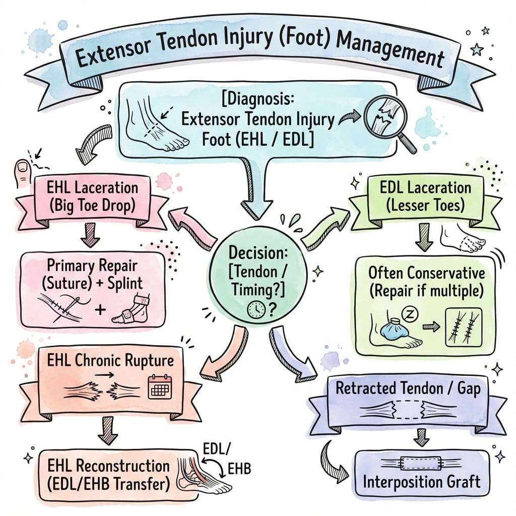
Laceration or Rupture | Zone Classification | Early Repair | Protected Mobilization
- Tibialis anterior is the most important extensor - always repair if injured
- Zone classification guides repair technique and prognosis (8 zones from muscle belly to toe)
- Early repair (within 2 weeks) gives best outcomes - delayed repair more difficult
- Protected mobilization balances tendon healing with preventing adhesions
- EDL tendons to lesser toes may not require repair if extensor digitorum brevis intact
- “TA rupture causes foot drop - unopposed plantar flexion during swing phase
- “EHL injury prevents hallux IP extension - important for propulsion
- “Multiple tendon injuries common with lacerations across dorsum of foot
- “Repair strength depends on suture technique - modified Kessler or Krackow preferred
Eight zones from muscle belly to toe guide management. Zone 1-3 (muscle/MTJ/distal leg) best prognosis. Zone 4-5 (ankle/dorsal foot) moderate adhesion risk. Zone 6-8 (MTP/phalanges) high adhesion risk but limited excursion.
Tibialis anterior provides 80% of dorsiflexion power. Loss leads to foot drop gait. Always explore and repair if suspected injury. EHL can partially compensate but inadequate alone.
Primary repair (within 2 weeks) is ideal - direct end-to-end repair possible. Delayed primary (2-6 weeks) may need tendon advancement. Late reconstruction (more than 6 weeks) often requires graft or transfer.
Balance protection (prevent rupture) with early motion (prevent adhesions). Controlled dorsiflexion in first 3-4 weeks. Full weight bearing delayed until 6 weeks. Stiffness more common than re-rupture.
- Location
- Muscle belly and MTJ
- Injury Pattern
- Laceration or strain
- Management Priority
- Debride, approximate muscle. Good healing potential
- Location
- Distal leg
- Injury Pattern
- Laceration common
- Management Priority
- Primary repair with core and epitenon sutures
- Location
- Ankle and superior retinaculum
- Injury Pattern
- Laceration or closed rupture (TA)
- Management Priority
- Critical zone - repair essential for TA/EHL
- Location
- Dorsum of foot to MTP
- Injury Pattern
- Laceration, often multiple tendons
- Management Priority
- Repair all major tendons. EDL may not need repair if EDB intact
- Location
- MTP and digits
- Injury Pattern
- Mallet toe, laceration
- Management Priority
- Zone 6: repair. Zone 7-8: splinting often sufficient
TALETALE - The Four Main Extensors
Hook:Tell a TALE of the four extensors - TA is the hero of the story
EIGHTEIGHT ZONES - Foot Extensor Zones
Hook:EIGHT zones progress from proximal to distal - remember ankle (4) and MTP (6) are critical transitions
REPAIRREPAIR - Primary Repair Principles
Hook:REPAIR guides you from recognition through rehabilitation
Overview and Epidemiology
Extensor tendon injuries of the foot and ankle encompass a spectrum from acute lacerations to chronic degenerative ruptures. Unlike flexor tendons, extensor tendons are more superficial and vulnerable to laceration but have better healing potential due to abundant vascular supply and looser paratenon.
- Laceration - most common, often from glass, machinery, or sharp objects across dorsum of foot
- Closed rupture - tibialis anterior most susceptible, usually at musculotendinous junction or insertion
- Avulsion - terminal extensor avulsion causes mallet toe
- Attrition rupture - chronic inflammatory arthropathy or repetitive trauma
- Lacerations most common in young active males (20-40 years)
- Closed TA rupture typically older patients (40-60 years) with degenerative changes
- Multiple tendon injuries common with extensive lacerations
- EHL injuries often associated with TA injuries due to anatomical proximity
Better healing potential than flexor tendons because: (1) More robust vascular supply from paratenon, (2) Less critical gliding requirements, (3) Lower tensile loads. However, still require formal repair for optimal function, especially tibialis anterior.
- Most powerful dorsiflexor (80% of dorsiflexion strength)
- Critical for toe clearance in swing phase
- Loss causes foot drop and altered gait
- Always requires repair
- Extends hallux IP and MTP joints
- Important for terminal stance and push-off
- Loss impairs propulsion and causes clawing
- Repair strongly recommended
- Extends lesser toes at MTP and IP joints
- Extensor digitorum brevis provides backup extension at MTP
- Repair when feasible but functional loss less severe
- Accessory dorsiflexor and everter
- Least important of the four
- Repair not always necessary
Anatomy and Biomechanics

The extensor compartment of the leg contains four muscles and tendons:
1. Tibialis Anterior (TA)
- Origin: Lateral tibial condyle, proximal lateral tibia, interosseous membrane
- Insertion: Medial cuneiform and base of first metatarsal
- Function: Dorsiflexion (primary), inversion (secondary)
- Nerve: Deep peroneal nerve (L4, L5)
- Unique features: Largest cross-sectional area, most medial tendon at ankle
2. Extensor Hallucis Longus (EHL)
- Origin: Middle anterior fibula, interosseous membrane
- Insertion: Base of hallux distal phalanx
- Function: Hallux IP and MTP extension, weak dorsiflexion
- Nerve: Deep peroneal nerve (L5, S1)
- Unique features: Passes between TA and EDL at ankle
3. Extensor Digitorum Longus (EDL)
- Origin: Lateral tibial condyle, proximal fibula, interosseous membrane
- Insertion: Middle and distal phalanges of lesser toes (2-5)
- Function: Toe extension at MTP and IP joints, weak dorsiflexion
- Nerve: Deep peroneal nerve (L5, S1)
- Unique features: Divides into four slips on dorsum of foot
4. Peroneus Tertius
- Origin: Distal anterior fibula (part of EDL complex)
- Insertion: Base of 5th metatarsal (dorsal aspect)
- Function: Dorsiflexion, eversion
- Nerve: Deep peroneal nerve (L5, S1)
- Unique features: Often absent (10-15% of population)
- Tendons pass deep to superior and inferior extensor retinaculum
- Order from medial to lateral: TA, EHL, EDL, Peroneus Tertius ("THE DEPT")
- Deep peroneal nerve and anterior tibial artery pass deep to inferior retinaculum
- Retinaculum prevents bowstringing and provides mechanical advantage
- Tendons diverge as they progress distally
- Extensor digitorum brevis (EDB) originates from calcaneus, inserts on EDL tendons
- EDB provides MTP extension even if EDL divided
- EHL crosses superficial to dorsalis pedis artery


Vascular supply:
- Paratenon provides diffuse blood supply
- Better vascularity than flexor tendons
- Anterior tibial artery gives small branches
- Critical to preserve during repair
Zone 4 (ankle/retinaculum) is critical because: (1) Tendons confined in tight compartments under retinaculum, (2) TA and EHL most vulnerable here, (3) Adhesions to retinaculum common, (4) Deep peroneal nerve at risk during exploration.
- Tibialis anterior: 300-500N during normal gait
- EHL: 50-100N during push-off
- EDL: 30-50N per toe
- Peak loads occur during heel strike and toe-off
- TA: 20-30mm excursion during gait cycle
- EHL: 15-20mm excursion
- EDL: 10-15mm excursion
- Limited excursion compared to hand extensors
- Swing phase: Extensors contract to lift foot (dorsiflexion) and clear toes
- Heel strike: Extensors eccentrically control plantarflexion
- Midstance: Extensors stabilize ankle
- Terminal stance: EHL maintains hallux extension for push-off
Classification Systems
Zone Classification (an anatomical proximal-to-distal system, analogous to the hand extensor zones)
The foot and ankle extensor tendons are divided into 8 zones from proximal to distal:
Zone 1: Muscle Belly
- Location: Proximal and mid leg
- Injury: Usually laceration or direct trauma
- Characteristics: Excellent healing potential, muscle can reapproximate
- Treatment: Debridement, loose approximation of muscle and fascia
- Prognosis: Excellent
Zone 2: Musculotendinous Junction (MTJ)
- Location: Distal leg
- Injury: Closed rupture more common (especially TA)
- Characteristics: Transition zone with variable strength
- Treatment: Direct repair with nonabsorbable suture
- Prognosis: Good but risk of re-rupture higher than pure tendinous zones
Zone 3: Distal Leg (Tendinous)
- Location: Above ankle, before retinaculum
- Injury: Laceration, closed rupture of TA
- Characteristics: Full tendon, good healing
- Treatment: End-to-end repair with core and epitenon sutures
- Prognosis: Excellent with appropriate repair
Zone 4: Ankle and Retinaculum
- Location: Beneath superior and inferior extensor retinaculum
- Injury: Laceration or closed rupture
- Characteristics: Critical zone - tendons confined, adhesion risk
- Treatment: Careful repair, consider retinaculum release if tight
- Prognosis: Good but adhesions to retinaculum can limit motion
Tibialis anterior most commonly ruptures at the MTJ (Zone 2) in middle-aged patients with degenerative changes. Often occurs during eccentric loading (deceleration or downhill walking). Presents with sudden pain and foot drop.
Zone 4 is the critical zone at the ankle.
- Clean laceration: Sharp object, minimal contamination - best for primary repair
- Contaminated laceration: Dirty wound, delayed presentation - washout and delayed repair
- Extensive laceration: Multiple tendons, nerve/vessel injury - staged reconstruction
- Acute rupture: Sudden event, distinct timeline - primary repair possible
- Chronic rupture: Gradual onset or delayed presentation - reconstruction often needed
- Degenerative rupture: Underlying tendinopathy, older patients - poor tissue quality
- Partial laceration (less than 50%): May not require repair if tendon continuity maintained
- Complete laceration: Always requires repair
- Complete rupture with retraction: May require tendon advancement or graft
Clinical Assessment
- Mechanism: Sharp object, glass, machinery
- Timing: Important for deciding primary vs delayed repair
- Contamination: Assess tetanus status and infection risk
- Hand dominance and occupation (for functional demands)
- Onset: Sudden pop vs gradual weakness
- Precipitating activity: Eccentric loading, sports activity
- Previous symptoms: Antecedent tendinopathy or pain
- Medical history: Diabetes, inflammatory arthropathy, fluoroquinolone use
Physical examination
- Wound: Location, extent, contamination if acute laceration
- Resting position: Foot drop (plantarflexed) if TA ruptured
- Swelling: Hematoma or edema at injury site
- Gait: Foot drop, slapping gait, toe drag during swing phase
- Contour: Loss of anterior ankle contour with TA rupture
- Gap: Palpable defect in tendon continuity
- Tenderness: Along tendon course
- Crepitus: Suggests tendinopathy or partial tear
- Active dorsiflexion: Ask patient to dorsiflex foot against gravity
- Grading: 0 = no movement, 1 = flicker, 2 = movement with gravity eliminated, 3 = against gravity, 4 = against resistance, 5 = normal
- Inversion strength: TA also inverts foot
- Inability to heel walk: Pathognomonic for TA dysfunction
- Hallux IP extension: Ask patient to extend great toe IP joint against resistance
- MTP extension: Test separately (EHB can compensate at MTP)
- Loss indicates EHL injury
- Lesser toe extension: Test each toe individually
- MTP vs IP extension: EDB extends MTP, so preserved MTP with weak IP suggests EDL injury
- Intact EDB can mask EDL injury
- With patient prone or sitting, squeeze anterior compartment
- Normal: Should see dorsiflexion
- Positive (no dorsiflexion): Suggests TA rupture
- Passive plantarflexion should make toes extend (if extensors intact)
- Passive dorsiflexion should make toes flex
- Loss of effect suggests complete tendon disruption
- Manual resistance to dorsiflexion and toe extension
- Grade strength using MRC scale
- Compare to contralateral side
Deep peroneal nerve runs with anterior tibial artery beneath inferior extensor retinaculum. Assess: (1) Sensation in first web space, (2) EDB function (extends toes at MTP), (3) Dorsalis pedis pulse. Injuries to nerve/artery require urgent vascular surgery consultation.
Examination findings by tendon:
- Active Test
- Dorsiflexion, inversion
- Loss of Function
- Foot drop, slapping gait
- Compensation
- EHL weak dorsiflexion (inadequate)
- Active Test
- Hallux IP extension
- Loss of Function
- Weak push-off, claw hallux
- Compensation
- EHB at MTP only
- Active Test
- Lesser toe extension
- Loss of Function
- Weak toe extension
- Compensation
- EDB at MTP preserved
- Active Test
- Dorsiflexion, eversion
- Loss of Function
- Minimal functional loss
- Compensation
- TA dorsiflexes, peroneals evert
Differential diagnosis of foot drop / weak dorsiflexion:
A palpable tendon gap with focal anterior ankle weakness points to a tendon injury, but several non-tendinous causes mimic it. Distinguishing them prevents an unnecessary operation.
- Key Discriminator
- Palpable gap, loss of anterior ankle contour, isolated DF weakness
- Sensation
- Normal
- Investigation
- Ultrasound or MRI shows tendon gap
- Key Discriminator
- Weak DF AND eversion AND EHL/toe extension; positive Tinel at fibular neck
- Sensation
- Loss over dorsum and first web space
- Investigation
- Nerve conduction studies, MRI of nerve
- Key Discriminator
- Back/leg pain, dermatomal sensory change, may have hip abductor weakness (L5)
- Sensation
- L5 dermatome change
- Investigation
- MRI lumbar spine, EMG
- Key Discriminator
- Exercise-induced pain and weakness that resolves with rest
- Sensation
- Transient first web space numbness
- Investigation
- Compartment pressure testing
- Key Discriminator
- Spasticity, hyperreflexia, pyramidal pattern weakness
- Sensation
- Variable, central pattern
- Investigation
- Brain imaging, neurology review
The single most useful bedside discriminator: an isolated dorsiflexion deficit with preserved eversion and intact sensation in the first web space favours a tibialis anterior tendon lesion. Add weak eversion, weak toe/hallux extension, and first web space numbness and you are dealing with a common (or deep) peroneal nerve problem instead.
DROPDROP FOOT - TA Rupture Clinical Features
Hook:DROP FOOT describes the clinical picture when TA is ruptured
Investigations
Clinical diagnosis is usually sufficient for acute lacerations with obvious tendon injury. Imaging helps in closed ruptures, delayed presentations, and surgical planning.
- All trauma to exclude fracture
- Chronic injuries to assess for bony avulsion
- Mallet toe to identify terminal phalanx avulsion
- AP, lateral, and oblique foot
- AP and lateral ankle if injury near ankle
- Usually normal in tendon injury
- May show avulsion fragment (insertion injuries)
- Soft tissue swelling or gas if infected
- Dynamic assessment
- Can visualize gap and retraction
- Assess tendon continuity and quality
- Real-time comparison to contralateral side
- Inexpensive and readily available
- Complete rupture: Gap with retracted tendon ends
- Partial tear: Hypoechoic defect, thickening
- Tendinopathy: Thickened, hypoechoic, loss of fibrillar pattern
- Operator dependent
- Limited for deep structures
- Difficult with extensive swelling
- Closed rupture with uncertain diagnosis
- Chronic injuries for surgical planning
- Multiple tendon involvement suspected
- Associated soft tissue or bone injury
- T1: Anatomical detail
- T2/STIR: Edema, fluid in tendon sheath
- Proton density: Tendon morphology
- Acute rupture: High signal, gap, retraction, surrounding edema
- Chronic rupture: Tendon ends, scar tissue, muscle atrophy
- Partial tear: Increased signal within tendon
- Tendinopathy: Thickening, intermediate signal
- Gold standard for soft tissue
- Multiplanar imaging
- Can assess muscle quality (atrophy, fatty infiltration)
Acute open lacerations - imaging not needed, go to OR for exploration and repair. Closed rupture or delayed presentation - MRI or ultrasound useful to: (1) Confirm diagnosis, (2) Assess gap size, (3) Evaluate tendon quality, (4) Identify retraction, (5) Plan reconstruction.
- Limited role
- Useful for bony avulsion injuries
- Assesses fracture displacement
- Not typically used
- May differentiate tendon vs neurological cause of weakness
Management Algorithm
Within 2 weeks of injury
- History and examination - establish diagnosis
- Wound assessment - clean vs contaminated
- Neurovascular status - deep peroneal nerve and dorsalis pedis
- Radiographs - exclude fracture
- Decision:
- Clean laceration + intact skin = Primary repair in OR
- Contaminated wound = Washout, delayed primary repair
- Closed rupture = Early surgical repair (TA/EHL), conservative vs surgery (EDL)
If missed acutely or delayed presentation
- Tendon ends still identifiable
- May require tendon advancement or recession
- Less predictable than primary repair
- Consider MRI for surgical planning
Chronic injuries
- Tendon ends retracted, scarred
- Direct repair usually not possible
- Options: Tendon graft, tendon transfer, arthrodesis (for mallet toe)
- Reconstruction vs acceptance of deficit depends on functional demands
- Partial lacerations less than 50% with continuity preserved
- EDL injuries with intact EDB (lesser toes)
- Peroneus tertius injuries
- Mallet toe (closed injuries)
- Medical comorbidities precluding surgery
- Low functional demands
- Immobilization in boot or cast with ankle in neutral to slight dorsiflexion
- Duration: 3-4 weeks for partial injuries
- Serial examination to ensure no progression
- Physiotherapy for strengthening after immobilization
- Partial tears: 70-80% good function without surgery
- EDL to lesser toes: Minimal functional deficit if EDB intact
- Mallet toe: 85-90% good outcomes with splinting
- Complete TA laceration or rupture - absolute indication
- Complete EHL laceration or rupture - strong relative indication
- EDL lacerations - repair when feasible
- Multiple tendon injuries
- Associated nerve or vascular injury
- Failed conservative management
- Restore tendon continuity
- Restore length-tension relationship
- Achieve stable repair allowing early mobilization
- Minimize adhesions
- Restore function
- Advantages
- Direct repair, no retraction, best outcomes
- Disadvantages
- May need to delay if contaminated
- Best For
- Clean lacerations
- Advantages
- Allows wound optimization, still repairable
- Disadvantages
- Some retraction, more difficult repair
- Best For
- Contaminated wounds after washout
- Advantages
- Allows full assessment, planned reconstruction
- Disadvantages
- Often needs graft or transfer, worse outcomes
- Best For
- Missed injuries, chronic ruptures
Surgical Technique
- Mark tendon course on skin with ankle in neutral
- Identify injury zone from history and examination
- Plan incision - curvilinear or longitudinal, avoid skin creases
- Consent for possible nerve injury, adhesions, weakness, re-rupture
- Supine positioning
- Thigh tourniquet (thigh level for ankle/foot injuries)
- Bump under ipsilateral hip if needed for positioning
- Image intensifier available but rarely needed
- Loupe magnification helpful for distal repairs
- Zone 3-4 (leg/ankle): Longitudinal incision centered over tendon
- Zone 5 (dorsum foot): Curvilinear to avoid crossing joints at right angles
- Zone 6-8 (toes): Longitudinal over dorsum of toe
- Extend incision as needed to retrieve retracted tendon ends
- Incise skin and subcutaneous tissue
- Identify and protect superficial nerves (superficial peroneal branches on dorsum)
- Identify deep peroneal nerve - runs with anterior tibial artery beneath retinaculum
- Locate tendon ends - use passive ankle/toe motion to aid identification
- Retrieve retracted ends - may need proximal and distal extensions
- Debride minimal amount of nonviable tendon (preserves length)
- Assess tendon quality - healthy tendon should be white and glistening
- Retinaculum may need to be partially released to access tendons
- Preserve retinaculum for later repair (prevents bowstringing)
- Identify anterior tibial artery and deep peroneal nerve
- Multiple tendons may be injured - identify all structures
Proximal retraction is common, especially with TA. Techniques to find tendon: (1) Passive motion - plantarflex ankle and watch for tendon movement, (2) "Milking" muscle belly proximally, (3) Extended incision if needed, (4) Contralateral side for reference anatomy.
This completes the exposure phase of the operation.
Reconstructing an Irreparable EHL (Second-Toe EDL Transfer)
When the EHL cannot be brought together end-to-end - a gap from retraction, tissue loss or a chronic laceration - the surgical section otherwise offers only hallux IP fusion. But active hallux extension can usually be restored rather than sacrificed, and the Wong evidence card names the technique without the body ever developing it.
- Why not just fuse. A simple hallux IP arthrodesis removes deformity but leaves the toe with no active dorsiflexion, weakening toe clearance and push-off. A bridging reconstruction keeps the hallux moving.
- Second-toe EDL transfer (deep transfer). The extensor digitorum longus slip to the second toe is divided and woven into the distal EHL stump, rerouting a nearby expendable extensor to power hallux dorsiflexion. In Wong et al's series this was used whenever the EHL ends were not opposable and restored active hallux dorsiflexion in 19 of 20 patients (mean FAAM about 94%), avoiding any need for allograft. The second toe tolerates the donor loss because the extensor digitorum brevis still extends it at the MTP.
- Free graft as the alternative bridge. A free tendon graft (plantaris or a toe-extensor slip) woven across the gap is the other graft-based option. The separate EHL-to-tibialis-anterior transfer solves the opposite problem - an irreparable tibialis anterior - where the whole EHL is sacrificed to restore dorsiflexion and the hallux is then fused.
- How to choose. Prefer a reconstruction that preserves active hallux extension (second-toe EDL transfer or a bridging graft) in any patient who needs push-off; reserve IP fusion for the genuinely low-demand patient or when no donor is available.
Q: The EHL is lacerated with a gap that will not close end-to-end - how do you preserve active hallux extension? A: Reconstruct rather than fuse. A deep transfer of the extensor digitorum longus slip from the second toe into the distal EHL stump reroutes an expendable extensor to power the hallux (Wong: active hallux dorsiflexion restored in 19 of 20, about 94% FAAM, no allograft; the second toe is still extended at the MTP by extensor digitorum brevis). A free tendon graft is the alternative bridge. Reserve hallux IP fusion for the low-demand patient or when no donor is available.
Chronic Tibialis Anterior Rupture: Interposition Graft and Gastrocnemius Recession
The Sammarco series (the largest reported) found that once a tibialis anterior rupture is more than a few weeks old, direct end-to-end repair is usually impossible - an interposition graft was needed in most cases and a gastrocnemius recession may be required to balance the repair. The body names grafting but never explains why the calf must sometimes be lengthened.
- Why direct repair fails late. The ruptured tibialis anterior retracts and its muscle loses excursion, so the ends cannot be brought together at a physiological length. Bridging the gap needs an interposition graft.
- The interposition graft. A free tendon graft (hamstring/gracilis, a toe extensor, a tibialis posterior slip, or allograft) is Pulvertaft-woven into the proximal muscle-tendon unit and the distal stump/medial cuneiform, restoring a continuous dorsiflexion vector. In Sammarco's series an intercalated graft was needed in about two-thirds of cases, and outcomes (AOFAS hindfoot about 55 to 94) were good regardless of age or delay.
- Why a gastrocnemius recession. With the tibialis anterior out of action, the unopposed gastroc-soleus pulls the ankle into a fixed equinus contracture. If the tight calf is left alone, the reconstruction is fighting a plantarflexion deformity and will over-tension or stretch out. A gastrocnemius (or gastroc-soleus) recession lengthens the posterior calf so the reconstructed dorsiflexor can hold the foot at neutral without excessive load - it protects the graft.
- Tensioning. Set the graft with the ankle in neutral dorsiflexion and confirm the foot rests plantigrade once the calf has been released.
Q: A tibialis anterior rupture presents 3 months late with a fixed equinus - what does reconstruction involve beyond bridging the gap? A: Two extra steps. Late ruptures retract and cannot be repaired directly, so an interposition free tendon graft (hamstring/toe-extensor/allograft, Pulvertaft-woven into the proximal unit and the distal stump/cuneiform) bridges the gap - needed in about two-thirds of Sammarco's cases. And because the unopposed gastroc-soleus has pulled the ankle into a fixed equinus, a gastrocnemius recession is often added to lengthen the tight calf so the reconstruction sits at neutral without over-tensioning. Tension the graft with the foot plantigrade.
Complications
- Incidence: 5-10% with modern repair techniques
- Risk factors: Inadequate repair strength, premature weight bearing, poor compliance
- Presentation: Sudden pain, loss of dorsiflexion, palpable gap
- Management: Revision repair if early (within 2 weeks), reconstruction if late
- Prevention: Adequate suture technique (Krackow preferred), protected mobilization protocol
- Incidence: 2-5% (higher with contaminated wounds)
- Risk factors: Contamination, delayed treatment, diabetes, immunosuppression
- Presentation: Wound erythema, drainage, fever, pain
- Management: Wound culture, antibiotics, washout if deep infection
- Prevention: Prophylactic antibiotics, meticulous wound care, early coverage
- Incidence: 5-10%
- Risk factors: Poor skin quality, tension, infection, poor vascularity
- Management: Local wound care, delayed closure, skin graft if needed
- Prevention: Tension-free closure, avoid incisions over bony prominences
- Deep peroneal nerve: Numbness in first web space, weakness of EDB
- Superficial peroneal nerve: Numbness on dorsum of foot
- Incidence: 2-5% iatrogenic injury
- Management: Observation (most neuropraxia recover), neurolysis if needed
- Prevention: Careful identification and protection during dissection
- Anterior tibial artery: Rare but catastrophic
- Presentation: Loss of dorsalis pedis pulse, foot ischemia
- Management: Immediate vascular surgery consultation
- Prevention: Identify artery during dissection, careful retraction
- Incidence: Rare (less than 1%)
- Risk factors: Extensive trauma, prolonged tourniquet, post-op hematoma
- Presentation: Severe pain, tense anterior compartment, pain with passive stretch
- Management: Urgent fasciotomy
- Prevention: Monitor high-risk patients, avoid overly tight dressings
Re-rupture is devastating complication. Prevention requires: (1) Strong repair - Krackow or Kessler core suture plus epitenon, (2) Protected mobilization - Non-weight bearing for 4 weeks, (3) Patient education - compliance with restrictions critical, (4) Graduated return - progressive loading over 8-12 weeks.
- Incidence: 20-30% have some limitation
- Risk factors: Zone 4 injuries, prolonged immobilization, extensive dissection
- Presentation: Limited ankle dorsiflexion, painful motion
- Management: Aggressive physiotherapy, tenolysis if severe and disabling
- Prevention: Gentle tissue handling, early protected mobilization, retinaculum release
- Incidence: 10-20% have some residual weakness
- Causes: Elongation at repair site, muscle atrophy, adhesions
- Presentation: Reduced dorsiflexion strength compared to contralateral
- Management: Strengthening exercises, orthotic support, acceptance
- Prevention: Adequate tensioning at repair, early mobilization, strengthening
- Incidence: 5-10%
- Causes: Neuroma, adhesions, tendinopathy, complex regional pain
- Management: Physiotherapy, nerve blocks, neuroma excision if identified
- Prevention: Protect sensory nerves, gentle tissue handling
- Incidence: 10-15%
- Mechanism: Gap formation, suture pull-through, inadequate repair
- Presentation: Weakness despite intact tendon on imaging
- Management: Plication or reconstruction if severe
- Prevention: Strong repair technique, appropriate tensioning
- Incidence: 5-10% develop fixed deformity
- Presentation: DIP flexion deformity, nail deformity
- Management: Splinting, arthrodesis if symptomatic and rigid
- Prevention: Adequate extension splinting duration (6 weeks minimum)
- Difficulty with stairs (especially descending)
- Tripping risk due to subtle weakness
- Slapping gait may persist
- Claw hallux
- Reduced push-off power
- Shoe wear issues
- Occurs with EDL over-pull or FDL tightness
- Causes metatarsalgia, shoe fitting issues
- May require flexor tenotomy or transfer
Postoperative Care and Rehabilitation
- Below-knee back-slab or boot
- Ankle in neutral dorsiflexion
- Non-weight bearing with crutches
- Elevate limb above heart level to reduce swelling
- Keep dressing clean and dry
- First dressing change at 48-72 hours
- Inspect wound for signs of infection
- Suture removal at 10-14 days
- Simple analgesia (paracetamol, NSAIDs)
- Avoid excessive opioid use
- Cryotherapy (ice packs) for swelling
- Convert to walking boot (removable)
- Continue non-weight bearing
- Begin gentle passive dorsiflexion with boot removed (therapist-supervised)
- No active dorsiflexion yet (protect repair)
- Maintain ankle ROM in plantarflexion
- Prevent adhesions without stressing repair
- Reduce swelling and inflammation
- Avoid active dorsiflexion
- Avoid resisted exercises
- No weight bearing
- Begin partial weight bearing in boot at 4 weeks
- Progress to full weight bearing by 6 weeks
- Wean from boot as tolerated (typically 6 weeks)
- Gentle active dorsiflexion against gravity (no resistance)
- Ankle circles (plantarflexion, dorsiflexion, inversion, eversion)
- Toe flexion and extension exercises
- Intrinsic muscle strengthening
- Passive dorsiflexion stretch (gastrocnemius and soleus)
- Avoid aggressive stretching
- Restore active ROM
- Prevent Achilles tendon contracture
- Begin muscle re-education
- Resistance band dorsiflexion - start with light resistance
- Progressive strengthening of TA, EHL, EDL
- Proprioception exercises (balance board)
- Heel walking progression
- Gait re-education
- Stair training
- Single-leg balance
- Sport-specific drills (if applicable)
- Restore strength to 80% of contralateral
- Normal gait pattern
- Return to daily activities
- Full pain-free ROM
- Strength at least 80-90% of contralateral
- Normal gait without limp
- Sport-specific testing passed
- Plyometrics
- Agility drills
- Sport-specific training
- Running progression
Begin partial weight bearing in boot. Start gentle active dorsiflexion. No resistance.
Full weight bearing without boot. Active ROM exercises. Light resistance begins.
Progressive resistance exercises. Proprioception training. Return to work (sedentary).
Return to unrestricted activities of daily living. Light sports and recreation.
Full return to competitive sports if criteria met. Continue strengthening program.
- Longer protection (may extend immobilization to 6 weeks)
- Critical for gait function
- More conservative return to sport
- Can progress faster (less critical loads)
- Earlier active motion possible
- Functional demands guide progression
- Extension splinting for 6 weeks continuous
- Then night splinting for additional 4-6 weeks
- Buddy taping during mobilization
Balance is key in extensor tendon rehab: (1) Too aggressive = re-rupture, elongation, failure, (2) Too conservative = adhesions, stiffness, weakness. Modern approach favors protected early motion - passive/active-assisted ROM early, graduated resistance later. Goal: optimal healing AND function.
- Return to baseline function
- Full or near-full strength
- Minimal pain or stiffness
- Mild weakness or stiffness
- Return to most activities
- Occasional discomfort
- Persistent weakness
- Chronic pain or stiffness
- Activity limitations
- May need revision or salvage
Rehabilitation is complete when functional goals are achieved and patient satisfied with outcome.
Guidelines, Registries & Global Practice
Global epidemiology:
- Tibialis anterior rupture is rare - the literature consists of small series and case reports rather than population data (largest surgical series 19 tendons).
- Two demographic peaks: young active patients with traumatic lacerations (glass, machinery, lawn mowers) and older patients (50-70 years) with atraumatic/degenerative rupture, often at the musculotendinous junction.
- Open extensor lacerations cluster in occupational and agricultural settings; dorsal foot lacerations frequently involve multiple tendons.
- Risk factors for atraumatic rupture: degenerative tendinopathy, diabetes, inflammatory arthropathy, corticosteroid injection and fluoroquinolone exposure.
Guideline landscape (no dedicated society guideline):
- Position on extensor tendon injury
- No injury-specific guideline; covered under general soft-tissue trauma. Endorses early diagnosis, repair of complete disruptions, and early controlled mobilization.
- Position on extensor tendon injury
- No specific BOAST; principles of the Open Fractures and Wound Management BOASTs apply to contaminated dorsal lacerations (early washout, antibiotics, soft-tissue cover).
- Position on extensor tendon injury
- Provides tendon repair principles - atraumatic handling, core plus epitenon suture, balanced tensioning, protected early motion.
- Position on extensor tendon injury
- No formal consensus; European practice mirrors individualized, demand-based decision-making.
- There is no implant or arthroplasty registry relevant here - these are soft-tissue repairs, not tracked by NJR/AJRR/AOANJRR-type registries. Outcome evidence therefore comes from institutional series, which limits the strength of any recommendation.
- Well-resourced settings: MRI/ultrasound for closed or delayed presentations; allograft and microsurgical nerve repair available; structured physiotherapy-led rehabilitation.
- Limited-resource settings: Clinical diagnosis predominates; autograft (hamstring, peroneus, FHL) preferred over costly allograft; reliance on casting and self-directed rehabilitation; later presentation increases the proportion needing reconstruction rather than direct repair.
- Document neurovascular status (deep peroneal nerve, dorsalis pedis) before surgery.
- Consent must cover infection, iatrogenic nerve injury, re-rupture, adhesions/stiffness and residual weakness.
- Counsel that delayed presentation worsens prognosis and may convert a simple repair into a graft reconstruction.
Exam Viva Scenarios
Practise clinical reasoning and management decisions out loud
“A 32-year-old construction worker presents to ED 4 hours after stepping through a glass window. He has a 6cm laceration across the anterior ankle. On examination, he cannot dorsiflex the ankle against gravity and there is a palpable gap. Neurovascular status is intact. What is your assessment and management?”
“A 58-year-old recreational runner presents with 3-week history of anterior ankle pain and weakness after feeling a 'pop' while running downhill. Examination reveals weak dorsiflexion (3/5 power) and a palpable gap 5cm above the ankle. MRI confirms complete TA rupture at the musculotendinous junction with 4cm gap. He wants to return to running. How would you manage this patient?”
“A 25-year-old presents with a lawn mower injury causing extensive laceration across the dorsum of the foot at the mid-foot level. Wound is contaminated with grass and dirt. On examination, there is a 10cm laceration with obvious injury to multiple structures. She cannot dorsiflex the ankle or extend any toes. Pulses are intact but sensation in the first web space is diminished. How do you proceed?”
Four Main Tendons (TALE)
- Tibialis Anterior - strongest dorsiflexor, ALWAYS repair
- Extensor Hallucis Longus - hallux IP extension, repair recommended
- Extensor Digitorum Longus - lesser toe extension, repair when feasible
- Peroneus Tertius - weakest, accessory dorsiflexor/everter, repair optional
Zone Classification (8 Zones)
- Zone 1-2: Muscle belly and MTJ - good healing, TA rupture common at MTJ
- Zone 3: Distal leg - primary repair with core plus epitenon
- Zone 4: Ankle/retinaculum - CRITICAL zone, adhesion risk high
- Zone 5: Dorsum foot to MTP - multiple tendon injuries common
- Zone 6-8: MTP and phalanges - mallet toe at Zone 8
TA Rupture Clinical Features (DROP FOOT)
- Dorsiflexion weak or absent (0-2/5 power)
- Foot drop and slapping gait
- Palpable gap anterior ankle
- Cannot heel walk or lift foot against gravity
- MTJ rupture most common (Zone 2)
Surgical Repair Technique
- Core suture: Krackow (strongest, 50-60N) or Kessler (30-40N)
- Add running epitenon (adds 15-20% strength)
- Ankle in neutral dorsiflexion for repair
- Primary repair ideal within 2 weeks
- Delayed primary 2-6 weeks, reconstruction more than 6 weeks
Rehabilitation Protocol
- 0-4 weeks: Non-weight bearing, gentle passive ROM
- 4-6 weeks: Progress to full weight bearing, active ROM
- 6-12 weeks: Strengthening with resistance, proprioception
- 3-6 months: Return to sport if criteria met (80-90% strength)
Key Complications
- Re-rupture 5-10% - prevented by adequate suture technique and compliance
- Adhesions 20-30% (especially Zone 4) - early motion helps prevent
- Infection 2-5% - higher with contaminated wounds
- Deep peroneal nerve injury - assess first web space sensation and EDB
- Weakness 10-20% - some residual deficit expected
Exam Traps to Avoid
- Not documenting neurovascular exam (deep peroneal nerve critical)
- Offering conservative management for active patient with TA rupture
- Using inadequate suture technique (simple sutures insufficient)
- Too aggressive rehab (risk re-rupture before 4 weeks)
- Not recognizing Zone 4 as critical zone for adhesions
High Yield Exam Points
- TA provides 80% of dorsiflexion - ALWAYS repair
- Zone 4 (retinaculum) worst outcomes due to adhesions
- Krackow suture strongest technique (50-60N breaking strength)
- Early protected motion superior to prolonged immobilization
- EDL repair optional if EDB intact (lesser toe function preserved)
- Surgical repair achieves 85% strength vs 60% conservative for TA
- Deep peroneal nerve runs with anterior tibial artery at ankle
Evidence Base
The literature on foot/ankle extensor tendon injury is low-level (case series, Level IV, and one comparative study). There are no randomized trials. Recommendations rest on small cohorts and biomechanical reasoning, so individualize by patient demand and tendon involved.
Surgical Repair of Acute and Chronic Tibialis Anterior Tendon Ruptures
- AOFAS hindfoot score improved 55.5 to 93.6 points
- Direct repair only in 7 of 19 - the rest needed interposition tendon graft
- 5/5 dorsiflexion strength regained in 15 of 19 cases
- Outcomes independent of age, sex or comorbidity; delayed cases more complex
Tibialis Anterior Ruptures: Operative versus Nonoperative Outcomes
- 8 operative versus 8 nonoperative, no statistically significant outcome difference
- Difference is confounded by selection bias (age/demand bimodality)
- Authors still recommend repair/reconstruction in young high-demand patients
- Nonoperative care is appropriate for low-demand elderly patients
Anterior Tibial Tendon Rupture: Individualized Reconstruction
- All 7 operatively reconstructed patients gained function and strength
- Reconstruction technique was individualized (repair, advancement, transfer)
- AFO improved function in 2 of the nonoperative patients
- Operative reconstruction recommended for suitable (active) patients
Repair of Acute Extensor Hallucis Longus Tendon Injuries
- Primary repair achieved in 80% of EHL injuries
- Active hallux dorsiflexion restored in 19 of 20 patients
- Mean FAAM ADL and Sports scores both 94.2%
- EDL-to-hallux transfer is a graft-free option when ends are not opposable
Open Extensor Hallucis Longus Lacerations: Zone-Based Treatment
- All 17 patients healed without infection or symptomatic neuroma
- Complete lacerations repaired and protected 6 weeks (K-wire plus cast)
- Partial lacerations did well with conservative care and early motion
- AOFAS hallux pain score 40/40 in all; mean function 42.1/45
Hallux Extensor/Flexor Tendon Injuries: Decision Framework
- Treatment is dictated by mechanism, site, timing and associated injuries
- Not every hallux tendon injury requires operative repair
- Define surgical goals (pain, motion, deformity) before operating
- Satisfactory results are expected when goals are clearly set
- No specific guideline for extensor tendon injuries (included in general soft tissue trauma)
- Recommend early diagnosis and repair for complete tendon disruptions
- Support early controlled mobilization protocols
- Primary repair within 2 weeks for complete TA and EHL disruptions
- Core suture technique (Krackow or Kessler) plus epitenon
- Protected early mobilization starting at 2-4 weeks
- EDL repairs recommended when feasible but not critical for lesser toes
- MRI or ultrasound for diagnosis of closed ruptures
- Some authors advocate repair of all EDL tendons
- Others suggest selective repair (only if EDB absent or patient high-demand)
- Consensus: Repair when feasible but low priority if EDB intact
- Traditional: 6 weeks strict immobilization
- Modern: Early protected motion starting at 2-4 weeks
- Consensus shifting toward early motion based on biomechanical and clinical data
- Some advocate up to 6 months for TA reconstruction
- Others suggest diminishing returns after 6 weeks
- Consensus: Best outcomes with repair within 6 weeks; reconstruction possible up to 6 months in motivated patients
References
-
Scaduto AA, Cracchiolo A. Lacerations and ruptures of the flexor or extensor hallucis longus tendons. Foot Ankle Clin. 2000;5(3):725-736. PMID: 11232406.
-
Markarian GG, Kelikian AS, Brage M, et al. Anterior tibialis tendon ruptures: an outcome analysis of operative versus nonoperative treatment. Foot Ankle Int. 1998;19(12):792-802. PMID: 9872465. doi:10.1177/107110079801901202.
-
Sammarco VJ, Sammarco GJ, Henning C, Chaim S. Surgical repair of acute and chronic tibialis anterior tendon ruptures. J Bone Joint Surg Am. 2009;91(2):325-332. PMID: 19181976. doi:10.2106/JBJS.G.01386.
-
Wong JC, Daniel JN, Raikin SM. Repair of acute extensor hallucis longus tendon injuries: a retrospective review. Foot Ankle Spec. 2014;7(1):45-51. PMID: 24334369. doi:10.1177/1938640013514271.
-
Al-Qattan MM. Surgical treatment and results in 17 cases of open lacerations of the extensor hallucis longus tendon. J Plast Reconstr Aesthet Surg. 2007;60(4):360-367. PMID: 17349589. doi:10.1016/j.bjps.2006.05.003.
-
Ouzounian TJ, Anderson R. Anterior tibial tendon rupture. Foot Ankle Int. 1995;16(7):406-410. PMID: 7550953. doi:10.1177/107110079501600705.
-
Bernstein RM. Spontaneous rupture of the tibialis anterior tendon. J Foot Surg. 1985;24(5):354-357.
-
Anagnostakos K, Bachelier F, Fürst OA, Kelm J. Rupture of the tibialis anterior tendon: a case report and literature review. Arch Orthop Trauma Surg. 2006;126(1):59-62.
-
Trouillas J, Fessy MH. Rupture of the anterior tibial tendon: 5 cases. Rev Chir Orthop Reparatrice Appar Mot. 2003;89(3):262-267.
-
Larsen E, Lauridsen F. Rupture of the tibialis anterior tendon. Injury. 1984;16(3):150-151.
-
Kashyap S, Prince R. Spontaneous rupture of the tibialis anterior tendon. Clin Orthop Relat Res. 1987;(216):159-161.
-
Christman RA. Foot and Ankle Radiology. Churchill Livingstone. 2003:425-430.
-
Kausch T, Rutt J, Schuler P. Ultrasonographic findings in tendon diseases of the ankle. Foot Ankle Surg. 2004;10(2):73-78.
-
Mengiardi B, Pfirrmann CW, Vienne P, et al. Anterior tibial tendon abnormalities: MR imaging findings. Radiology. 2005;235(3):977-984.