Ficat-Arlet Staging of Femoral Head Osteonecrosis
The single most important clinical threshold in the Ficat-Arlet system is the transition from Stage II to Stage III. In Stage II the femoral head is still spherical and the subchondral plate is intact — joint-preserving procedures have a realistic chance of success. In Stage III the subchondral plate has fractured (the crescent sign) and the head is starting to flatten — the prognosis for any procedure short of arthroplasty falls sharply. Examiners expect you to name the stage, state whether the head is spherical, and then link it to the appropriate procedure.
What Is Being Staged: Aetiology and Risk Factors
You cannot stage a disease you cannot recognise, and the examiner will expect you to reel off the causes of femoral head osteonecrosis before you ever reach a stage. The causes divide cleanly into traumatic (a mechanical insult to the blood supply) and non-traumatic (a systemic or local insult to the same vessels). Corticosteroids and alcohol together account for the majority of non-traumatic cases worldwide. For the full pathology, epidemiology and management of the disease itself see avascular necrosis of the hip.
- Causes
- Displaced femoral neck fracture; hip dislocation; slipped capital femoral epiphysis
- Mechanism / exam point
- Direct disruption of the retinacular (lateral epiphyseal) vessels — the dominant blood supply to the head; risk rises with displacement and delay to reduction
- Causes
- High-dose or prolonged steroids (transplant, SLE, asthma, chemotherapy adjunct)
- Mechanism / exam point
- The commonest non-traumatic cause; dose- and duration-related; thought to act via marrow fat-cell hypertrophy, fat embolism and impaired osteoblast function
- Causes
- Chronic excess alcohol consumption
- Mechanism / exam point
- Second commonest non-traumatic cause; similar fatty-marrow and microvascular mechanisms; dose-related
- Causes
- Sickle cell disease; thrombophilia / hypofibrinolysis; myeloproliferative disease
- Mechanism / exam point
- Microvascular occlusion by sickled cells or thrombus; sickle cell is the dominant cause in sub-Saharan Africa and the Caribbean
- Causes
- Gaucher disease; hyperlipidaemia
- Mechanism / exam point
- Marrow infiltration (Gaucher cells) or fat embolism raising intraosseous pressure and occluding sinusoids
- Causes
- Caisson disease (decompression sickness in divers and tunnel workers)
- Mechanism / exam point
- Intraosseous nitrogen-bubble emboli occluding the subchondral sinusoids
- Causes
- Radiation; chemotherapy; pancreatitis; pregnancy; HIV; smoking; idiopathic
- Mechanism / exam point
- A substantial minority remain idiopathic; aetiology does not change the Ficat-Arlet stage but does affect bilaterality and progression risk
A SEPTIC hip that is not infectedCauses of avascular necrosis
Hook:ASEPTIC — the necrotic hip is 'aseptic' (not infected): Alcohol, Steroids/Sickle cell, Erlenmeyer/Gaucher, Pancreatitis/Pregnancy, Trauma, Idiopathic, Caisson.
Why the Head Dies: Blood Supply and Pathophysiology
Understanding why the femoral head is so vulnerable is a classic basic-science viva that examiners hang off this topic. The femoral head has a precarious, largely end-arterial blood supply, so a single vascular insult produces ischaemia that the head cannot compensate for. (For the anatomy in full see hip blood supply.)
- The deep branch of the medial femoral circumflex artery (MFCA) is the dominant supply. It gives rise to the lateral epiphyseal (superior retinacular) vessels, which run beneath the synovial reflection along the posterosuperior neck and supply the great majority of the weight-bearing femoral head.
- The lateral femoral circumflex artery contributes to the anteroinferior head, and the artery of the ligamentum teres (a branch of the obturator/medial circumflex) supplies only a small foveal region and is insufficient to sustain the head alone in adults.
- Because these are terminal vessels with limited anastomosis, a displaced femoral neck fracture (tearing the retinacular vessels), a dislocation, raised intraosseous pressure (fatty marrow, Gaucher cells), or microvascular occlusion (sickle cells, nitrogen or fat emboli, thrombus) all converge on the same end point: ischaemia of the subchondral weight-bearing bone.
The final common pathway is the same whatever the cause: ischaemia kills osteocytes and marrow, the body mounts a repair response (creeping substitution) in which osteoclastic resorption of dead bone outpaces new bone formation, the subchondral bone is transiently weakened, and under continued load it fractures (the crescent sign) and then collapses. The Ficat-Arlet stages are simply snapshots of this sequence — and the reason early (pre-collapse) intervention works is that it acts before the resorption-weakening-fracture cascade becomes irreversible.
The reason core decompression works in early disease maps directly onto this pathophysiology: drilling the necrotic segment lowers the raised intraosseous pressure, interrupts the ischaemic cycle, and creates a channel for revascularisation and creeping substitution before the subchondral plate has fractured. Once the crescent sign appears, the mechanical defect cannot be reversed by decompression alone — which is exactly why the stage dictates the operation.
The Ficat-Arlet Classification
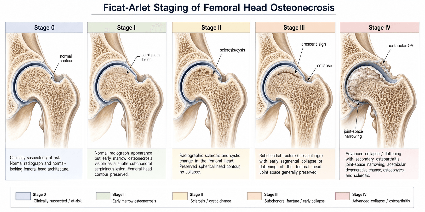
The Ficat-Arlet system was described by Ficat and Arlet in the 1960s and 1970s as a radiographic staging system for idiopathic avascular necrosis of the femoral head. It was originally designed before MRI was available, so early stages relied on bone scans and functional bone marrow exploration. Modern practice adds MRI to detect Stage 0 and Stage I disease that plain radiographs miss entirely.
- Radiographic Appearance
- Normal radiographs; abnormal bone scan or MRI (low signal on T1)
- Femoral Head Contour
- Normal and spherical
- Pain
- May be asymptomatic or mild groin pain
- Key Feature
- Diagnosis only on MRI or bone scan — plain films are normal
- Radiographic Appearance
- Normal radiographs; MRI shows oedema and necrosis (double-line sign on T2)
- Femoral Head Contour
- Normal and spherical
- Pain
- Groin or thigh pain, often activity-related
- Key Feature
- Normal plain films with positive MRI; the earliest clinically detectable stage
- Radiographic Appearance
- Sclerosis, cystic changes, or osteopenia in the femoral head; head still spherical
- Femoral Head Contour
- Spherical but with internal changes
- Pain
- Moderate pain on weight-bearing
- Key Feature
- The head holds its shape — this is the last stage where joint-preserving surgery is reliable
- Radiographic Appearance
- Subchondral fracture line (crescent sign); early flattening of the superior pole
- Femoral Head Contour
- Beginning to lose sphericity
- Pain
- Significant pain, reduced range of motion
- Key Feature
- The crescent sign — a radiolucent line beneath the subchondral plate — is pathognomonic
- Radiographic Appearance
- Flattened head with secondary osteoarthritis; joint-space narrowing, acetabular involvement
- Femoral Head Contour
- Flattened, mushroomed, or fragmented
- Pain
- Severe pain, marked stiffness, functional limitation
- Key Feature
- End-stage disease — arthroplasty is the standard treatment
Nothing • Normal shape • Notch in the plate • Nothing left of the headThe five stages in order (0–IV)
Hook:Each stage starts with N — the head goes from Nothing-visible to Nothing-left as necrosis progresses to collapse.
The crescent sign is a radiolucent crescent-shaped line beneath the subchondral cortex of the femoral head, best seen on the anteroposterior radiograph. It represents gas or synovial fluid in the subchondral fracture plane. It is the radiographic hallmark of Stage III and signifies that the structural integrity of the weight-bearing surface has failed. Once this appears, joint-preserving surgery is unlikely to succeed.
Staging Investigations
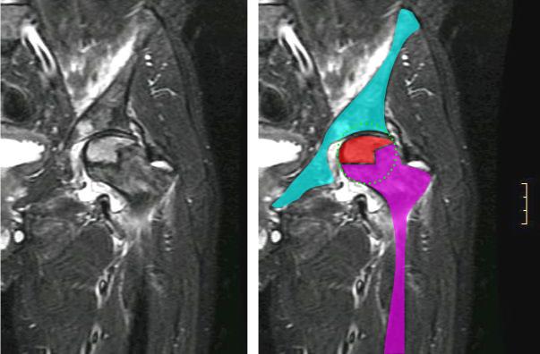
Accurate Ficat-Arlet staging requires a combination of plain radiographs and MRI. Bone scan has been superseded by MRI for early detection but may still have a role when MRI is unavailable or contraindicated.
- Plain Radiographs
- Normal
- MRI
- Low signal on T1 in the necrotic segment; high signal on T2 with double-line sign at the boundary
- Bone Scan
- Cold spot or decreased uptake in the femoral head
- CT
- May show subtle trabecular changes before X-ray
- Plain Radiographs
- Normal
- MRI
- Necrotic area with characteristic geographic pattern; serpiginous low-signal border on T1; double-line sign on T2
- Bone Scan
- Decreased or absent tracer uptake
- CT
- Not routinely required
- Plain Radiographs
- Sclerosis, cysts, osteopenia — head still round
- MRI
- Geographic necrotic area with reactive interface; oedema in surrounding marrow; head contour preserved
- Bone Scan
- Hot spot at reactive margin around cold necrotic core
- CT
- May better define the extent of necrosis and any subchondral changes
- Plain Radiographs
- Crescent sign; early flattening of the superior pole
- MRI
- Subchondral fracture line with marrow oedema; early head flattening
- Bone Scan
- Intense uptake from reactive bone around the fracture
- CT
- Best for defining the exact extent of subchondral fracture and collapse
- Plain Radiographs
- Flattened head; joint-space narrowing; osteophytes; acetabular changes
- MRI
- Extensive marrow changes; head deformity; secondary OA changes in acetabulum
- Bone Scan
- Not routinely needed — diagnosis is obvious on X-ray
- CT
- Occasionally useful for pre-operative planning of arthroplasty
X-ray shows the shape, MRI shows the death, CT shows the crackImaging mnemonic for staging
Hook:Three modalities, three jobs: X-ray for contour, MRI for viability, CT for the fracture line.
The double-line sign on T2-weighted MRI is pathognomonic for osteonecrosis. It consists of a low-signal inner border (sclerotic bone) and a high-signal outer border (granulation tissue or hypervascular fibrous tissue) at the junction between necrotic and living bone. It is present in the majority of early-stage lesions and confirms the diagnosis when the plain radiograph is normal.
A normal-radiograph hip with marrow oedema on MRI is not automatically early AVN; two mimics change management completely:
- Transient osteoporosis of the hip (bone marrow oedema syndrome): typically a middle-aged man or a woman in late pregnancy, with diffuse marrow oedema of the head AND neck (high T2/STIR, low T1) but no focal geographic necrotic segment and no double-line sign. It is self-limiting — it resolves over months with protected weight-bearing and analgesia (a migratory/regional form exists) — so it should NOT be core-decompressed as if it were AVN.
- Subchondral insufficiency fracture (SIF): an older, osteoporotic patient (often a woman) with acute pain and no preceding necrosis; MRI shows a subchondral low-signal line roughly parallel to the cortex with surrounding oedema, but without the demarcating serpiginous necrotic interface of AVN (here any necrosis is secondary to the fracture). It can progress to rapid collapse, so it needs recognition and offloading.
The discriminator is the well-demarcated geographic necrotic segment with a double-line sign — present in AVN, absent in transient osteoporosis, and not the primary lesion in SIF.
Treatment by Stage
The Ficat-Arlet stage is the primary determinant of treatment. The fundamental question is whether the femoral head is structurally salvageable or whether collapse has progressed to the point where arthroplasty is the only reliable option.
- Head Status
- Normal contour, necrosis confined to marrow
- Joint-Preserving Options
- Core decompression (CD) alone; protected weight-bearing; address risk factors (steroids, alcohol)
- Arthroplasty
- Not indicated
- Prognosis of Preservation
- Best outcomes for CD — up to 70-80 percent success in small lesions
- Head Status
- Spherical with sclerosis or cysts
- Joint-Preserving Options
- Core decompression with or without bone grafting (vascularised fibular graft, tantalum rod); rotational osteotomy in selected cases
- Arthroplasty
- Consider for large lesions (greater than 50 percent head involvement) or failed preservation
- Prognosis of Preservation
- Moderate — success 50-70 percent with CD plus grafting; lesion size is the key modifier
- Head Status
- Subchondral fracture (crescent sign), early flattening
- Joint-Preserving Options
- Joint-preserving surgery is unreliable; osteotomy occasionally attempted in young patients with small lesions
- Arthroplasty
- Total hip arthroplasty is standard; hemiarthroplasty in elderly, low-demand patients
- Prognosis of Preservation
- Poor for preservation — most patients progress to arthroplasty within 2-3 years
- Head Status
- Collapsed head with secondary OA
- Joint-Preserving Options
- Not appropriate
- Arthroplasty
- Total hip arthroplasty; resurfacing in selected young, active patients with good bone quality
- Prognosis of Preservation
- Arthroplasty outcomes are good but longevity is a concern in very young patients
Core decompression is the most effective joint-preserving procedure in early-stage osteonecrosis (Stages I-II), but it must be performed before collapse. Once the crescent sign appears (Stage III), decompression alone has a very high failure rate. In a young patient with bilateral disease, early detection and decompression of the pre-collapse side can prevent a second arthroplasty.
Pre-collapse drill, Post-collapse replaceTreatment by stage
Hook:Drill the round head, replace the flat head — the crescent sign is the dividing line.
Beyond the stage-driven algorithm, three adjuncts are worth knowing for the exam. First, risk-factor modification is treatment: minimising corticosteroids (with the prescribing team) and stopping alcohol can halt progression and protect the contralateral hip, and any joint-preserving operation is undermined if ongoing steroid exposure continues. Second, pharmacological and biological adjuncts — bisphosphonates (to reduce osteoclastic resorption during the weakening phase) and, increasingly, augmentation of core decompression with bone marrow aspirate concentrate or other cell therapy — have shown promise in early-stage disease but the evidence remains limited and they are adjuncts, not replacements for sound staging. Third, in the young patient facing arthroplasty, the conversation is about implant longevity and bearing choice: AVN patients are typically decades younger than osteoarthritis patients, historically had higher failure rates, and now do well with modern cementless stems and hard bearings — hip resurfacing is reserved for selected young, active patients with good bone stock and no large necrotic segment in the residual head.
The Crescent Sign — Recognition and Significance
The crescent sign is the radiographic signature of Stage III and the single most important imaging finding that shifts management from joint preservation to arthroplasty. It was first described by Ficat and Arlet and is present on plain radiographs in most Stage III lesions.
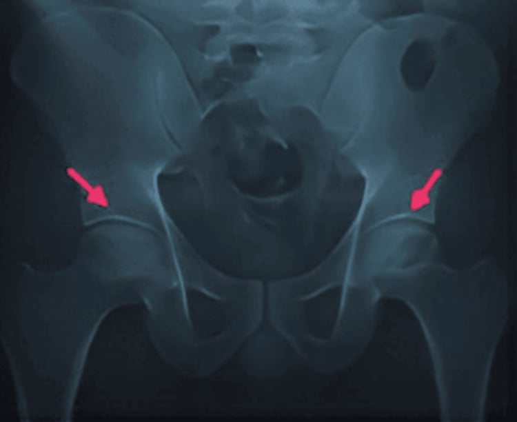
What it is: A curved, radiolucent line beneath the subchondral cortex of the femoral head, most commonly at the superior weight-bearing dome. It represents a fracture plane through the necrotic subchondral bone, filled with gas or joint fluid under pressure.
How to detect it:
- Best seen on the anteroposterior pelvis radiograph; the frog-lateral view may show it in a different plane.
- Look carefully at the superior margin of the femoral head — it appears as a thin dark crescent just beneath the dense subchondral cortex.
- CT is more sensitive than plain radiography and can demonstrate the subchondral fracture line before it becomes radiographically apparent.
- MRI shows it as a low-signal line through the subchondral bone on T1 with surrounding marrow oedema on T2.
Why it matters:
- It signifies that the structural support of the articular cartilage has failed — the necrotic bone cannot bear load and has fractured.
- Once the crescent sign appears, the femoral head will progressively flatten under continued weight-bearing.
- Joint-preserving procedures performed after the crescent sign appears have a significantly higher failure rate because the structural defect cannot be reversed.
If a patient with known osteonecrosis develops a sudden increase in pain with a new radiolucent line beneath the subchondral plate, suspect progression from Stage II to Stage III. Obtain an urgent MRI and restrict weight-bearing while you plan definitive treatment. The crescent sign on a plain radiograph is a surgical sign — it tells you that arthroplasty, not decompression, is the correct operation.
Lesion Size and Location — Modifiers Within Each Stage
The Ficat-Arlet stage alone does not predict outcomes perfectly. The size of the necrotic segment and its location within the femoral head are critical modifiers that affect prognosis at every stage.
- Head Involvement
- Less than 30 percent of the head (medial or central location)
- Prognosis at Stage I-II
- Good — core decompression success 70-80 percent
- Prognosis at Stage III-IV
- Relatively better but arthroplasty still indicated
- Head Involvement
- 30 to 50 percent of the head
- Prognosis at Stage I-II
- Moderate — core decompression with bone grafting; success 50-65 percent
- Prognosis at Stage III-IV
- Arthroplasty indicated; outcomes satisfactory
- Head Involvement
- Greater than 50 percent of the head (lateral or extensive location)
- Prognosis at Stage I-II
- Poor even at early stages — consider vascularised fibular graft or early arthroplasty
- Prognosis at Stage III-IV
- Arthroplasty — outcomes satisfactory but younger age remains a concern
Lateral lesion classification (Kerboul angle): The lateral extent of the necrotic segment is measured on MRI by combining the arc of necrosis seen on the coronal and sagittal planes. A combined angle greater than 200 degrees (large lateral lesion) carries a much higher risk of collapse and is the strongest imaging predictor of failure of joint-preserving surgery, even in Ficat-Arlet Stage II.
Two patients with the same Ficat-Arlet Stage II may have completely different prognoses depending on lesion size. A small medial lesion in a 40-year-old has an excellent chance of responding to core decompression; a large lateral lesion (greater than 200 degrees Kerboul angle) in the same patient is at high risk of collapse regardless of the procedure. Always estimate the lesion size on MRI when counselling a patient on joint-preserving surgery.
The two quantification methods the prognosis hinges on are worth being able to describe:
- Kerboul combined necrotic angle: measure the arc of the necrotic segment subtended at the centre of the femoral head on the mid-coronal image and add it to the arc on the mid-sagittal image — the combined angle. A combined angle under about 190 degrees is low-risk, while over about 200 to 240 degrees predicts collapse, even in a pre-collapse (Stage II) hip. Originally measured on AP and lateral radiographs (Kerboul), it is now applied on MRI.
- Japanese Investigation Committee (JIC) types classify by the location of the necrotic area on the mid-coronal slice relative to the weight-bearing surface and the acetabular edge: Type A (medial third or less — low collapse risk), Type B (central, up to the medial two-thirds), Type C1 (occupies more than the medial two-thirds but stays medial to the lateral acetabular edge), and Type C2 (extends laterally beyond the acetabular edge — the highest collapse risk).
The recurring theme is that it is the lateral, weight-bearing extent of the necrosis (a large Kerboul angle, a JIC C2 lesion) that drives collapse — which is why two hips at the same Ficat-Arlet stage can have opposite prognoses.
Limitations and Modern Context
- The Ficat-Arlet system was developed before MRI, so the original Stages 0 and I were defined by bone scan and functional bone marrow biopsy. Modern MRI has largely replaced bone scan for early detection, but the stage names and definitions remain unchanged.
- The ARCO (Association Research Circulation Osseous) classification adds MRI-based sub-staging and lesion quantification to the Ficat-Arlet framework, and is preferred in some centres. ARCO stages I, II, III, and IV roughly correspond to Ficat-Arlet I through IV, with ARCO Stage 0 equating to Ficat-Arlet 0. The 2019 revised ARCO simplified the system to four stages and made the separation of pre-collapse (stage II) from collapse (stage III, subdivided IIIA early and IIIB late by the degree of depression) the central decision point — the same preserve-versus-replace logic as Ficat-Arlet.
- The Steinberg (University of Pennsylvania) classification is the principal alternative used in North America and in much of the research literature (it is the system the Castro meta-analysis used). Its key advantage over Ficat-Arlet is that it quantifies the disease: it has seven stages (0–VI) and, crucially, each stage is subdivided A (mild, under 15 percent of the head involved), B (moderate, 15–30 percent), and C (severe, over 30 percent), formalising the lesion-size effect that Ficat-Arlet captures only qualitatively.
- Inter-observer reliability is moderate for the four-type system, particularly for distinguishing early Stage II from late Stage II (the degree of sclerosis and cystic change is subjective). The distinction between late Stage II and early Stage III (before the crescent sign is radiographically apparent) is the most difficult.
- The system does not incorporate lesion size, lateral involvement, or aetiology. A steroid-induced lateral lesion occupying 70 percent of the head has a vastly different prognosis from a small post-traumatic medial lesion, yet both may be classified as Ficat-Arlet Stage II.
- Ficat-Arlet applies only to the femoral head. Other sites of osteonecrosis (knee, talus, scaphoid, humeral head) are staged with different systems.
Guidelines, Registries and Global Practice
- AAOS Clinical Practice Guideline (US) on osteonecrosis of the femoral head recommends core decompression or core decompression with bone grafting for pre-collapse disease (Ficat-Arlet Stages I-II) and total hip arthroplasty for post-collapse disease (Stages III-IV). The guideline notes the limited evidence for many joint-preserving procedures and emphasises shared decision-making.
- NICE (UK) does not have a specific guideline for osteonecrosis but NICE interventional procedures guidance covers core decompression and vascularised bone grafting as treatment options requiring specialist referral.
- BOA standards (UK) recommend that suspected osteonecrosis be referred to a hip preservation or arthroplasty specialist; staging with MRI is mandatory before any surgical decision.
- ARCO (Association Research Circulation Osseous) international classification builds on the Ficat-Arlet system by incorporating MRI-based lesion quantification and is widely used in research across Europe and Asia.
- Japanese Investigation Committee (JIC) classification divides osteonecrosis by the location of the necrotic segment (Type A, B, C1, C2) and is used extensively in Japan and parts of East Asia alongside Ficat-Arlet staging.
- Global epidemiology: Steroid use and excessive alcohol consumption remain the leading causes worldwide. Sickle cell disease is the dominant aetiology in sub-Saharan Africa and parts of the Caribbean and Middle East. Idiopathic osteonecrosis accounts for a significant proportion in all populations.
Exam Viva
Practise clinical reasoning and management decisions out loud
“A 38-year-old man presents with a three-month history of deep left groin pain that worsens with weight-bearing and is relieved by rest. He has a history of prolonged high-dose corticosteroid use for a renal transplant. Anteroposterior radiographs of the pelvis are normal. What is the most likely diagnosis, how would you investigate it, and how would you classify and manage it?”
“A 45-year-old woman with systemic lupus erythematosus on long-term corticosteroids has worsening bilateral hip pain. Radiographs show a crescent sign in the right hip with early flattening of the superior pole and a sclerotic, cystic left femoral head that remains spherical. How would you stage each hip, and how would you manage her?”
The five stages (0–IV; original Ficat-Arlet was I–IV, Stage 0 added later)
- Stage 0: normal X-ray, abnormal bone scan or MRI — preradiographic
- Stage I: normal X-ray, MRI positive — necrotic but no structural change
- Stage II: sclerosis, cysts, or osteopenia on X-ray — head still spherical
- Stage III: crescent sign, subchondral fracture, early flattening — structural failure
- Stage IV: collapsed head with secondary osteoarthritis — end-stage
The crescent sign
- Radiolucent line beneath the subchondral cortex of the femoral head, best seen on AP radiograph
- Represents a fracture plane through necrotic subchondral bone filled with gas or fluid
- Pathognomonic of Stage III and marks the transition from salvageable to unsalvageable
- Once the crescent sign appears, core decompression has a very high failure rate
Treatment by stage
- Stages 0-II (pre-collapse): core decompression, with or without bone grafting; address risk factors
- Stage III (crescent): arthroplasty is standard — joint preservation is unreliable
- Stage IV (collapse with OA): total hip arthroplasty — outcomes comparable to OA
- Lesion size matters at every stage: small medial (good prognosis) versus large lateral (poor prognosis)
Key exam pearls
- The Ficat-Arlet system pivots on one question: is the femoral head still round?
- MRI double-line sign = T2 high-signal outer rim (granulation tissue) and low-signal inner rim (sclerosis)
- Steroid and alcohol use are the leading causes; sickle cell disease in specific populations
- Always stage both hips — bilateral disease is common and each hip may be at a different stage
- Know the modern alternatives: ARCO and Steinberg/Pennsylvania (both quantify lesion size) and the JIC (location-based) systems
- Causes (ASEPTIC): Alcohol, Steroids/Sickle cell, Erlenmeyer/Gaucher, Pancreatitis/Pregnancy, Trauma, Idiopathic, Caisson - steroids and alcohol are commonest
Evidence Base
The evidence here tells a consistent story with one important caveat. Ficat's original description (Ficat 1985) is the foundational classification, but it is a pre-MRI, descriptive paper — Level 4. The treatment evidence (Mont, Castro) shows that core decompression beats non-operative care, but the benefit is concentrated in the earliest, pre-collapse disease — and the caveat is that Castro's meta-analysis found the difference reached statistical significance for Steinberg stage I only, with stage II essentially a coin-toss. So the honest exam line is "core decompression helps in early pre-collapse hips, most clearly the very earliest," not "core decompression works for all pre-collapse disease." The ARCO review (Hines 2021) is an expert-consensus update, not new primary data, and its key message reinforces the topic's theme: lesion size and location predict collapse independent of which staging label you use. Note throughout that these systems show only moderate inter-observer reliability — a reason to quantify the lesion on MRI rather than rely on the stage name alone.
Idiopathic bone necrosis of the femoral head. Early diagnosis and treatment.
- Described the four-stage radiographic classification system for avascular necrosis of the femoral head
- Established the prognostic significance of the crescent sign as a marker of subchondral fracture and impending collapse
- Demonstrated that joint-preserving surgery was most successful when performed before the crescent sign appears
Core decompression versus nonoperative management for osteonecrosis of the hip.
- Core decompression was most effective in early-stage (pre-collapse) osteonecrosis with small to medium lesions
- Success rates declined sharply once collapse had occurred (Ficat-Arlet Stage III or greater)
- The procedure worked by reducing intraosseous pressure and allowing revascularisation of the necrotic segment
Core decompression and conservative treatment for avascular necrosis of the femoral head: a meta-analysis.
- Meta-analysis of 22 core decompression studies versus 8 conservatively treated cohorts, staged by the Steinberg (University of Pennsylvania) system
- Core decompression success rates were 84 percent, 63 percent and 29 percent for Steinberg stages I, II and III respectively, falling sharply once the head collapses
- On chi-square analysis core decompression was statistically superior to conservative treatment for stage I hips ONLY (stage II was 63 versus 61 percent — not significant)
- The authors called for large randomised, stage-stratified trials, which are still lacking
Osteonecrosis of the Femoral Head: an Updated Review of ARCO on Pathogenesis, Staging and Treatment.
- Updated ARCO classification incorporates MRI-based lesion quantification and extends the Ficat-Arlet framework with standardised sub-staging
- Lesion size and location within the femoral head are the strongest imaging predictors of progression to collapse, independent of the staging system used
- Joint-preserving procedures (core decompression with or without bone grafting or cell therapy) have the highest success rates in early stages (ARCO I-II) with small lesions and limited lateral/weight-bearing involvement