Retrograde and Precarious
- The Medial Femoral Circumflex Artery (MFCA) is the primary blood supply to the adult femoral head.
- Blood flow is RETROGRADE: From the base of the neck up to the head.
- The Lateral Epiphyseal Artery (from the MFCA) supplies the majority of the weight-bearing dome.
- Intracapsular fractures disrupt the ascending retinacular vessels, risking Avascular Necrosis (AVN).
- The artery of the Ligamentum Teres (from Obturator) is negligible in adults but important in children.
- “The Posterior Superior retinacular arteries are the most critical group.
- “In children, the physis acts as a barrier, isolating the epiphysis from metaphyseal supply.
- “Displaced intracapsular fractures carry a substantial AVN risk (~15-30% in young adults even with anatomic fixation).
- “The MFCA runs between the Pectineus and Iliopsoas (or Quad Femoris) posteriorly.
Overview
What this anatomy decides. The femoral head is supplied by vessels that run outside the bone, on the surface of a neck that breaks — which is why one anatomical fact drives so much clinical practice, and why each consequence has its own page. Failure of that supply is avascular necrosis of the hip; the injury that most often causes it is a neck of femur fracture, and when the vessels fail without the bone uniting the result is femoral neck non-union. The same supply is what surgical hip dislocation (Ganz) is built to protect, what hip dislocation and acetabular fracture surgery must not tear, and what makes the paediatric hip vulnerable in Perthes disease and SUFE.
Retrograde and exposed. The supply to the femoral head is unusual because it is retrograde: the vessels travel up the femoral neck to reach the head at the articular margin. This precarious arrangement means that any fracture of the femoral neck is likely to sever these vessels, leaving the head without a valid blood supply.
Neurovascular
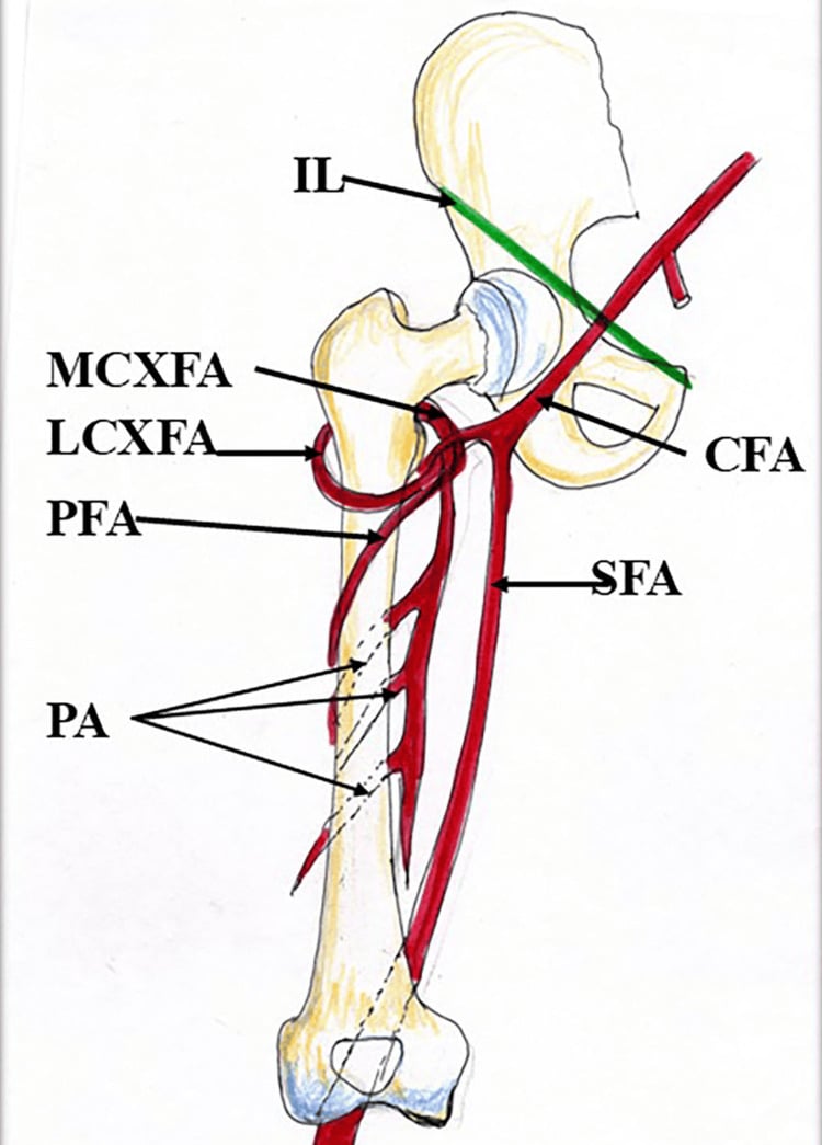
The sources. The femoral artery gives off the profunda femoris, and from the profunda come the two circumflex femoral arteries that supply the neck and head. The obturator artery, from the internal iliac, reaches the head through the ligamentum teres, and the inferior gluteal artery, also from the internal iliac, makes a minor contribution to the posterior capsule.
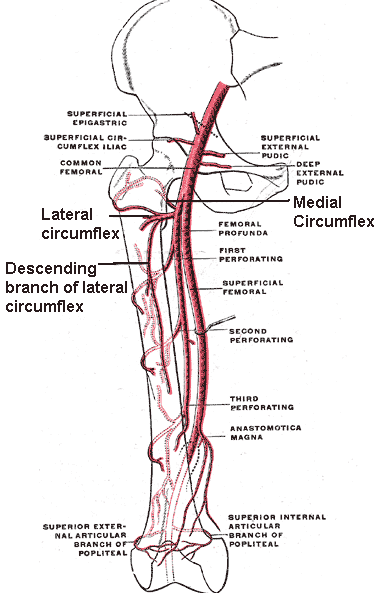
Medial femoral circumflex artery (MFCA). It usually arises from the profunda femoris, posteriorly, but can come directly from the common femoral. It winds medially around the femur, passing between pectineus and iliopsoas, then runs superior to adductor brevis and inferior to obturator externus, and the main trunk continues deep to quadratus femoris. Its terminal branch is the lateral epiphyseal artery, which reaches the head through the retinacular system below.
This is the critical vessel: 80% of the head's supply, covering the posterior, superior and lateral aspects of the head, which is the weight-bearing zone.
Lateral femoral circumflex artery (LFCA). It leaves the profunda femoris laterally and runs laterally deep to sartorius and rectus femoris. It has three branches:
- Ascending - anastomoses with the MFCA at the base of the neck
- Transverse - contributes to the cruciate anastomosis
- Descending - to the knee
Its share of the head is minor: anterior retinacular branches supply the anterior inferior aspect of the neck. Where the ascending branch meets the MFCA at the base of the neck the two form the extracapsular arterial ring of Crock.
F-L-I-MCruciate Anastomosis
Hook:The Anastomosis at the level of the Lesser Trochanter ('Film').
The ascending cervical (retinacular) arteries. These arise from the extracapsular ring, pierce the capsule at the base of the neck and run up the neck primarily outside the bone, under the synovial reflection, in the retinacula of Weitbrecht. They enter the bone at the articular cartilage margin, in the subcapital region, and become the intra-osseous supply. There are three groups:
- Posterior-superior - 2-4 vessels, branches of the MFCA; large and critical
- Posterior-inferior - branches of the MFCA
- Anterior - branches of the LFCA; smaller
Whatever the grouping, the anterior, inferior and medial contributions are small; the posterior-superior group is the main supply, and the one to protect or avoid.
Artery of the ligamentum teres. It comes from the posterior branch of the obturator artery (or from the medial circumflex) and travels within the ligament from the acetabular notch to the fovea; it is sometimes called the medial epiphyseal artery. In infants it is a vital supply and in children a variable one. In adults it is often patent but supplies only a small area around the fovea, which is insufficient to rescue the head if the neck vessels are cut.
Vascular Rings
Thinking of the supply as three rings in series, from the base of the neck to the head, makes the vulnerability at each level easy to follow.
1. The extracapsular ring sits at the base of the neck, formed by the anastomosis of the MFCA posteriorly and the LFCA anteriorly, so it draws on both medial and lateral sources. It is the base station for everything above it: if a basicervical fracture disrupts it, the supply fails.
2. The ascending (retinacular) system runs on the surface of the neck, inside the capsule, and is the vulnerable link. Subcapital fractures almost universally disrupt it, and intracapsular tamponade from a haemarthrosis can compress these vessels even when they are not torn. Preserving them is the key to head viability.
3. The subsynovial intra-articular ring lies just below the cartilage junction and is the point of entry into bone. From here on the vessels are end-arteries with no further collaterals.
Pathology: AVN
The sequence. Ischaemia kills osteocytes within 12-24 hours. The body then attempts to revascularise and remodel the necrotic bone by creeping substitution, laying new woven bone on the dead trabeculae. If resorption exceeds formation during that remodelling phase the weakened subchondral bone collapses, seen as the crescent sign, and the result is articular incongruity and secondary osteoarthritis.
The clock. The point of no return is often debated. Prompt reduction, variously put at 6 or 24 hours, is advocated for young patients; whether a hard cut-off is justified is taken up under Controversies.
Why the hip. Three features explain why:
- A retrograde supply with no collaterals
- An intracapsular space that can be pressurised
- High weight-bearing loads, which lead to collapse
The scaphoid, talus and humeral head share the retrograde arrangement.
VITTAVN Pathophysiology
Hook:Mechanisms of cell death.
Classification Relevance
Garden. The Garden classification of femoral neck fractures correlates directly with vascular integrity, and it is the predicted vascular survival that drives the fix-versus-replace decision:
- Grade I (valgus impacted) - trabeculae angulated, minimal displacement; vessels likely intact and the risk of AVN low (less than 15%)
- Grade II (complete, undisplaced) - the break is complete but anatomically reduced; vessels intact or kinked
- Grade III (complete, partially displaced) - vessels tethered or torn; the posterior retinaculum may still be intact
- Grade IV (complete, fully displaced) - the head is dissociated; vessels torn and the risk of AVN high
The caveat is that the Garden grade has only moderate interobserver reliability, and many surgeons collapse it to a binary undisplaced-versus-displaced decision in practice.
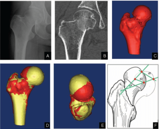
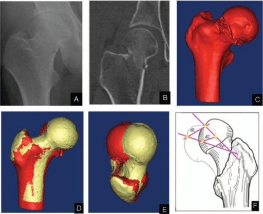
In children the AVN risk tracks the fracture level (and so the vessels divided), graded by the Delbet classification — the paediatric counterpart to Garden, with AVN risk falling as the fracture moves distally away from the retinacular entry point:
- Type I — transepiphyseal (through the physis, with or without dislocation of the epiphysis): highest AVN risk (up to roughly 80-100 percent when the epiphysis is dislocated), as the epiphyseal vessels are stripped.
- Type II — transcervical (mid-neck): the commonest type; high AVN risk (roughly 40-50 percent).
- Type III — cervicotrochanteric / basicervical (base of neck): lower AVN risk (roughly 20-30 percent).
- Type IV — intertrochanteric (extracapsular): lowest AVN risk (under about 10 percent), as the retinacular supply is largely spared.
The principle mirrors the adult: the more proximal the fracture, the more of the ascending retinacular supply is lost. Anatomic reduction, stable fixation and decompression of the capsular haematoma are advocated to limit AVN.
Clinical Assessment
The displaced neck fracture. A shortened and externally rotated leg means the fracture is displaced (Garden III or IV), and the rotation is the reason the posterior retinaculum is likely torn.
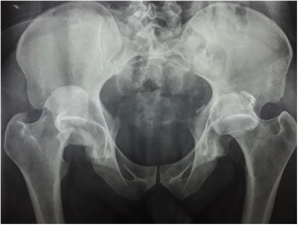
Posterior dislocation. A dashboard injury forces the head out of the acetabulum posteriorly, which tensions or tears the MFCA and the ligamentum teres. The hip is reduced immediately to restore flow, by relieving tamponade and kinking.
Investigations
Bone scan (technetium-99m). Uptake depends on blood flow and osteoblastic activity, so a cold spot in the early phase indicates an avascular head and a hot spot in the late phase indicates revascularisation and healing. It is useful for predicting head viability after a fracture but rarely changes acute management.
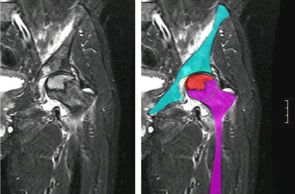
MRI. The gold standard for the diagnosis of AVN and the most sensitive modality, it can detect ischaemia within 24-48 hours. The signs are a serpentine line marking the demarcation between dead and living fatty marrow, the double-line sign on T2, and marrow oedema, which precedes collapse.
Differential Diagnosis of Femoral Head Marrow Signal / Osteonecrosis
When a young or middle-aged patient presents with hip pain and abnormal femoral head marrow on MRI, the following entities mimic or overlap with post-traumatic AVN. Distinguishing them changes management.
- Typical Patient / Trigger
- After displaced #NOF or dislocation
- Key Distinguishing Feature
- Vascular disruption of retinacular vessels; double-line sign on T2
- Course
- Risk of segmental collapse (crescent sign)
- Typical Patient / Trigger
- Steroids, alcohol, sickle cell, SLE, caisson disease
- Key Distinguishing Feature
- Often bilateral; serpentine demarcation line
- Course
- May collapse if untreated
- Typical Patient / Trigger
- Middle-aged men, pregnant women (3rd trimester)
- Key Distinguishing Feature
- Diffuse marrow oedema WITHOUT focal subchondral line
- Course
- Self-limiting, resolves in months
- Typical Patient / Trigger
- Elderly, osteoporotic, low bone mass
- Key Distinguishing Feature
- Low-signal subchondral line parallel to endplate, oedema
- Course
- Can mimic and progress to collapse
- Typical Patient / Trigger
- Fever, raised inflammatory markers, child or immunocompromised
- Key Distinguishing Feature
- Joint effusion, synovitis, restricted aspiration
- Course
- Surgical emergency
Management Strategy
The decision. Two questions separate the rows of the table: is this head worth preserving, and can this patient survive a failed fixation and a second operation?
- Injury
- Undisplaced / impacted
- Strategy
- Urgent fixation (cannulated screws) - the head is viable and worth keeping
- Injury
- Displaced
- Strategy
- Reduction and fixation to preserve the head - reduction QUALITY is the dominant modifiable predictor, not the clock
- Injury
- Undisplaced
- Strategy
- Fixation (screws or DHS) - the head is not the limiting factor
- Injury
- Displaced
- Strategy
- Arthroplasty (hemi or total) - accept the head is lost rather than risk a second operation
Read "young" and "older" physiologically, not off a birthday. A hard age line is the compressed version of this table and it is the wrong one: the cut-off is genuinely contested through the 50-65 band, where pre-injury mobility, cognition, bone quality and the patient's own priorities decide - see Controversies below.
Decompression and reduction. In young patients, releasing the capsule to let out the haematoma is thought to reduce intracapsular pressure and improve flow, though the evidence is mixed. Anatomic reduction is essential to un-kink the retinacular vessels, and that un-kinking is the primary rationale for urgent surgery.
Surgical Technique
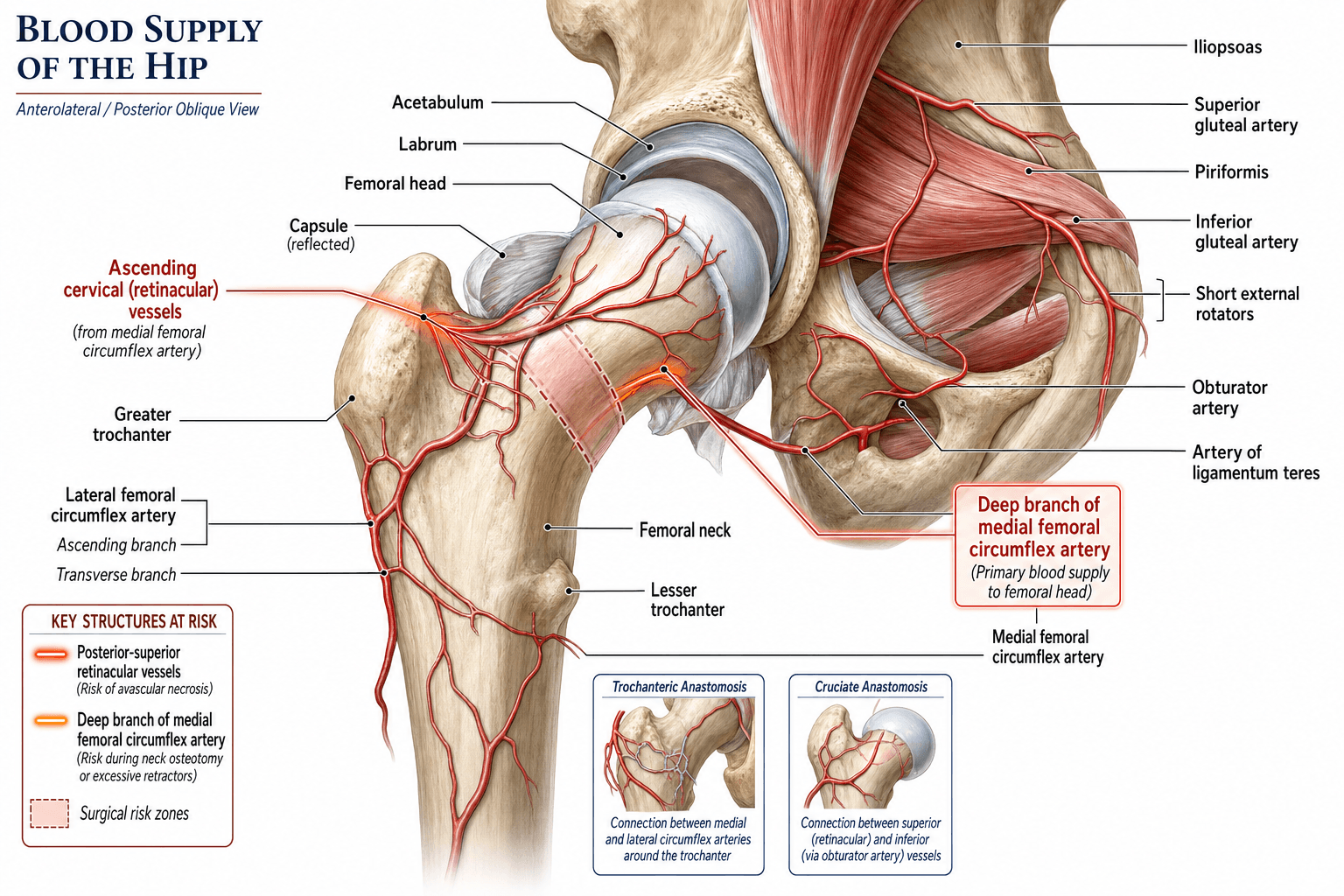
Cannulated screws. The goal is compression without rotation, and the configuration is an inverted triangle: one screw inferior, two superior. The inferior screw passes through the dense bone of the calcar. The posterior-superior screw is the risk zone: if its threads penetrate the posterior cortex they catch the lateral epiphyseal vessels where they run in the synovial reflection (Weitbrecht's retinacula) and tear them. Careful lateral viewing confirms that the threads are strictly intracapsular and intra-osseous.
Stay central or inferior-posterior where safe.
The posterior approach. The MFCA runs superior to quadratus femoris and deep to the piriformis tendon. Taking quadratus femoris down too proximally, or extending the piriformis release into the capsule, can damage the main trunk, and the result is complete AVN of the head.
Surgical dislocation (Ganz). Through a trochanteric flip (digastric slide) - see surgical hip dislocation (Ganz) - the head is dislocated anteriorly while the MFCA is kept intact posteriorly with the obturator externus and quadratus femoris muscle flap. Done correctly it gives a full view of the head and acetabulum without AVN risk, and it demonstrates that the MFCA is tethered to the external rotators.
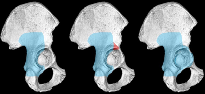
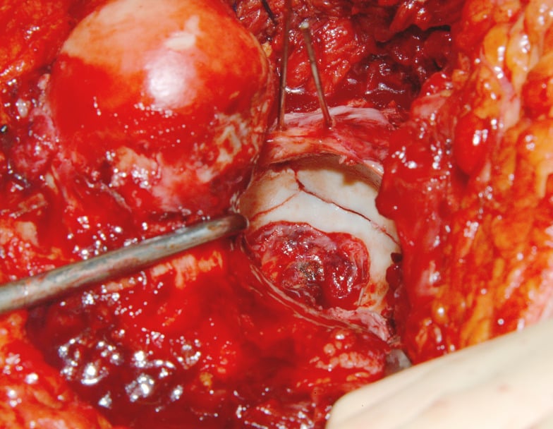
The MFCA dictates safety in the posterior and Ganz surgical-dislocation approaches (keep obturator externus intact). The direct anterior approach (DAA) uses the internervous Hueter interval — superficially between sartorius (femoral nerve) and tensor fasciae latae (superior gluteal nerve), deep between rectus femoris and gluteus medius — and has its own vascular landmark:
- The ascending branch of the lateral femoral circumflex artery (LFCA) crosses the distal part of the interval and is the classic "DAA bleeder"; it is identified and ligated or cauterised early to keep the field dry.
- Because the DAA works anteriorly, it does not disturb the posterosuperior retinacular vessels, so it is vascularly kind to the femoral head — a theoretical advantage, although any dislocation is still through the (less critical) anterior capsule.
- The lateral femoral cutaneous nerve runs near the interval and is the commonest complication (meralgia paraesthetica) — a neurological, not vascular, hazard, but worth pairing with the LFCA point.
Complications
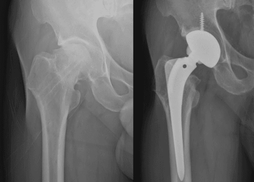
Avascular necrosis declares itself as late segmental collapse, which is painful and requires total hip replacement; the collapse can occur up to 2-3 years after the injury. Non-union follows when vascular failure prevents healing. Hardware failure, screws backing out or cutting out, is the consequence of poor bone stock or non-union.
Rehabilitation Protocol
After fixation the weight-bearing prescription is contested: touch or partial weight bearing to protect the reduction and the blood supply has its advocates, but most allow weight bearing as tolerated in stable constructs. After replacement the patient bears full weight immediately, because vascularity is no longer an issue.
Prognosis
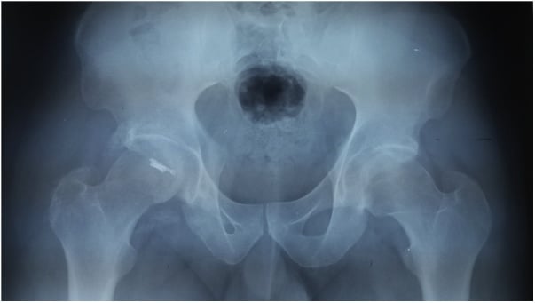
Displaced fractures in young adults carry a non-union rate of roughly 10-30% and an AVN rate of roughly 15-30% even with anatomic fixation; the Upadhyay RCT reported overall AVN of 16.3%. For undisplaced fractures, non-union is approximately 5% and AVN under 10%. In children the risk follows the Delbet level set out above.
Guidelines, Registries & Global Practice
Global Epidemiology
- Hip fractures are projected to reach 4.5-6 million annually worldwide by 2050, with the burden shifting toward Asia.
- Intracapsular (femoral neck) fractures account for roughly half of all proximal femoral fractures; in adults under 50 they are usually high-energy and carry a disproportionate AVN/non-union risk.
- AVN after displaced neck fracture in young adults is reported at roughly 15-30% even with optimal treatment; non-union at 10-30%.
Side-by-Side Guideline Comparison
- AAOS (US)
- Arthroplasty over internal fixation
- NICE / BOA-BOAST (UK)
- Arthroplasty (THR if mobile, independent, not cognitively impaired)
- AO Foundation
- Replacement when head viability is doubtful
- AAOS (US)
- Anatomic reduction + fixation to preserve head
- NICE / BOA-BOAST (UK)
- Reduction and fixation; minimise delay to theatre
- AO Foundation
- Urgent anatomic reduction; preserve retinacular vessels
- AAOS (US)
- Surgery within 24-48h reduces complications
- NICE / BOA-BOAST (UK)
- Surgery on day of or day after admission (governance target)
- AO Foundation
- Treat young displaced fractures urgently
Registry Evidence
- NJR (UK), AOANJRR (Australia), AJRR (US), SHAR (Sweden): Track arthroplasty done for failed fixation/AVN; total hip replacement generally outperforms hemiarthroplasty in younger, active patients for function and revision in displaced neck fractures.
- National Hip Fracture Databases (UK NHFD and similar): "time to theatre" is a quality metric, but the strongest modifiable driver of head salvage remains reduction quality, not the clock alone (see Upadhyay).
High- vs Limited-Resource Practice Variation
- High-resource: MRI for early AVN, dedicated trauma lists, choice of THR vs fixation, and surgical-dislocation expertise for select head/neck pathology.
- Limited-resource: Cannulated screws or DHS predominate because of cost and implant availability; muscle-pedicle bone grafting (e.g. quadratus femoris pedicle) is still used in some centres to biologically augment fixation in young patients.
Paediatric Note
- The artery of the ligamentum teres is important in infants; septic arthritis of the hip in this group causes a pressure tamponade that can infarct the epiphysis and is a surgical emergency.
Controversies & Areas of Uncertainty
Does timing really matter? The "hip attack" / 6-hour dogma is challenged by the Upadhyay RCT, which found delay beyond 48 hours did not increase AVN or non-union in young adults. Reduction quality and posterior comminution were the dominant predictors. Most still operate urgently, but the evidence for a hard time cut-off is weak.
Capsulotomy, aspiration or nothing. Intracapsular tamponade is real, but whether decompressing the haematoma improves head survival is unproven. It is biologically plausible and low-risk, so many surgeons do it for young patients despite the absence of high-level evidence.
Implant choice and vascular insult. FAITH showed higher AVN with the sliding hip screw than with parallel cancellous screws, raising the question of whether reaming and a single large central device add vascular or thermal insult to an already compromised head.
THR versus fixation in the 50-65 group. The "young" cutoff is contested. Below this band, head preservation is favoured; above it, registries and trials increasingly favour arthroplasty (often THR) for displaced fractures. The middle band remains an individualised decision.
MCQ Practice Points
Q: Which artery provides the majority of blood supply to the adult femoral head? A: Medial Femoral Circumflex Artery (Deep Branch).
Q: Where are the retinacular vessels located relative to the capsule? A: Intracapsular but Extrasynovial. (They run on the neck under the synovial lining).
Q: At what age is the artery of the ligamentum teres most significant? A: Infancy (under 4 years).
Q: The Cruciate Anastomosis involves which vessels? A: First Perforator, Medial Circumflex, Lateral Circumflex, Inferior Gluteal. (Not Superior Gluteal).
Q: Which movement is most likely to tear the MFCA? A: Posterior Dislocation.
Q: The Artery of the Ligamentum Teres is a branch of which vessel? A: Obturator Artery. (Posterior branch).
Viva Scenarios
Practise clinical reasoning and management decisions out loud
“Describe the precise course of the Medial Femoral Circumflex Artery.”
“You are fixing a femoral neck fracture with cannulated screws. Why do you avoid the posterior superior quadrant for screw placement?”
“A 30-year-old cyclist falls and sustains a displaced intracapsular femoral neck fracture. It is 10 PM. What is your plan?”
“A 38-year-old on long-term steroids for lupus has bilateral groin pain. MRI shows a serpentine subchondral line in both femoral heads. How does the vascular mechanism here differ from a fractured hip, and how does that change management?”
Anatomy
- Main: MFCA (Deep Branch)
- Minor: LFCA (Anterior)
- Retinacula: Posterior-Superior dominant
- Lig Teres: Negligible in adults
Vascular Rings
- 1. Extracapsular (Base of Neck)
- 2. Retinacular (Ascending on Neck)
- 3. Subsynovial (Head Entry Point)
- Ring of Crock = MFCA + LFCA Anastomosis
Clinical
- Fracture: Garden III/IV = AVN Risk
- Approach: Preserve Quadratus Femoris
- Emergent: Young Displaced #NOF
- Screw Hazard: Post-Sup Quadrant
Evidence Base
Normal Vascular Anatomy of the Femoral Head
- Classic injection-radiograph study of the adult femoral head vasculature
- Described the lateral epiphyseal vessels (from the MFCA) as supplying the bulk of the weight-bearing superolateral head
- Showed the foveal (ligamentum teres) artery supplies only a small medial zone in the adult
Distribution and Anastomoses of Arteries Supplying the Head and Neck of the Femur
- Angiographic and dissection study mapping the extracapsular ring and ascending cervical (retinacular) vessels
- Confirmed the posterosuperior retinacular group as the principal source to the epiphysis
- Defined the anastomotic redundancy that fails after subcapital fracture
Anatomy of the Medial Femoral Circumflex Artery and its Surgical Implications
- Dissection of 24 cadaver hips with latex injection
- The deep branch of the MFCA crosses posterior to obturator externus and anterior to the conjoint tendon, perforating the capsule at gemellus superior
- Obturator externus protected the deep branch from rupture during dislocation in any direction after complete capsulotomy
Surgical Dislocation of the Adult Hip Without the Risk of AVN
- 213 hips over 7 years using a trochanteric-flip osteotomy with anterior dislocation
- External rotators left intact so the MFCA is protected by obturator externus
- No case developed avascular necrosis on follow-up
Changing Patterns of Proximal Femoral Vascularity
- Described age-related evolution of proximal femoral blood supply from infancy to skeletal maturity
- The physis acts as a barrier isolating the epiphysis from metaphyseal vessels until closure
- Ligamentum teres contribution is significant in infancy and involutes with growth
Delayed Internal Fixation of Femoral Neck Fractures in Young Adults (RCT)
- 102 patients aged 15-50 with displaced (Garden III/IV) fractures randomised to closed vs open reduction
- No difference in union or AVN between closed and open reduction; overall AVN was 16.3%
- Delay of more than 48 hours did not increase union failure or AVN; posterior comminution, poor reduction and malplaced screws drove non-union
FAITH: Sliding Hip Screw vs Cancellous Screws (RCT)
- 1108 patients aged 50+ randomised to sliding hip screw vs cancellous screws
- No overall difference in 24-month reoperation (20% vs 22%)
- Avascular necrosis was higher with the sliding hip screw (9% vs 5%; HR 1.91); smokers and displaced/base-of-neck fractures may favour the SHS