Garden Classification vs Pauwels: Femoral Neck Fractures and Why It Changes the Operation
The Garden classification and Pauwels angle for clinicians: the stages, how reliable they are, and how each one steers fixation or arthroplasty of the hip.
By OrthoVellum Editorial TeamPublished 12 min read
Educational content for clinicians, not medical advice. Editorial policy

Key points
- Garden grades displacement on the AP radiograph (stages I–IV), but the four stages are poorly reproducible; grouping I and II together as undisplaced, and III–IV as displaced, is what observers reliably agree on.
- Pauwels grades the angle of the fracture line to the horizontal (under 30°, 30–50°, over 50°), and so the shear the fixation has to resist. Its interobserver agreement is poor.
- Undisplaced fractures are fixed at any age. Displaced fractures are replaced in older patients and reduced and fixed in the physiologically young (under about 60–65).
- Read the lateral film. In a FAITH secondary analysis, posterior tilt of 20° or more nearly doubled later arthroplasty after fixation of Garden I–II fractures (22.4% vs 11.9%).
- A vertical fracture is the biomechanical case for a fixed-angle construct, but FAITH found no overall reoperation advantage for the sliding hip screw, and more avascular necrosis with it.
On this page9 sections
Every intracapsular hip fracture gets a Garden stage on the trauma board, and most get a Pauwels grade in the viva. The useful question is what either label changes in theatre. The short answer: the Garden classification is used in practice as a two-way split, undisplaced (I–II) or displaced (III–IV), and that split, together with the patient's age and physiology, decides between fixation and arthroplasty. Pauwels describes the shear on the fracture line and shapes the construct once you have decided to fix. This post works through both systems, how far they can be trusted, and the decision they feed. The full clinical picture is on the neck of femur fracture topic page.
What are the four Garden stages?
Garden published the system in 1961, in a paper about low-angle fixation of the femoral neck. It grades the fracture on the AP radiograph by the degree of displacement and the alignment of the trabecular lines, and it correlates with the risk of avascular necrosis because displacement is what tears the retinacular vessels.
| Stage | The fracture | Trabecular lines on the AP | Practical group |
|---|---|---|---|
| I | Incomplete; head tilted into valgus (impacted) | Continuous across the fracture | Undisplaced |
| II | Complete; no displacement; head in neutral | Head lines align with the acetabulum and the neck | Undisplaced |
| III | Complete; partial displacement; head rotated but still in some contact with the neck | Head lines out of line with the neck | Displaced |
| IV | Complete; total displacement; head dissociated from the neck | Head lines align with the acetabulum but not with the neck | Displaced |
Reading stage IV. Once the head is completely free of the neck it sits in the acetabulum, so its trabeculae line up with the acetabulum's; it is the neck and shaft that have moved. In stage III the head is still in some contact with the neck and has been rotated, so its trabeculae are out of line with the neck.
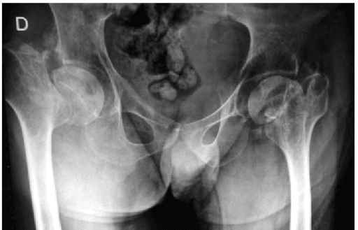
The reason the stage matters is vascular. The main supply to the femoral head comes from the lateral epiphyseal vessels, branches of the medial femoral circumflex artery that run along the posterosuperior neck inside the capsule (see blood supply of the hip). Displacement disrupts them, and there is no periosteal sleeve at this level to help the fracture heal. That combination is why a displaced fracture in an older patient is replaced rather than fixed.
Why is Garden collapsed to undisplaced versus displaced?
Because the four-stage system is not reproducible enough to carry four different decisions, while the two-group version is.
- Thomsen (Int Orthop 1996): six observers classified 96 fractures and all six agreed on only 14 (15%), kappa 0.39. Reduced to undisplaced versus displaced, agreement rose to kappa 0.68. Telling stage II from stage III remained the problem.
- Cazzato (BMC Musculoskelet Disord 2022): six surgeons, 150 fractures, AP and lateral films. Garden interobserver kappa ranged 0.28–0.73 (mean 0.49); the 2018 AO/OTA system was worse (mean 0.30). More trauma experience did not appear to improve agreement, and no system was shown to be superior.
- Cai (BMC Surg 2022): eight observers, 886 older patients, radiographs and CT. Four-stage agreement on radiographs was kappa 0.18–0.43; a three-group version (I+II, III, IV) reached 0.76–0.90.
The weak boundary, II against III, is exactly where the operation changes in an older patient. When a fracture sits on it, the useful response is to look harder at both views, not to argue over the number. The binary split is not a simplification for exam convenience; it is the part of the classification the evidence supports.
Does Garden I really exist?
Garden I is taught as an incomplete fracture, and it is still examined that way. Two imaging studies challenge it.
Cai and colleagues identified 52 patients whom the eight observers jointly classified as Garden I. Of these, 38 had an arthroplasty, and every one of them had a complete fracture at operation. The authors concluded that in older adults all undisplaced fractures may be Garden II, with no true Garden I.
Fu and colleagues reconstructed 120 consecutive undisplaced fractures (60 Garden I, 60 Garden II) in three dimensions from CT and found that many called incomplete on the AP were not. Impacted Garden I fractures had a mean femoral head centre displacement of 6.22 mm and rotation of 17.8°, no different from Garden II (7.16 mm, 18.8°). Only the genuinely incomplete fractures differed, with a mean rotation of 4.9°.
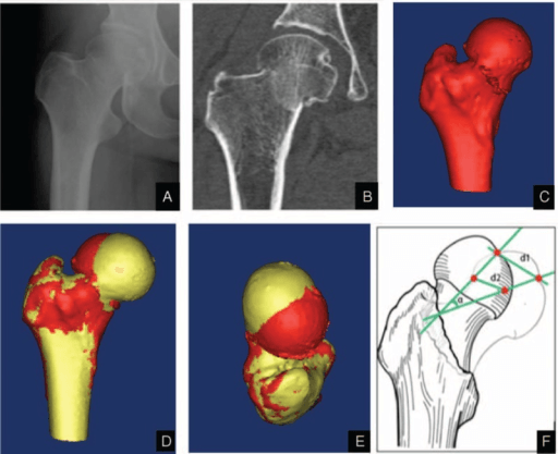
The error runs towards under-treatment. Treat an apparent Garden I in an older patient as a fracture that may be complete and rotated, and do not let the word "incomplete" argue you out of fixation. An impacted fracture left alone can displace.
What does Garden miss on the lateral film?
Garden is an AP classification. Two things that predict failure are seen only on the lateral.
Posterior comminution predicts loss of reduction even when the AP looks acceptable. Posterior tilt of the head predicts failure of fixation in fractures that are undisplaced on the AP.
Okike and colleagues tested tilt in a preplanned secondary analysis of the FAITH trial: 555 patients with Garden I–II fractures and an adequate pre-operative lateral. Tilt of 20° or more (67 patients, 12.1%) was associated with subsequent arthroplasty within 24 months in 22.4%, against 11.9% with less tilt (hazard ratio 2.22, 95% CI 1.24–4.00). Age 80 or over was the other independent factor. The authors' wording is that primary arthroplasty may be considered for undisplaced fractures with tilt of 20° or more, especially in older patients. It is a prognostic level IV analysis, not a randomised comparison of fixation against arthroplasty.
What is the Pauwels angle?
Pauwels (1935) classified femoral neck fractures by the angle of the fracture line to the horizontal on the AP radiograph. The steeper the line, the more body weight acts along the fracture as shear rather than across it as compression.
| Type | Angle to the horizontal | Forces across the fracture | Stability |
|---|---|---|---|
| I | Under 30° | Mostly compressive | Relatively stable |
| II | 30–50° | Mixed compression and shear | Moderate |
| III | Over 50° (vertical) | Predominantly shear | Very unstable |
Two cautions travel with the table. Other thresholds circulate in textbooks; Bartoníček went back to the original and gave the correct values as up to 30°, 30–50° and 50° or more. And the failure rates often attached to the types (under 5%, 10–15% and 30–40% with standard screws) are conventional teaching rather than figures from Pauwels's own work.
Measuring it. Draw a horizontal reference along the superior margin of the sacral ala or across the ischial tuberosities, then a line along the fracture plane. Measure on the AP; the lateral shows sagittal displacement, not the Pauwels angle. CT shows a vertical fracture line better than plain films and is worth having when you are planning fixation in a young patient.
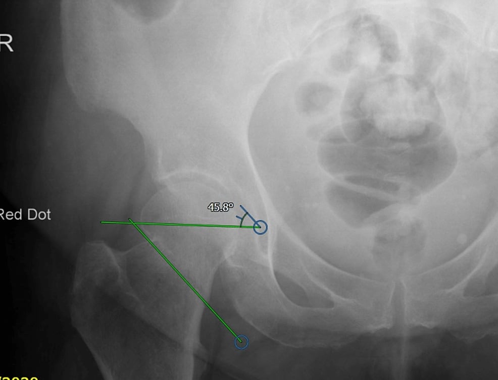
Reliability is worse than Garden's. Interobserver kappa for the three Pauwels types is only about 0.18–0.38 across studies, and Turgut and colleagues found both Garden and AO more reliable than Pauwels, with surgical experience making no significant difference. Small amounts of pelvic or hip rotation move the measured angle. The shear biomechanics are sound; the II-versus-III boundary is not. In a viva, quote the angle you measured, call the fracture vertical and high-shear, and justify the construct from the mechanics rather than defending a grade the literature cannot agree on.
Garden vs Pauwels: what each one tells you
The two systems answer different questions, and one does not stand in for the other: an undisplaced fracture can still have a steep fracture line.
| Garden | Pauwels | |
|---|---|---|
| Measures | Displacement and trabecular alignment | Fracture-line angle to the horizontal |
| Predicts | Head blood supply, AVN and nonunion risk | Shear across the fracture, fixation failure |
| Decision it informs | Fix or replace | Which fixation construct |
| Reproducibility | Four stages fair to moderate; two groups acceptable | Poor (kappa about 0.18–0.38) |
Do not confuse either with the Garden alignment index, which judges the reduction, not the fracture. On the AP the compression trabeculae of the head should meet the medial femoral cortex at about 160°; on the lateral they should be roughly colinear, about 180°. An acceptable reduction sits between 155° and 180° on both views.
How does the classification change the operation?
Displacement and physiology decide it. An undisplaced fracture is fixed at any age; a displaced fracture in an older patient is replaced; a displaced fracture in a physiologically young patient is reduced and fixed to keep the native head.
| Fracture and patient | Usual operation | Why |
|---|---|---|
| Undisplaced (Garden I–II), any age | Three cannulated screws in an inverted triangle, or a sliding hip screw | Retinacular supply often intact; fixation lets the fracture heal |
| Undisplaced, posterior tilt 20° or more, older patient | Consider primary arthroplasty | Later arthroplasty 22.4% vs 11.9% in the FAITH tilt analysis |
| Displaced (Garden III–IV), older patient | Hemiarthroplasty or total hip replacement | AVN and nonunion up to 30–50% after fixation |
| Displaced, physiologically young (under about 60–65) | Urgent anatomic reduction and fixation, ideally within 6 hours | Preserve the head; a 30% AVN risk is preferable to a THR at 40–50 |
| Vertical (Pauwels III), young | Fixed-angle construct: sliding hip screw with a derotation screw, or a fixed-angle device | Parallel screws give no resistance to shear |
| Basicervical (AO/OTA 31-B3) | Sliding hip screw | Extracapsular; lower AVN risk but higher risk of fixation failure |
Choosing the arthroplasty. A cemented total hip replacement is traditionally chosen for the patient who walks outdoors independently, is cognitively intact and is expected to live more than about 4 years, or who has pre-existing arthritis. NICE's 2023 wording is broader and more hedged: consider total hip replacement (opens in a new tab) rather than hemiarthroplasty for people who walked independently out of doors with no more than a stick, have no condition that makes the procedure unsuitable, and are expected to stay independent in daily activities beyond 2 years. A hemiarthroplasty suits the frail patient with limited mobility or cognitive impairment. Hold the functional argument against HEALTH: in 1,495 independently ambulating patients aged 50 or over with a displaced fracture, secondary procedures at 24 months were 7.9% after THA and 8.3% after hemiarthroplasty, dislocation was 4.7% against 2.4%, and the authors called the functional gain from THA clinically unimportant. The technique is set out in the cemented hemiarthroplasty operative guide and the patient selection in total hip arthroplasty indications.
The Pauwels III instruction, held loosely. "Vertical fracture, use a DHS" is a biomechanical argument. The clinical counterweight is FAITH: 1,108 patients aged 50 or over with low-energy fractures were randomised to a sliding hip screw or cancellous screws. Reoperation at 24 months was 20% against 22% (HR 0.83, 95% CI 0.63–1.09), and avascular necrosis was more common with the sliding hip screw (9% vs 5%, HR 1.91). The suggestion that smokers and displaced or base-of-neck fractures might do better with a sliding hip screw comes from subgroups of a trial with a null primary result. Use the mechanics to choose the construct, as in the DHS fixation operative guide, and do not promise that it changes the reoperation rate.
Age means physiological age. The cut-off of about 60–65 describes the physiologically young patient, not a date of birth. The reason to keep the native head is what the alternative costs: a total hip replacement at 40 or 50 means uncemented implants, wear-resistant bearings and the expectation of several revisions over a lifetime.
What happens when fixation fails?
Both classifications reappear here. Nonunion follows 10–30% of femoral neck fractures and is more common after displaced (Garden III–IV) and vertical (Pauwels III) fractures, poor reduction and posterior comminution. In patients aged 60 or under, a meta-analysis of 1,558 fractures from 41 studies found a pooled reoperation rate of 18.0% after fixation of isolated neck fractures, with overall pooled rates of 14.3% avascular necrosis and 9.3% nonunion.
The salvage decision turns on age and whether the head is alive. In a young patient with a viable head, a valgus intertrochanteric osteotomy tilts the head so that the vertical fracture line becomes more horizontal and is loaded in compression: Pauwels's principle used in reverse. A dead or collapsing head, an older patient or a failed osteotomy means arthroplasty. The detail is on the femoral neck nonunion and avascular necrosis of the hip pages, and the bone-health work that should follow any fragility fracture is covered in our post on secondary fracture prevention.
In the exam
Examiners want the classification named, its weakness acknowledged and a decision justified from displacement, physiology and mechanics. For the memory side of the problem, see how to memorise orthopaedic classifications.
Frequently asked questions
What are the four Garden stages of a femoral neck fracture?
Garden I is an incomplete, valgus-impacted fracture with the trabeculae continuous. Garden II is complete but undisplaced. Garden III is complete and partially displaced, with the head trabeculae out of line with the neck. Garden IV is completely displaced: the head is dissociated from the neck and its trabeculae line up with the acetabulum. In practice I–II are grouped as undisplaced and III–IV as displaced.
What is the difference between the Garden and Pauwels classifications?
Garden describes displacement, which predicts the state of the femoral head blood supply and decides between fixation and arthroplasty. Pauwels describes the angle of the fracture line to the horizontal, which predicts the shear force across the fracture and influences which fixation construct to use. They measure different things, so one does not predict the other: an undisplaced fracture can still have a steep fracture line.
Is the Garden classification reliable?
Only partly. With all four stages, six observers in one study agreed on just 15% of fractures (kappa 0.39), and a 2022 study found mean interobserver kappa 0.49. When the system is reduced to undisplaced versus displaced, agreement improves to kappa 0.68, and in an 886-patient radiograph and CT study a three-group version reached 0.76–0.90. Stage II versus III is the weak boundary.
Should every undisplaced femoral neck fracture be fixed?
Fixation is the standard for Garden I–II fractures at any age, but not every undisplaced fracture does well with it. In a secondary analysis of the FAITH trial, posterior tilt of 20° or more on the lateral radiograph was associated with later arthroplasty in 22.4% of patients against 11.9% with less tilt, and age 80 or over was also a risk factor. The authors suggest primary arthroplasty may be considered in that group.
How is the Pauwels angle measured?
On the AP radiograph, draw a horizontal reference line, for example along the superior margin of the sacral ala or across the ischial tuberosities, then a line along the fracture plane. The angle between them is the Pauwels angle. Under 30° is type I, 30–50° type II, and over 50° type III. Small amounts of hip or pelvic rotation change the measured angle, which is part of why the grading is poorly reproducible.
References
- Garden RS. Low-angle fixation in fractures of the femoral neck. J Bone Joint Surg Br 1961;43-B(4):647-663. DOI (opens in a new tab)
- Bartoníček J. Pauwels' classification of femoral neck fractures: correct interpretation of the original. J Orthop Trauma 2001;15(5):358-360. DOI (opens in a new tab)
- Thomsen NO, Jensen CM, Skovgaard N, et al. Observer variation in the radiographic classification of fractures of the neck of the femur using Garden's system. Int Orthop 1996;20(5):326-329. DOI (opens in a new tab)
- Cazzato G, Oliva MS, Masci G, et al. Femoral neck fracture: the reliability of radiologic classifications. BMC Musculoskelet Disord 2022;22(Suppl 2):1063. DOI (opens in a new tab)
- Cai Z, Zhang Z, Ren L, et al. Does Garden type I incomplete femoral neck fracture really exist in older adults? BMC Surg 2022;22(1):276. DOI (opens in a new tab)
- Fu X, Xu GJ, Li ZJ, et al. Three-dimensional reconstruction modeling of the spatial displacement, extent and rotational orientation of undisplaced femoral neck fractures. Medicine (Baltimore) 2015;94(39):e1393. DOI (opens in a new tab)
- Turgut A, Kumbaracı M, Kalenderer Ö, et al. Is surgeons' experience important on intra- and inter-observer reliability of classifications used for adult femoral neck fracture? Acta Orthop Traumatol Turc 2016;50(6):601-605. DOI (opens in a new tab)
- Okike K, Udogwu UN, Isaac M, et al. Not all Garden-I and II femoral neck fractures in the elderly should be fixed: effect of posterior tilt on rates of subsequent arthroplasty. J Bone Joint Surg Am 2019;101(20):1852-1859. DOI (opens in a new tab)
- FAITH Investigators. Fracture fixation in the operative management of hip fractures (FAITH): an international, multicentre, randomised controlled trial. Lancet 2017;389(10078):1519-1527. DOI (opens in a new tab)
- HEALTH Investigators. Total hip arthroplasty or hemiarthroplasty for hip fracture. N Engl J Med 2019;381(23):2199-2208. DOI (opens in a new tab)
- Slobogean GP, Sprague SA, Scott T, Bhandari M. Complications following young femoral neck fractures. Injury 2015;46(3):484-491. DOI (opens in a new tab)
Study the full topic
Included in Pro
Go deeper with Pro
- Operative guide: cemented hemiarthroplasty for femoral neck fracture
- Operative guide: DHS fixation of the femoral neck
- Operative guide: cannulated screws and DHS for neck of femur fractures
Share this article
Cite this article
OrthoVellum Editorial Team. Garden Classification vs Pauwels: Femoral Neck Fractures and Why It Changes the Operation [Internet]. OrthoVellum; 2026 Oct 7 [cited 2026 Oct 7]. Available from: https://www.orthovellum.com/blog/garden-classification-femoral-neck-fractures
About this content
Prepared by the OrthoVellum Editorial Team from cited sources, under our editorial policy.
For education and exam preparation; not medical advice or a substitute for clinical judgement and local guidance.
Spotted an error? Tell us; every report is read.
Get new articles by email
New clinical explainers, landmark-trial summaries and exam guidance from the OrthoVellum blog. No account needed; unsubscribe from any email.