OrthoVellum
Knowledge Hub

Study

  • Topics
  • MCQs
  • ISAWE
  • Operative Surgery
  • Flashcards

Company

  • About Us
  • Editorial Policy
  • Contact
  • FAQ
  • Blog

Legal

  • Terms of Service
  • Privacy Policy
  • Cookie Policy
  • Medical Disclaimer
  • Copyright & DMCA
  • Refund Policy

Support

  • Help Center
  • Accessibility
  • Report an Issue
OrthoVellum

© 2026 OrthoVellum. For educational purposes only.

Not affiliated with the Royal Australasian College of Surgeons.

Neck of Femur Fracture

Back to Topics
Contents
0%

Neck of Femur Fracture

Comprehensive guide to femoral neck fractures and Garden classification for FRCS exam preparation

complete
Updated: 2026-01-19
High Yield Overview

NECK OF FEMUR FRACTURE

Hip Fracture Emergency | Garden Classification | Fix vs Replace

GardenClassification
Less than 48hSurgery timing
AVNRisk with displacement
30%1-year mortality

Garden Classification

Garden I
PatternIncomplete/impacted valgus
TreatmentScrew fixation
Garden II
PatternComplete, non-displaced
TreatmentScrew fixation
Garden III
PatternComplete, partial displacement
TreatmentArthroplasty (elderly)
Garden IV
PatternComplete, full displacement
TreatmentArthroplasty (elderly)

Critical Must-Knows

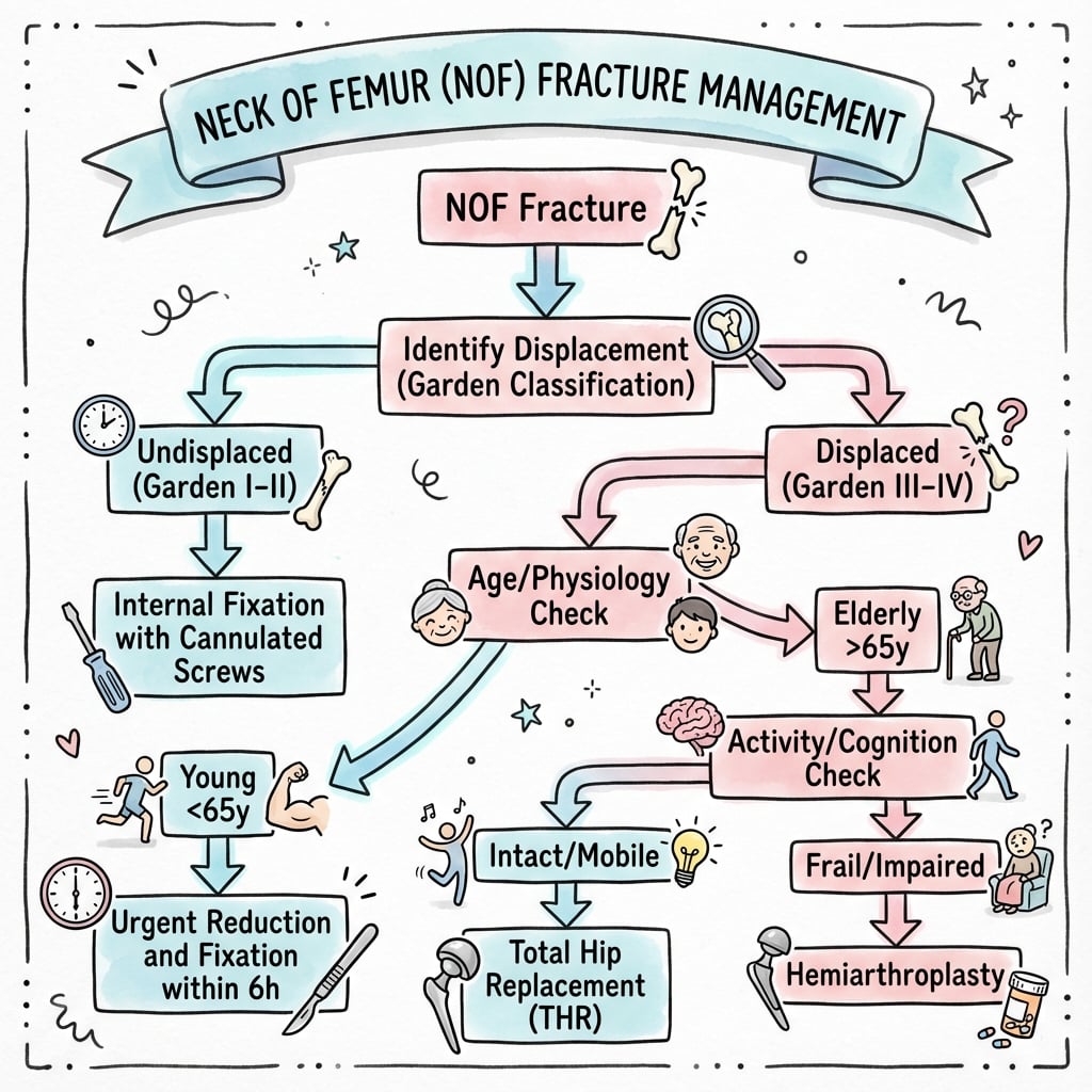
  • Garden I/II = undisplaced (fix). Garden III/IV = displaced (replace in elderly)
  • Intracapsular location = AVN risk (blood supply)
  • Surgery within 48 hours improves outcomes
  • Hemiarthroplasty vs THR: Depends on cognitive status, mobility, age
  • Young patients with displaced fractures: attempt reduction and fixation

Examiner's Pearls

  • "
    Blood supply: lateral epiphyseal vessels from MFCA
  • "
    Displaced fractures in elderly: arthroplasty (hemi or THR)
  • "
    Garden I may progress to displaced if not fixed
  • "
    Cannulated screws in inverted triangle configuration

Clinical Imaging

Imaging Gallery

5-panel (a-e) AP hip X-rays demonstrating different malunion patterns after parallel screw fixation: coxa breva, coxa valga, rotational malunion, translational malunion, coxa vara.
Click to expand
5-panel (a-e) AP hip X-rays demonstrating different malunion patterns after parallel screw fixation: coxa breva, coxa valga, rotational malunion, tranCredit: Satish BR et al. - Indian J Orthop via Open-i (NIH) - PMC3654468 (CC-BY 4.0)
AP pelvis radiograph showing bilateral Garden IV (completely displaced) femoral neck fractures with superior femoral head migration.
Click to expand
AP pelvis radiograph showing bilateral Garden IV (completely displaced) femoral neck fractures with superior femoral head migration.Credit: Mariani P et al. - Case Rep Orthop via Open-i (NIH) - PMC4258342 (CC-BY 4.0)
2-panel (a-b) hip radiographs with measurement annotations for preoperative planning: AP and lateral views showing calcar, canal, and shaft diameter measurements.
Click to expand
2-panel (a-b) hip radiographs with measurement annotations for preoperative planning: AP and lateral views showing calcar, canal, and shaft diameter mCredit: Yeo AK et al. - Patient Saf Surg via Open-i (NIH) - PMC4658799 (CC-BY 4.0)
AP pelvis X-ray showing neck of femur fracture on the right side
Click to expand
Anteroposterior radiograph of pelvis demonstrating a displaced right neck of femur fracture in an 80-year-old female following a fall. This is a common presentation requiring urgent surgical intervention within 36-48 hours.Credit: OrthoVellum
AP pelvis showing bilateral Garden IV femoral neck fractures
Click to expand
AP pelvis radiograph showing BILATERAL completely displaced (Garden IV) femoral neck fractures with superior migration of both femoral heads. This rare presentation demonstrates the classic appearance of Garden IV fractures - complete loss of trabecular continuity with the femoral heads displaced from the acetabula. Bilateral fractures may occur in patients with severe osteoporosis, seizure disorders, or high-energy trauma.Credit: Mariani P et al. - Case Rep Orthop (CC-BY 4.0)
2-panel preoperative hip measurements for surgical planning
Click to expand
2-panel (a-b) preoperative radiographic measurements for femoral neck fracture surgical planning: (a) AP hip view with calcar diameter, canal diameter, and shaft diameter measurements at 10cm below the lesser trochanter. (b) Lateral hip view with corresponding canal and shaft diameter measurements. These measurements guide implant selection for internal fixation or arthroplasty - canal diameter determines stem size, and calcar diameter indicates bone quality for fixation.Credit: Yeo AK et al. - Patient Saf Surg (CC-BY 4.0)

Critical Neck of Femur Points

Garden Classification

I/II = Undisplaced - screw fixation. III/IV = Displaced - arthroplasty in elderly (AVN risk too high). Simpler to remember: undisplaced = fix, displaced = replace.

Blood Supply

Lateral epiphyseal vessels from medial femoral circumflex artery (MFCA) are main supply to femoral head. Displaced fractures disrupt these = AVN.

Time to Surgery

Surgery within 36-48 hours reduces mortality, pressure sores, complications. Delay increases mortality. This is a surgical emergency.

THR vs Hemi

THR: Cognitively intact, independently mobile, expected survival greater than 4 years. Hemiarthroplasty: Cognitive impairment, limited mobility, frail elderly. THR has better function but higher dislocation risk.

At a Glance

Neck of femur fractures are orthopaedic emergencies requiring surgery within 36-48 hours to reduce mortality and complications. The Garden classification guides treatment: Garden I-II (undisplaced) undergo cannulated screw fixation in inverted triangle configuration, while Garden III-IV (displaced) in elderly patients require arthroplasty. Blood supply via the medial femoral circumflex artery (MFCA) is disrupted in displaced fractures, leading to high AVN risk. THA is indicated for cognitively intact, independently mobile patients with expected survival greater than 4 years; hemiarthroplasty for frail, cognitively impaired, or limited mobility patients. Young patients (under 60 years) with displaced fractures receive urgent reduction and internal fixation to preserve the femoral head. 1-year mortality is approximately 30% in elderly populations.

Mnemonic

FIX-REPLACEGarden Simplified

I
Impacted/Incomplete
SCREWS
II
Complete non-displaced
SCREWS
III
Displaced partial
REPLACE
IV
Displaced complete
REPLACE

Memory Hook:I/II = FIX with screws. III/IV = REPLACE (arthroplasty)!

Mnemonic

MICTHR Indications

M
Mobile
Independently mobile - walks outdoors
I
Intact cognition
Cognitively intact - can follow precautions
C
Continued survival
Life expectancy greater than 4 years

Memory Hook:MIC check before THR - Mobile, Intact, Continued survival!

Mnemonic

MFCA-LEVNOF Blood Supply

M
Medial Femoral
Main artery - branch of profunda femoris
C
Circumflex Artery
Wraps around femoral neck posteriorly
L
Lateral
Lateral epiphyseal vessels arise from MFCA
E
Epiphyseal
Main supply to femoral head
V
Vessels
Disrupted in displaced fractures - causes AVN

Memory Hook:MFCA gives LEV - disrupt it and head dies!

Overview and Anatomy

Neck of femur (NOF) fractures are common in elderly with osteoporosis. They are a major cause of morbidity and mortality.

Anatomy

NOF fractures are intracapsular - within the hip joint capsule. This has two implications:

  1. Blood supply at risk: Lateral epiphyseal vessels (from MFCA) run along the neck and are disrupted by displacement → AVN.
  2. No periosteal healing: Intracapsular fractures rely on endosteal healing.

Blood Supply

Medial femoral circumflex artery (MFCA) gives rise to lateral epiphyseal vessels which are the main supply to the femoral head. Displaced fractures disrupt these.

Classification of hip fractures showing femoral neck, trochanteric, and subtrochanteric regions
Click to expand
Anatomical classification of hip fractures. Femoral neck fractures (intracapsular) include subcapital, transcervical, and basicervical patterns. Trochanteric (intertrochanteric) and subtrochanteric fractures are extracapsular with different treatment implications.Credit: OrthoVellum

Pathophysiology

Mechanism of Injury

Neck of femur fractures occur through two main mechanisms:

  1. Low-energy falls in elderly osteoporotic patients (most common)
  2. High-energy trauma in younger patients (road traffic accidents, falls from height)

Blood Supply Disruption

The femoral head receives blood supply primarily from the medial femoral circumflex artery (MFCA), a branch of the profunda femoris. The MFCA gives rise to the lateral epiphyseal vessels which run along the posterior-superior aspect of the femoral neck within the capsule.

In displaced fractures, these vessels are disrupted, leading to:

  • Avascular necrosis (AVN): 20-30% in young patients with displaced fractures
  • Higher rates with delayed reduction (greater than 6 hours)
  • Retrograde blood flow from ligamentum teres contributes minimally

Why Intracapsular Location Matters

  1. No periosteal sleeve: Intracapsular fractures lack periosteal contribution to healing
  2. Synovial fluid washout: Disrupts hematoma formation needed for healing
  3. Increased nonunion risk: 10-30% depending on fracture pattern

Classification Systems

Neck of femur fractures can be classified by multiple systems, each providing different prognostic and treatment information.

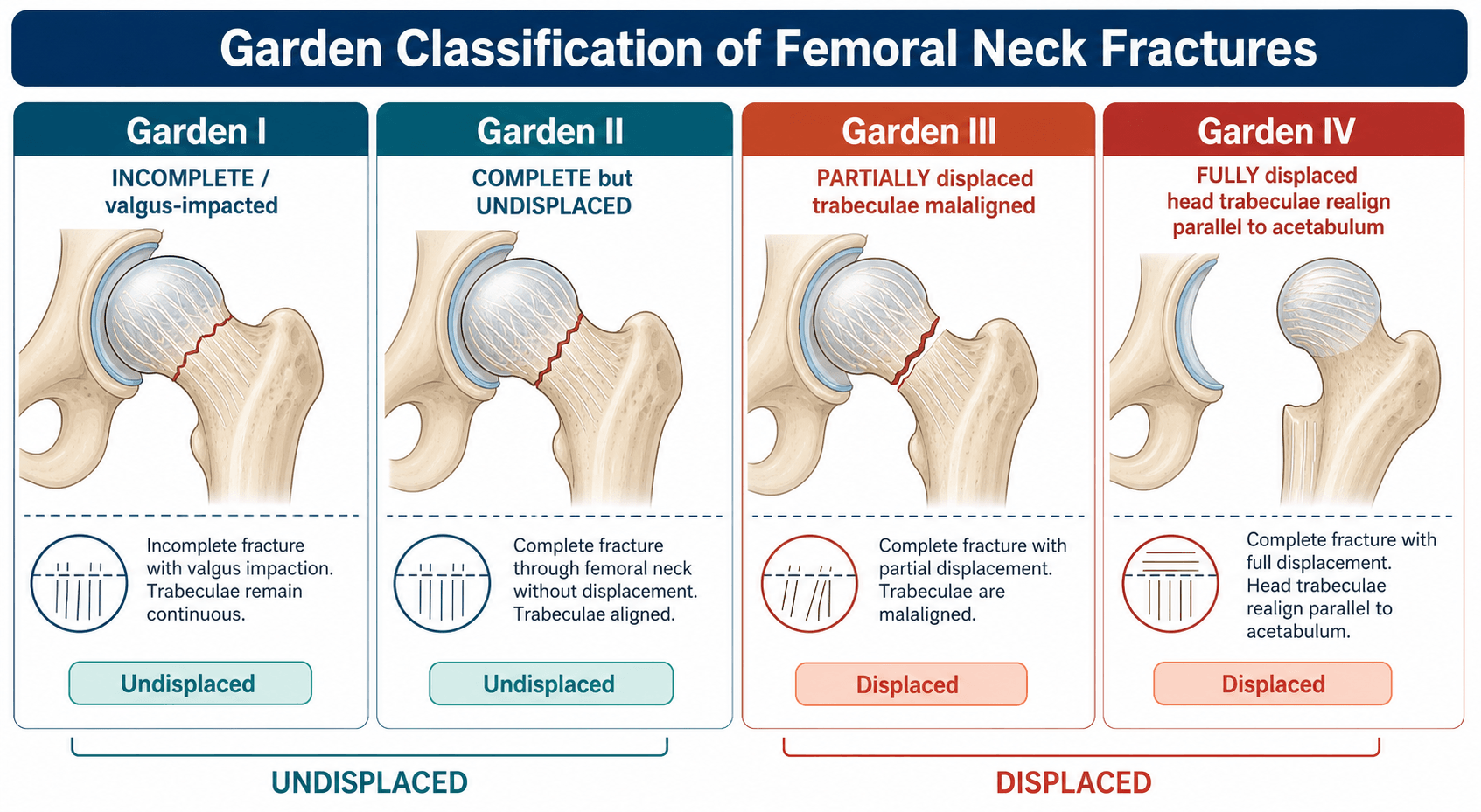
Garden Classification (1961)

Based on degree of displacement and alignment of trabecular lines on AP radiograph. Correlates with AVN risk and guides treatment.

Garden I - Incomplete/Impacted Valgus:

  • Fracture line visible but incomplete
  • Femoral head tilted into valgus (impacted position)
  • Trabecular lines remain continuous across fracture
  • AVN risk: 0-10%
  • Treatment: Cannulated screw fixation

Garden II - Complete, Non-Displaced:

  • Complete fracture line visible
  • No displacement - trabecular lines of head align with acetabulum and neck
  • Head remains in neutral position
  • AVN risk: 10-20%
  • Treatment: Cannulated screw fixation

Garden III - Complete, Partially Displaced:

  • Complete fracture with partial displacement
  • Trabecular lines of head misaligned with neck
  • Head partially rotated but still some contact with neck
  • AVN risk: 20-30%
  • Treatment: Arthroplasty (elderly), urgent reduction and fixation (young)

Garden IV - Complete, Fully Displaced:

  • Complete fracture with total displacement
  • Trabecular lines of head align with acetabulum but not with neck
  • Head fully dissociated from neck
  • AVN risk: 30-100%
  • Treatment: Arthroplasty (elderly), urgent reduction and fixation (young)

Clinical Simplification: Garden I-II (undisplaced) = FIX. Garden III-IV (displaced) = REPLACE (in elderly).

Reliability: Inter-observer agreement only moderate (kappa 0.4-0.6). Many surgeons simplify to just displaced vs undisplaced.

Pauwels Classification (1935)

Measures the angle of the fracture line to the horizontal on AP radiograph. Predicts shear forces and fixation failure risk.

Pauwels Type I: Fracture angle less than 30 degrees

  • Mostly compressive forces across fracture
  • Relatively stable
  • Good healing potential
  • Low failure rate with screw fixation (less than 5%)

Pauwels Type II: Fracture angle 30-50 degrees

  • Mixed compression and shear forces
  • Moderate stability
  • Intermediate failure rate (10-15%)

Pauwels Type III: Fracture angle greater than 50 degrees (vertical)

  • Predominantly shear forces across fracture
  • Very unstable
  • High failure rate with standard screw fixation (30-40%)
  • Requires augmented fixation: Sliding hip screw with derotation screw, or fixed-angle device

Clinical Significance: Pauwels III fractures in young patients are challenging - high failure rate necessitates stronger constructs than simple cannulated screws.

AO/OTA Classification

Comprehensive fracture description system from the AO Foundation/Orthopaedic Trauma Association.

31-B1: Subcapital fracture with slight displacement (Garden I-II equivalent)

  • B1.1: Impacted valgus (Garden I)
  • B1.2: Non-impacted (Garden II)
  • B1.3: Varus impacted (rare, very unstable)

31-B2: Transcervical (midcervical) fracture

  • Through middle of femoral neck
  • Less common than subcapital
  • AVN risk similar to subcapital

31-B3: Basicervical fracture

  • At base of femoral neck (junction with intertrochanteric region)
  • Extracapsular (outside joint capsule)
  • Lower AVN risk but higher fixation failure risk
  • Often requires sliding hip screw rather than screws alone

Clinical Use: More detailed than Garden but less commonly used clinically. Useful for research and registries.

Delbet Classification

Specific classification for pediatric femoral neck fractures (rare, less than 1% of pediatric fractures).

Type I: Transepiphyseal (through growth plate)

  • Highest AVN risk (100%)
  • Surgical emergency

Type II: Transcervical

  • High AVN risk (50%)
  • Urgent fixation required

Type III: Cervicotrochanteric (base of neck)

  • Moderate AVN (25%)
  • Surgical treatment preferred

Type IV: Intertrochanteric

  • Low AVN risk (less than 10%)
  • Best prognosis

Significance: Pediatric NOF fractures are surgical emergencies due to very high AVN risk. All require urgent reduction and fixation.

Clinical Presentation

History

Elderly Patient (Low-Energy Fall):

  • Pain in groin, hip, or referred to knee
  • Unable to weight-bear following fall from standing height
  • Falls at home (bathroom, bedroom) or on street/stairs
  • May report hearing/feeling "snap" or "pop" at time of injury
  • Risk factors: Osteoporosis, age greater than 65 years, previous fragility fracture, low BMI, sedentary lifestyle

Young Patient (High-Energy Trauma):

  • Road traffic accident, fall from height, sports injury
  • Severe pain, inability to move leg
  • May have polytrauma with other injuries

Examination

Inspection:

  • Patient lying still, reluctant to move affected leg
  • Classic posture: Leg shortened, externally rotated (typically 90 degrees), adducted
  • Look for: swelling, bruising (may be minimal acutely), deformity

Palpation:

  • Tenderness over groin/hip
  • Pain on log-rolling leg (specific test - gentle internal/external rotation)
  • Check peripheral pulses (dorsalis pedis, posterior tibial)
  • Neurovascular exam: Sciatic nerve function (foot dorsiflexion/plantarflexion, sensation)

Movement:

  • Active movement: Patient cannot straight leg raise or flex hip
  • Passive movement: Painful with any attempt
  • Do NOT stress fracture - risk displacement

Special Considerations

Impacted Fractures (Garden I):

  • May be able to weight-bear partially (patient walked after fall)
  • Less obvious deformity
  • Diagnosis can be missed - have high index of suspicion

Occult Fractures:

  • Pain but normal X-rays initially
  • MRI or CT needed to confirm
  • Treat as fracture until excluded

Investigations

Imaging

Plain Radiographs (First-line):

  • AP pelvis: Shows both hips for comparison, assess acetabulum
  • Lateral hip: Cross-table lateral (avoid moving patient excessively)
  • Look for: Fracture line, displacement, Garden classification, Shenton's line disruption

CT Scan:

  • Indications: Occult fracture (suspected clinically but X-ray negative), complex fracture patterns, pre-operative planning for young patients
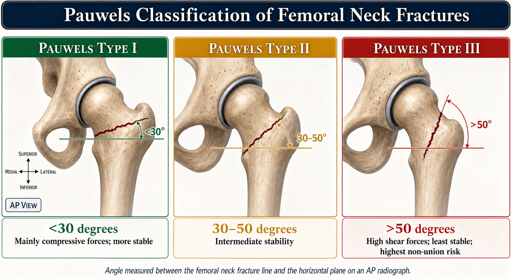
  • Better delineation of fracture pattern, posterior comminution, vertical fracture angle (Pauwels classification)

MRI:

  • Gold standard for occult fractures
  • High sensitivity (99%) and specificity (95%)
  • Shows bone marrow edema, undisplaced fracture lines not visible on X-ray
  • Indicated when: clinical suspicion high but X-ray normal, patient unable to mobilize post-fall

Laboratory Tests

Pre-operative Bloods:

  • FBC: Baseline hemoglobin (anemia common in elderly, may need transfusion)
  • U&E: Renal function for anesthesia, contrast studies
  • Coagulation: INR if on warfarin, platelet count if on antiplatelet agents
  • Group and Save/Cross-match: For anticipated blood loss in arthroplasty
  • CRP/ESR: Baseline (may be elevated due to fracture, useful if later infection suspected)

Cardiac Assessment:

  • ECG: All patients (high-risk population for cardiac disease)
  • Troponin: If chest pain or ECG changes (myocardial injury common post-fracture)
  • Echocardiogram: If significant cardiac history or murmur detected

Bone Health Investigations (Post-acute):

  • DEXA scan: Assess bone mineral density (osteoporosis)
  • Vitamin D levels: Often deficient, correct post-operatively
  • Calcium, phosphate, PTH: If secondary causes of osteoporosis suspected
  • Thyroid function: Hyperthyroidism causes bone loss

Medical Optimization

Essential Pre-op Assessment:

  • Cardiac clearance: Anesthesia review, optimization of cardiac medications
  • Anticoagulation management: Reverse warfarin if needed (Vitamin K, PCC), hold DOACs appropriately
  • Fluid resuscitation: Elderly often dehydrated, correct hypovolemia
  • Pain control: Fascia iliaca block (regional anesthesia reduces opioid requirement)

Management

📊 Management Algorithm
Neck of Femur (NOF) Fracture Management Algorithm Flowchart
Click to expand
Comprehensive management algorithm for Neck of Femur Fractures. Note the critical distinction between undisplaced (fixation) and displaced (arthroplasty in elderly, urgent fixation in young) fractures.Credit: OrthoVellum

Treatment: Internal fixation with cannulated screws (typically 3 screws in inverted triangle configuration).

Rationale: Blood supply is preserved. Screw fixation stabilizes the fracture and allows healing.

Caution: Garden I (impacted valgus) can displace if not fixed. Most surgeons recommend fixation.

Post-op: Protected weight-bearing initially. Monitor for AVN and nonunion.

Treatment: Arthroplasty (hemiarthroplasty or total hip replacement).

Rationale: Displaced fractures have high AVN and nonunion rates (up to 30-50%). Fixation in elderly has poor outcomes.

Hemiarthroplasty vs THR:

  • Hemiarthroplasty (unipolar or bipolar): Frail, limited mobility, cognitive impairment, shorter life expectancy, lower dislocation risk.
  • THR (cemented): Cognitively intact, independently mobile (walk outdoors), expected survival greater than 4 years. Better function but higher dislocation risk.

NICE guidelines recommend THR for independently mobile patients.

Young patients (less than 60-65 years old) with displaced NOF fractures:

Treatment: Attempt urgent reduction and internal fixation within 6 hours to minimize AVN risk.

Rationale: Preserving the native hip is important. Even though AVN risk is still present (20-30%), avoiding arthroplasty at a young age is preferable.

Technique: Closed or open reduction. 3 cannulated screws or sliding hip screw.

Counsel: High risk of complications (AVN, nonunion). May need arthroplasty later.

Time to Surgery

Hip fracture is a surgical emergency. Surgery within 36-48 hours is associated with lower mortality, fewer medical complications, shorter hospital stay. Delays significantly increase mortality.

Complications

Early Complications - First Days After Surgery

Mortality:

  • 30-day mortality: 6-8%
  • 1-year mortality: 25-30%
  • Higher in elderly, frail, multiple comorbidities
  • Delayed surgery (greater than 48 hours) increases mortality significantly

Medical Complications:

  • Pressure sores: Prolonged immobility pre-operatively
  • Pneumonia: Aspiration, reduced mobility, general anesthesia
  • DVT/PE: High risk in elderly immobile patients
  • UTI: Catheterization, dehydration
  • Delirium: 20-50% of elderly post-op (pain, medications, unfamiliar environment)
  • Myocardial infarction: Surgical stress, cardiac demand

Surgical Complications:

  • Bleeding: Arthroplasty (300-500mL blood loss), may require transfusion
  • Infection: Superficial (2-3%) or deep periprosthetic (less than 1%)
  • Nerve injury: Sciatic (posterior approach, less than 1%), femoral (anterior, rare)
  • Vascular injury: Femoral vessels (anterior approach - rare but catastrophic)

Intermediate Complications - Weeks to Months Post-Surgery

Specific to Internal Fixation:

  • Avascular necrosis (AVN):
    • 20-30% in displaced fractures (young patients with fixation)
    • 10% in undisplaced fractures
  • Nonunion: 10-30% (depends on displacement and fixation quality)
  • Implant failure: Screw cut-out (backing out through femoral head)
  • Malunion: Varus collapse, shortening
5-panel malunion types after femoral neck fracture fixation
Click to expand
5-panel (a-e) AP hip radiographs demonstrating different malunion patterns following parallel screw fixation of femoral neck fractures: (a) Coxa breva - neck shortening with loss of offset; (b) Coxa valga - valgus neck-shaft angle; (c) Rotational malunion - abnormal femoral head rotation; (d) Translational malunion - lateral or medial head displacement; (e) Coxa vara - varus collapse with loss of neck-shaft angle. These complications result from inadequate reduction, poor bone quality, or early weight-bearing.Credit: Satish BR et al. - Indian J Orthop (CC-BY 4.0)

Specific to Arthroplasty:

  • Dislocation: 2-10%
    • Higher in THR than hemiarthroplasty
    • Posterior approach greater than anterior
  • Periprosthetic fracture:
    • Intraoperative (during stem insertion)
    • Post-operative (fall on prosthesis)
  • Aseptic loosening: More common with uncemented
  • Acetabular erosion: Hemiarthroplasty (femoral head erodes acetabulum → groin pain)

Late Complications - Months to Years Post-Surgery

Functional Outcomes:

  • Loss of independence: 40% don't return to pre-fracture mobility
  • Chronic pain: Hip, groin, or thigh pain (10-20%)
  • Reduced quality of life: Loss of function, institutionalization

Fracture-Related:

  • AVN (late presentation): Femoral head collapse, severe pain, arthritis
  • Post-traumatic arthritis: Cartilage damage → degenerative change
  • Leg length discrepancy: More common with arthroplasty
  • Heterotopic ossification: Bone formation in soft tissues (more with THR)

Secondary Fragility Fractures:

  • Contralateral hip fracture: 5-10% within 5 years
  • Vertebral fractures, wrist fractures: Untreated osteoporosis
  • Prevention CRITICAL: Bisphosphonates, calcium, Vitamin D, falls programs

Surgical Techniques

Internal Fixation for Undisplaced or Young Patients

Indications:

  • Undisplaced fractures (Garden I-II) all ages
  • Displaced fractures (Garden III-IV) in physiologically young patients (less than 60-65 years)

Patient Positioning:

  • Supine on fracture table with gentle traction
  • Image intensifier for AP and lateral views

Reduction Technique (if displaced):

  • Leadbetter maneuver: Gentle longitudinal traction, flexion 15-30°, abduction 20-30°, internal rotation 15°
  • Check fluoroscopy (Pauwels' lines less than 160° lateral, less than 180° AP)
  • Acceptable criteria: Less than 20° angulation, less than 2mm displacement
  • If closed reduction fails → Open reduction (Watson-Jones or Smith-Petersen approach)

Guidewire Placement:

  • 3 wires in inverted triangle configuration
  • Inferior wire along calcar (inferior neck)
  • Two superior wires divergent
  • Parallel in sagittal plane
  • All within 5mm of subchondral bone
  • Critical: Avoid joint penetration (check lateral view)

Screw Insertion:

  • Partially threaded cannulated screws (6.5mm or 7.3mm diameter)
  • Threads cross fracture site
  • Compression by tightening screws
  • Inferior screw provides calcar support (prevents varus collapse)

Biomechanics:

  • Inverted triangle = best rotational stability
  • Parallel configuration = compression
  • Screws span fracture and engage far cortex

Post-operative:

  • Touch weight-bearing initially, advance as tolerated

Capsulotomy:

  • Controversial for AVN prevention
  • FAITH trial: No benefit demonstrated
  • Consider if delayed presentation (greater than 12 hours)

Sliding Hip Screw for Vertical Fractures

Indications:

  • Pauwels Type III fractures (greater than 50°, high shear forces)
  • Basicervical fractures (31-B3)
  • Failed initial screw fixation
  • Large or osteoporotic patients

Advantages over Cannulated Screws:

  • Greater fixation strength for vertical fractures
  • Allows controlled compression along lag screw
  • Derotation screw prevents rotation
  • Better for osteoporotic bone

Operative Technique:

  1. Approach: Lateral approach to proximal femur
  2. Guide wire: Center on AP/lateral, tip 5mm from subchondral bone
  3. Reaming: Over guide wire
  4. Lag screw: Insert (typically 80-110mm length)
  5. Side plate: Fix with cortical screws
  6. Derotation screw: Above lag screw
  7. Compression: Tighten lag screw

Critical Technical Points:

  • Tip-apex distance less than 25mm (reduces cut-out risk)
  • Center-center position in femoral head (AP and lateral)
  • Adequate screw length (within 5mm of subchondral bone)

For Displaced Fractures in Elderly (Low Demand)

Surgical Approaches:

1. Posterior (Moore/Southern) - Most common

  • Between gluteus maximus fibers
  • External rotators divided (piriformis, obturators)
  • Excellent acetabular exposure
  • Must repair external rotators (reduces dislocation 10% → 2-3%)
  • Sciatic nerve at risk (less than 1%)

2. Anterolateral (Hardinge)

  • Split gluteus medius
  • Lower dislocation risk (2-3%)
  • More muscular trauma, limp risk

3. Anterior (Smith-Petersen)

  • Internervous plane
  • Lowest dislocation (less than 1%)
  • Difficult femoral exposure

Implant Selection:

Head Type:

  • Bipolar (Bateman, Christiansen): Most common - dual articulation
  • Unipolar (Austin Moore, Thompson): Less used - higher acetabular erosion

Fixation:

  • Cemented preferred (Level 1 evidence - BMJ 2022):
    • Lower mortality
    • Less pain, better mobility
    • Lower periprosthetic fracture risk
    • BCIS risk 0.5-1%
  • Uncemented: Rarely used (higher fracture risk 5-8%)

Cementing Technique:

  1. Pulsatile lavage
  2. Cement restrictor
  3. Retrograde fill
  4. Pressurization

Operative Steps:

  1. Lateral position, posterior approach
  2. Capsulotomy, identify fracture
  3. Femoral neck osteotomy (remove head)
  4. Femoral preparation: Broach to size
  5. Cement technique
  6. Stem insertion, cement curing
  7. Trial reduction (check stability, leg length)
  8. Final head insertion
  9. Repair external rotators and capsule
  10. Closure

Challenges:

  • Fracture comminution → difficult version/leg length control
  • Weak bone → intraoperative fracture risk
  • Calcar deficiency → subsidence risk (use calcar-replacing stems)
X-ray showing right hip hemiarthroplasty
Click to expand
Post-operative AP pelvis radiograph demonstrating a cemented hemiarthroplasty on the right hip. The prosthesis replaces the femoral head while articulating with the native acetabulum. Cemented fixation is preferred in elderly patients (Level 1 evidence) for lower mortality and better early mobility.Credit: OrthoVellum

For Active, Cognitively Intact Elderly

Indications (Choose THR over Hemiarthroplasty if):

  • Cognitively intact
  • Independently mobile (walks outdoors)
  • Life expectancy greater than 4 years
  • Pre-existing hip arthritis

Implant Selection:

Femoral Stem:

  • Cemented preferred for osteoporotic bone
  • Common stems: Exeter, CPT, Charnley

Acetabular Cup:

  • Cemented preferred for elderly
  • Some use uncemented if good bone

Bearing Surface:

  • Metal or ceramic head on polyethylene (standard for elderly)
  • Head size: 32mm or 36mm (larger = less dislocation)

Dislocation Prevention:

  • Repair external rotators and capsule (reduces 10% → 2-3%)
  • Dual mobility cups: Very low dislocation (less than 1%), higher cost
  • Constrained liners: Last resort for recurrent dislocation

Operative Steps (Posterior Approach):

  1. Lateral position
  2. Posterior approach
  3. Capsulotomy
  4. Femoral neck osteotomy
  5. Acetabular preparation: Ream, trial, cement/press-fit cup
  6. Femoral preparation: Broach to size
  7. Cement technique: Clean canal, restrictor, retrograde cement, pressurization
  8. Stem insertion, cement curing
  9. Trial reduction (stability, leg length)
  10. Final head/liner insertion
  11. Capsular and external rotator repair
  12. Closure in layers

Advantages over Hemiarthroplasty:

  • Better pain relief
  • Better function (Oxford Hip Score)
  • No acetabular erosion
  • Suitable for active patients

Challenges:

  • Longer operative time
  • Higher dislocation risk if poor repair
  • More complex revision if fails

Biomechanics and Fixation Principles

Forces Acting on Femoral Neck

The femoral neck experiences complex loading during weight-bearing:

Compressive Forces (Primary):

  • Body weight transmitted from acetabulum to femoral head
  • Magnitude: 3-5x body weight (walking), up to 8x (running)
  • Concentrated on superomedial femoral head (weight-bearing zone)

Tensile Forces:

  • Along inferomedial cortex (calcar femorale)
  • Generated by moment arm of body weight
  • Resisted by thick cortical bone at calcar

Shear Forces:

  • Parallel to fracture line
  • Increase with fracture verticality (Pauwels angle)
  • Pauwels III (greater than 50°): Predominantly shear → high failure risk

Rotational Forces:

  • Torsion from muscle attachments (gluteus medius, iliopsoas)
  • Cause rotation and displacement of fracture fragments

Trabecular Architecture

The femoral neck contains two main trabecular systems:

Primary Compressive Trabeculae (Vertical):

  • Run from medial calcar to superior femoral head
  • Resist compressive loads during weight-bearing
  • Strongest trabecular system

Primary Tensile Trabeculae (Arcuate):

  • Arc from lateral cortex to inferior femoral head
  • Resist tensile forces along inferior neck
  • Important for fracture stability

Secondary Compressive Trabeculae:

  • From greater trochanter to femoral head
  • Provide additional support

Ward's Triangle:

  • Area of relative weakness (few trabeculae)
  • Located in femoral neck between trabecular systems
  • Common site of fracture initiation in osteoporotic bone

Cannulated Screw Biomechanics

Optimal Configuration:

  • 3 screws provide best stability (better than 2, minimal advantage with 4)
  • Inverted triangle: Superior resistance to rotational forces
    • Base of triangle anteriorly (lateral view)
    • Apex posteriorly
    • Spreads load across femoral head
  • Inferior screw along calcar: Critical for preventing varus collapse
  • Thread engagement: Partial threading allows interfragmentary compression

Screw Placement Principles:

  • All screws should be parallel (frontal and sagittal planes)
  • Tip-apex distance less than 5mm from subchondral bone
  • Avoid penetration into joint (check lateral view)
  • Spread screws maximally within head (wider spread = better rotation resistance)

Biomechanical Weakness:

  • No resistance to shear in vertical fractures (Pauwels III)
  • No sliding/compression mechanism (unlike DHS)
  • Relies entirely on fracture reduction quality and bone healing

Sliding Hip Screw Biomechanics

Advantages:

  • Dynamic compression: Lag screw slides through barrel, continuously compressing fracture
  • Greater purchase: Larger lag screw diameter (12-13mm vs 6.5-7.3mm cannulated)
  • Derotation screw: Prevents rotation
  • Better for vertical fractures: Resists shear forces better than screws alone

Biomechanical Principles:

  • Lag screw positioned center-center or inferior-posterior (AP/lateral views)
  • Tip-apex distance less than 25mm (predictor of cut-out)
    • Formula: (AP distance + lateral distance) x magnification factor
    • Greater than 25mm = 4x higher cut-out risk
  • Barrel angle typically 135° (matches anatomic angle)
  • Side plate distributes load to femoral shaft

Failure Modes:

  • Cut-out: Lag screw backs out through femoral head (most common)
    • Risk factors: Poor reduction, superior screw position, tip-apex greater than 25mm
  • Cut-through: Screw penetrates medially through head into pelvis
  • Z-effect: Lateral migration of screw, medial migration of neck

Biological vs Mechanical Stability

Absolute Stability (Goal for Intracapsular Fractures):

  • No motion at fracture site
  • Required for primary bone healing (direct osteonal remodeling)
  • Achieved by: Anatomic reduction + compression + adequate fixation
  • Intracapsular fractures MUST have absolute stability (no periosteal callus possible)

Relative Stability (Not Acceptable for NOF):

  • Controlled micromotion allowed
  • Permits secondary bone healing (callus formation)
  • Used for extracapsular fractures (pertrochanteric) but NOT intracapsular

Why Displaced Fractures Fail Fixation

Biological Factors:

  • Vascular disruption: Lateral epiphyseal vessels torn → AVN
  • Synovial fluid: Washes out hematoma, impairs healing
  • No periosteum: Intracapsular location prevents periosteal callus

Mechanical Factors:

  • Loss of calcar continuity: Medial support lost → varus collapse
  • Comminution: Posterior neck comminution prevents stable reduction
  • Vertical fracture angle: Pauwels III has high shear forces
  • Fracture gap: Even 2mm gap significantly impairs healing

Result: Combination of poor biology (AVN) + mechanical instability (collapse) = high failure rate in elderly (30-50%). This is why arthroplasty preferred for displaced fractures in elderly.

Evidence Base

THR vs Hemiarthroplasty - HEALTH Trial

1
Key Findings:
  • THR resulted in better functional outcomes at 24 months vs hemiarthroplasty
  • Secondary hip procedures: 5.8% THR vs 9.1% hemiarthroplasty
  • No difference in mortality between groups
  • Study included cognitively intact patients with displaced NOF fractures
Source: JAMA 2019

Timing of Surgery - HIP ATTACK Trial

1
Key Findings:
  • Accelerated surgery (median 6 hours) vs standard care (median 24 hours)
  • No difference in 90-day mortality (9% vs 9%)
  • No difference in major complications
  • Accelerated group had less delirium (4% vs 5%)
Source: Lancet 2020

Cemented vs Uncemented Hemiarthroplasty

1
Key Findings:
  • Cemented hemiarthroplasty associated with lower mortality
  • Less pain and better mobility compared to uncemented
  • Lower periprosthetic fracture rates with cemented
  • Bone cement implantation syndrome risk exists but low
Source: BMJ 2022

Internal Fixation Method - FAITH Trial

1
Key Findings:
  • No significant difference in reoperation rates: SHS 20.8% vs screws 17.5%
  • AVN rates similar between groups
  • Nonunion rates similar between groups
  • 2-year follow-up in 1108 patients
Source: JAMA 2017

Orthogeriatric Co-Management - Systematic Review

1
Key Findings:
  • Orthogeriatric co-management reduced 30-day mortality by 40% (OR 0.60)
  • Reduced delirium, pressure sores, and time to surgery
  • Lower length of hospital stay (mean reduction 2.5 days)
  • Higher rates of bone protection and falls prevention initiated
  • Cost-effective despite additional geriatrician input
Source: JAMA Surgery 2020

Fascia Iliaca Block for Analgesia - Cochrane Review

1
Key Findings:
  • Fascia iliaca block reduced pain scores by 2-3 points (VAS 0-10)
  • Reduced opioid requirement by 40-50% in first 24 hours
  • Lower delirium rates compared to systemic opioids alone
  • Safe, few complications (less than 1% hematoma, nerve injury)
  • Recommended in emergency department on arrival
Source: Cochrane Database 2021
Mnemonic

PAUWELS = Predict AVN Using Fracture AnglePauwels Classification

I
Less than 30°
Stable, compression forces, low failure (5%)
II
30-50°
Mixed forces, moderate failure (15%)
III
Greater than 50°
Vertical, shear forces, HIGH failure (40%) - needs DHS

Memory Hook:Pauwels III = VERTICAL = shear forces = needs stronger fixation (DHS not screws)!

Mnemonic

PALNOF Arthroplasty Approaches

P
Posterior
Most common - Higher dislocation (7%) - Repair capsule!
A
Anterolateral
Hardinge - Limp risk (Trendelenburg) - Lower dislocation (3%)
L
Lateral/Anterior
Smith-Petersen - Lowest dislocation (1%) - Hard exposure

Memory Hook:PAL approaches - Posterior most common but repair or dislocate!

Rehabilitation and Outcomes

Post-operative Mobilization

Early Mobilization (Day 1 Post-op):

  • Goal: Out of bed within 24 hours of surgery
  • Strong evidence that early mobilization reduces:
    • Pneumonia (40% reduction)
    • Pressure sores (50% reduction)
    • DVT/PE (30% reduction)
    • Delirium (20% reduction)

Weight-Bearing Status:

After Arthroplasty (Hemiarthroplasty or THR):

  • Weight-bearing as tolerated (WBAT) immediately
  • No need for protected weight-bearing (implant is load-bearing)
  • Encourage full weight-bearing as pain allows
  • Walking aids (frame, crutches) for balance support initially

After Internal Fixation (Undisplaced Garden I-II):

  • Touch weight-bearing for 6 weeks (some surgeons allow WBAT if stable fixation)
  • Progress to partial weight-bearing (50%) at 6 weeks (if X-ray shows healing)
  • Full weight-bearing at 12 weeks (if united)
  • Risk of displacement if full weight-bearing too early

After Internal Fixation (Displaced Garden III-IV in Young):

  • Strict non weight-bearing for 6-12 weeks
  • High risk of fixation failure, AVN, nonunion
  • Advance weight-bearing very cautiously based on serial X-rays

Hip Precautions (After Arthroplasty)

For Posterior Approach (Most Common):

  • Avoid:
    • Hip flexion greater than 90 degrees (no low chairs, toilet seats)
    • Hip adduction across midline (no crossing legs)
    • Internal rotation (avoid twisting inward)
  • Duration: 6 weeks (some surgeons recommend 12 weeks)
  • Risk: Posterior dislocation if precautions violated
  • Note: If external rotators repaired well, some surgeons omit precautions (evidence mixed)

For Anterior/Anterolateral Approach:

  • Avoid hip extension and external rotation
  • Less restrictive than posterior approach precautions
  • Lower dislocation risk overall

Phase 1 (Days 1-3 Post-op):

  • Bed mobility (rolling, sitting up)
  • Transfer training (bed to chair, toilet)
  • Gait re-education with walking aid
  • Active range of motion exercises
  • Isometric quadriceps and gluteal exercises

Phase 2 (Week 1-6):

  • Progressive weight-bearing (if fixation)
  • Strengthening exercises (hip abductors, extensors, quadriceps)
  • Balance and proprioception training
  • Stairs training
  • Aim for independent mobility with single-point stick

Phase 3 (Week 6-12):

  • Functional strengthening
  • Return to activities of daily living
  • Falls prevention strategies
  • Home hazard assessment and modification

Outcomes After NOF Fracture

Mortality:

  • 30-day mortality: 6-8%
  • 1-year mortality: 25-30%
  • 5-year mortality: 50%
  • Mortality higher than age-matched controls (excess mortality persists for 10 years)

Functional Outcomes:

  • 40% do NOT return to pre-fracture mobility level
  • 30% require increased care (from independent to assisted living or nursing home)
  • 25% remain non-ambulatory at 1 year
  • 20% have persistent pain (hip or thigh)
  • Better outcomes after arthroplasty than fixation (especially if fixation fails)

Quality of Life:

  • Significant reduction in quality of life scores (EQ-5D, SF-36)
  • Loss of independence is major factor
  • Depression common (30-40%)
  • Social isolation

Return to Independence:

  • Strong predictors of return to independence:
    • Pre-fracture independent mobility
    • Absence of dementia
    • Living at home (not nursing home)
    • Younger age (less than 85 years)
    • No major medical comorbidities
    • Early surgery (less than 48 hours)
    • Orthogeriatric co-management

Fixation vs Arthroplasty Outcomes:

  • Fixation (for undisplaced):

    • Lower surgical morbidity
    • Preserves anatomy
    • Risk of AVN (10%), nonunion (10-15%), reoperation (20%)
    • If fails, requires salvage arthroplasty (worse outcomes than primary)
  • Arthroplasty (for displaced):

    • Immediate stability, early mobilization
    • Lower reoperation rates (5-8%)
    • Better pain relief
    • Small risk dislocation (2-10%), infection (less than 1%)

Secondary Fracture Prevention

Osteoporosis Management (Essential):

  • DEXA scan: All patients post-fracture
  • Bisphosphonates: Alendronate 70mg weekly or zoledronic acid 5mg IV yearly
    • Evidence: 35% reduction in subsequent hip fractures
    • Start after surgical wounds healed (typically 2 weeks post-op)
    • Continue for 3-5 years minimum
  • Vitamin D and Calcium: Universal supplementation (Vitamin D 1000IU daily, Calcium 1200mg daily)
  • Falls prevention program: Mandatory
  • Fracture liaison service: Coordinates osteoporosis care

Falls Prevention:

  • Home hazards assessment: Remove trip hazards, improve lighting, install grab rails
  • Medication review: Reduce sedatives, antihypertensives if contributing
  • Vision assessment: Correct cataracts, update glasses
  • Balance and strength training: Ongoing physiotherapy
  • Vitamin D: Reduces falls risk independent of bone effect
  • Hip protectors: Controversial (poor compliance, limited evidence)

Multidisciplinary Hip Fracture Pathway:

Modern hip fracture care requires coordinated multidisciplinary approach:

  • Orthopaedic surgeon: Surgical management
  • Geriatrician/Orthogeriatrician: Medical optimization, delirium prevention
  • Anaesthetist: Regional anesthesia, perioperative optimization
  • Physiotherapist: Early mobilization, rehabilitation
  • Occupational therapist: ADL assessment, home modifications
  • Dietitian: Nutritional support (many elderly malnourished)
  • Pharmacist: Medication reconciliation, avoid inappropriate drugs
  • Fracture liaison coordinator: Osteoporosis treatment, follow-up
  • Social worker: Discharge planning, care needs

Hip Fracture Care Standards (ACSQHC Australia):

  1. Surgery within 48 hours (target greater than 85%)
  2. Orthogeriatric involvement within 72 hours
  3. DVT prophylaxis (LMWH or fondaparinux)
  4. Bone protection initiated before discharge
  5. Nutritional assessment and supplementation
  6. Pressure area care
  7. Early mobilization (day 1 post-op)
  8. Falls and bone health assessment
  9. Multidisciplinary rehabilitation
  10. Fracture liaison service follow-up

Special Populations

Mechanism: Usually high-energy trauma (RTA, fall from height)

Key Differences:

  • Preserve femoral head at almost any cost - avoid arthroplasty if possible
  • Even if AVN risk 30%, preferable to THR at age 40-50 (revision burden)
  • Urgent surgery (ideally less than 6 hours) to minimize AVN
  • Anatomic reduction mandatory - accept nothing less
  • Strong fixation: Consider DHS for vertical fractures (Pauwels III)
  • Prolonged follow-up: Monitor for AVN (can occur 2-5 years post-fixation)

If Fixation Fails:

  • Salvage options: Valgus osteotomy (if nonunion), arthroplasty (if AVN)
  • THR considerations in young: Uncemented implants, hard-on-hard bearings (ceramic-on-ceramic, highly cross-linked polyethylene), anticipate multiple revisions over lifetime

Challenges:

  • Cannot comply with precautions (high dislocation risk with THR)
  • Cannot comply with protected weight-bearing (not suitable for fixation of displaced fractures)
  • Poor functional outcomes (many do not mobilize post-operatively)
  • High mortality (50% at 1 year)

Management Approach:

  • Hemiarthroplasty preferred over THR (lower dislocation risk, simpler surgery, shorter operative time)
  • Consider constrained liner or dual mobility if very high dislocation risk
  • Optimize analgesia (fascia iliaca block, multimodal analgesia, avoid opioids if possible - increase delirium)
  • Aggressive delirium prevention: Familiar environment, avoid restraints, minimize medications, treat pain, encourage mobilization
  • Early involvement of geriatrician
  • Realistic goals: May not return to pre-fracture mobility, focus on comfort and basic transfers

Causes:

  • Metastatic disease: Breast, lung, kidney, thyroid, prostate most common
  • Myeloma: Common in elderly
  • Primary bone tumors: Rare (osteosarcoma, chondrosarcoma)

Clinical Clues:

  • Minimal trauma or atraumatic
  • Persistent pain before fracture (weeks to months)
  • Known primary malignancy
  • Constitutional symptoms (weight loss, night pain, fatigue)
  • Lytic or sclerotic lesion on X-ray

Investigations:

  • Staging CT: Chest, abdomen, pelvis
  • Bone scan or PET: Full skeletal survey
  • Myeloma screen: SPEP, UPEP, serum free light chains, bone marrow biopsy
  • Biopsy: If unknown primary (intraoperative or pre-operative CT-guided)

Management:

  • Almost always arthroplasty (fixation will fail - diseased bone won't heal)
  • Long-stem cemented prosthesis if metaphyseal or diaphyseal involvement
  • Consider THR (not hemiarthroplasty) if acetabular metastases present
  • Post-operative radiotherapy: Reduces local recurrence, pain
  • Bisphosphonates: Zoledronic acid for bony metastases
  • Oncology referral: Systemic treatment if appropriate

Common Scenarios:

  • Warfarin: For atrial fibrillation, mechanical heart valves, VTE
  • DOACs: Apixaban, rivaroxaban, dabigatran (increasingly common)
  • Dual antiplatelet therapy: Aspirin + clopidogrel/ticagrelor (post-coronary stent)

Management Principles:

  • Do NOT delay surgery waiting for natural clearance - increases mortality
  • Active reversal to achieve surgery within 48 hours

Warfarin Reversal:

  • Target INR less than 1.5 for surgery
  • Prothrombin complex concentrate (PCC): 25-50 units/kg IV (rapid reversal in 15-30 minutes)
  • Vitamin K: 5-10mg IV (slower reversal, 6-24 hours, but more sustained)
  • Avoid FFP: Requires large volumes (15-20mL/kg), slower reversal
  • Restart warfarin: 24-48 hours post-op when hemostasis secure, bridge with LMWH if high stroke risk (CHA2DS2-VASc greater than 4)

DOAC Management:

  • Half-life: Apixaban/rivaroxaban 8-12 hours, dabigatran 12-17 hours
  • Hold for 48 hours (2-3 half-lives) before surgery if renal function normal
  • Check anti-Xa levels (rivaroxaban, apixaban) or thrombin time (dabigatran) if available
  • Reversal agents if emergency:
    • Idarucizumab for dabigatran (specific reversal)
    • Andexanet alfa for apixaban/rivaroxaban (specific reversal, very expensive)
    • PCC as alternative (off-label but effective)
  • Restart: 24-48 hours post-op

Dual Antiplatelet Therapy (Post-Coronary Stent):

  • High bleeding risk if continue aspirin + clopidogrel
  • High stent thrombosis risk if stop both
  • Consult cardiology: Timing since stent, type of stent (bare metal vs drug-eluting)
  • General approach: Continue aspirin, hold clopidogrel/ticagrelor for 5-7 days before surgery if possible
  • If recent stent (less than 3 months): May need to operate on dual antiplatelets (accept higher bleeding risk - stent thrombosis is catastrophic)

Exam Viva Scenarios

Practice these scenarios to excel in your viva examination

VIVA SCENARIOStandard

Scenario 1: Displaced NOF in Elderly

EXAMINER

"An 80-year-old woman with osteoporosis falls at home and has a Garden IV neck of femur fracture. She was previously independently mobile and cognitively intact. How do you manage?"

EXCEPTIONAL ANSWER
This is a Garden IV (completely displaced) intracapsular femoral neck fracture in an elderly osteoporotic patient. The key decision is between hemiarthroplasty and total hip replacement. For displaced neck of femur fractures in the elderly, internal fixation has poor outcomes due to high rates of AVN and nonunion (because blood supply via the lateral epiphyseal vessels is disrupted). Therefore, arthroplasty is indicated. The choice between hemiarthroplasty and THR depends on the patient's pre-morbid status. This patient was **independently mobile and cognitively intact**. According to NICE guidelines, such patients benefit from **total hip replacement** (cemented) which provides better function, less erosion of acetabulum, and lower reoperation rates compared to hemiarthroplasty. However, THR has a slightly higher dislocation risk. If she had cognitive impairment or limited mobility, I would choose hemiarthroplasty. Regarding timing, hip fractures are surgical emergencies. **Surgery within 36-48 hours** is critical to reduce mortality, pressure sores, pneumonia, and DVT. I would optimize medically (correct anticoagulation, assess cardiac status) but not delay unnecessarily. Post-operatively, she would have a standardized hip fracture pathway including DVT prophylaxis, early mobilization, bone protection (bisphosphonates), and falls assessment.
KEY POINTS TO SCORE
Garden IV = displaced → arthroplasty
Independently mobile + cognitively intact = THR
Surgery within 36-48 hours
Bone protection and falls prevention
COMMON TRAPS
✗Offering internal fixation for displaced fracture in elderly
✗Delaying surgery unnecessarily
✗Not knowing THR vs hemiarthroplasty indications
LIKELY FOLLOW-UPS
"When would you choose hemiarthroplasty?"
"How would you manage this in a 45-year-old?"
VIVA SCENARIOChallenging

Scenario 2: NOF Fracture on Anticoagulation - Balancing Bleeding and Delay

EXAMINER

"A 78-year-old woman presents to the emergency department at 10pm with a Garden III displaced neck of femur fracture following a fall at home. She takes warfarin for atrial fibrillation. Her INR is 3.2 (therapeutic range 2.0-3.0 for AF). She is otherwise stable with no other injuries. The anaesthetist asks whether you want to reverse her warfarin and operate tonight, or wait for the INR to normalize naturally over the next 48 hours. What factors do you consider and how do you proceed?"

EXCEPTIONAL ANSWER
This is a common clinical dilemma balancing the risks of bleeding from operating on anticoagulated patients against the risks of delaying surgery for a hip fracture. The key principles are that hip fractures are surgical emergencies where surgery within 36-48 hours reduces mortality and morbidity, but operating on anticoagulated patients increases bleeding risk. For warfarin, the threshold INR for safe surgery is generally considered to be less than 1.5. This patient's INR of 3.2 is elevated and presents significant bleeding risk if we operate immediately. However, waiting 48 hours for natural reversal would take us beyond the optimal surgical window. My approach would be to actively reverse the warfarin to achieve surgery within the 36-48 hour window. I would discuss with the haematology team and anaesthetist, and my preferred reversal strategy would be to give Vitamin K (5-10mg IV slowly) and consider Prothrombin Complex Concentrate (PCC, previously known as Prothrombinex in Australia) which contains factors II, VII, IX, and X. PCC provides rapid reversal (within 15-30 minutes) allowing surgery to proceed urgently. The alternative of FFP requires large volumes and is less effective. I would aim to reverse to INR less than 1.5 and operate within 12-24 hours. After reversal, I need to consider the stroke risk from her atrial fibrillation. Warfarin can typically be restarted 24-48 hours post-operatively once surgical hemostasis is secure, bridging with therapeutic LMWH if needed depending on her CHA2DS2-VASc score. However, the immediate post-operative period carries thrombotic risk anyway, so standard DVT prophylaxis with LMWH would be started as per hip fracture protocols. The key is not to delay surgery beyond 48 hours waiting for natural warfarin reversal, as the increased mortality from surgical delay outweighs the bleeding risk from reversed anticoagulation. For patients on newer direct oral anticoagulants (DOACs like rivaroxaban, apixaban), the approach differs - these typically clear within 24-48 hours depending on renal function, and specific reversal agents (idarucizumab for dabigatran, andexanet alfa for factor Xa inhibitors) can be considered for urgent cases.
KEY POINTS TO SCORE
Hip fracture surgery within 36-48h reduces mortality - delay is harmful
Warfarin: Reverse with Vitamin K + PCC if INR greater than 1.5, target INR less than 1.5
PCC provides rapid reversal (15-30 min) vs FFP (large volumes, slower)
Restart anticoagulation 24-48h post-op when hemostasis secure, bridge with LMWH if high stroke risk
DOACs: Clear naturally 24-48h, specific reversal agents available for emergencies
COMMON TRAPS
✗Waiting 48 hours for natural warfarin reversal - misses optimal surgical window
✗Operating without reversal on INR 3.2 - significant bleeding risk
✗Not knowing PCC as rapid reversal agent for warfarin
✗Forgetting to restart anticoagulation post-operatively for stroke prevention
LIKELY FOLLOW-UPS
"What is the mechanism of PCC and how does it differ from FFP?"
"How would you manage a patient on apixaban with this fracture?"
"What is the CHA2DS2-VASc score and when do you bridge with therapeutic anticoagulation?"
VIVA SCENARIOCritical

Scenario 3: Young Patient Displaced NOF - Salvage Decision

EXAMINER

"A 42-year-old man is brought to your trauma center following a high-speed motorcycle accident. He has multiple injuries including a closed head injury (GCS 14), liver laceration being managed conservatively, and a Garden IV displaced neck of femur fracture. It is now 8 hours since injury. He is hemodynamically stable. X-rays show the femoral neck fracture is very vertical (Pauwels Type III) with significant posterior comminution. What are your management options and what factors influence your decision?"

EXCEPTIONAL ANSWER
This is a complex scenario involving a young patient with a displaced femoral neck fracture in the setting of polytrauma. The fundamental principle for displaced neck of femur fractures in young patients is to attempt urgent reduction and internal fixation to preserve the native femoral head, despite the high risk of complications (AVN 20-30%, nonunion 10-20%). However, several factors complicate this case. First, timing: we are now 8 hours post-injury. The critical window for reduction to minimize AVN risk is ideally within 6 hours. Studies show AVN rates increase significantly with delay beyond 6-12 hours as the disrupted blood supply (lateral epiphyseal vessels from MFCA) undergoes progressive thrombosis. At 8 hours, we are marginally outside the ideal window, but salvage is still worthwhile. Second, the polytrauma context: He has a closed head injury with GCS 14 (mild TBI) and liver laceration being observed. I need to coordinate with neurosurgery and general surgery. The liver laceration if stable does not preclude surgery. The head injury requires monitoring but is not severe enough to prevent operative intervention. Third, fracture pattern: The Pauwels Type III classification indicates a very vertical fracture (greater than 50 degrees to horizontal), which has much higher shear forces and therefore higher failure rates with standard screw fixation. Additionally, posterior comminution makes fixation more challenging and may indicate severe displacement with greater soft tissue disruption. My management approach would be to proceed urgently to the operating theatre for attempted closed reduction and internal fixation. For the reduction, I would use the technique of flexion, abduction, and internal rotation to disimpact and reduce the fracture, performed under image intensifier guidance. If closed reduction achieves anatomic alignment, I would proceed with internal fixation. For fixation in this scenario, given the Pauwels Type III vertical pattern and posterior comminution, standard three cannulated screws in inverted triangle may be insufficient. I would consider either a sliding hip screw with de-rotation screw (provides better resistance to shear forces in vertical fractures) or fixed-angle screw construct. Some surgeons advocate adding an anti-rotation screw or using a buttress plate on the lateral femoral neck. The key teaching point is that vertical fractures (Pauwels III) have significantly higher failure rates - quoted at 30-40% compared to 10-15% for horizontal fractures. If closed reduction fails to achieve acceptable alignment, I would proceed with open reduction via Watson-Jones or Smith-Petersen approach, ensuring I do not strip more soft tissues than necessary. Post-operatively, this patient faces a difficult road. I would counsel about the high likelihood of complications: AVN risk is probably 30-40% given the delay and severity, nonunion risk is elevated due to the vertical fracture and comminution, and he may well require conversion to total hip arthroplasty within 2-5 years. However, at age 42, preserving the native hip is still preferred over primary arthroplasty, as THR in young active patients has higher revision rates. I would follow him closely with serial X-rays at 6 weeks, 3 months, 6 months, and annually, looking for signs of AVN (femoral head collapse, sclerosis, cystic change) and nonunion (persistent fracture line, lack of bridging callus, screw cutout).
KEY POINTS TO SCORE
Young displaced NOF: Attempt urgent reduction and fixation despite high complication risk
6-hour window ideal for reduction to minimize AVN, but attempt worthwhile up to 12-24 hours
Pauwels Type III (vertical greater than 50°): Higher shear forces, 30-40% failure rate, consider SHS or buttress plate
AVN risk 20-30% baseline, higher with delay, posterior comminution, failed reduction
Young patient: Preserve native hip, accept high failure risk, counsel about likely conversion to THR
COMMON TRAPS
✗Offering primary arthroplasty to 42-year-old - should attempt fixation first
✗Using standard 3-screw fixation for Pauwels Type III - inadequate for vertical fracture
✗Not counseling about realistic high failure rates and need for subsequent THR
✗Delaying surgery due to polytrauma when patient is stable - time is critical for AVN prevention
LIKELY FOLLOW-UPS
"Describe the Pauwels classification and its biomechanical significance"
"What are the radiographic signs of established AVN of the femoral head?"
"If this patient develops AVN at age 45, what arthroplasty options would you consider?"
VIVA SCENARIOChallenging

Scenario 4: Pathological NOF Fracture - Occult Malignancy

EXAMINER

"A 68-year-old woman presents with a Garden III femoral neck fracture after a minor fall in the bathroom. On questioning, she mentions she has had worsening hip pain for the past 3 months that was present even before the fall. She attributes it to 'arthritis'. On examination, the fracture site is more tender than expected, and she appears cachectic. X-ray shows a displaced femoral neck fracture with some lytic changes in the intertrochanteric region. How do you approach this case?"

EXCEPTIONAL ANSWER
This presentation has several red flags that should immediately raise suspicion for a pathological fracture secondary to underlying malignancy rather than a simple osteoporotic fragility fracture. The key red flags are: (1) Prodromal hip pain for 3 months before the fracture - typical osteoporotic fractures are acute with minimal or no pre-fracture pain, whereas pathological fractures from metastatic disease often present with weeks to months of progressive pain before the fracture event. (2) Minimal trauma mechanism (bathroom fall) in the context of pre-existing pain suggests weakened bone. (3) Cachectic appearance - constitutional symptoms of malignancy (weight loss, anorexia). (4) Lytic changes visible on X-ray in the intertrochanteric region - this is highly suspicious for metastatic disease or myeloma. My immediate priority is to determine whether this is a pathological fracture and, if so, identify the primary malignancy, as this fundamentally changes surgical management. I would order urgent investigations: (1) Staging CT chest/abdomen/pelvis to look for a primary tumor (most common sources of bone metastases are breast, lung, kidney, thyroid, prostate - the 'BLT with a Kosher Pickle' mnemonic). (2) Myeloma screen: SPEP, UPEP, serum free light chains, calcium (hypercalcemia common in myeloma), and consider bone marrow biopsy if suspicion high. (3) Bone scan or PET-CT for full skeletal survey to identify other metastases. (4) Laboratory: FBC (anemia?), renal function, calcium, alkaline phosphatase (elevated in bone metastases), PSA if male. (5) Consider CT-guided biopsy of the lytic lesion if primary unknown (although in many cases I would obtain tissue intraoperatively during definitive fixation). Regarding surgical management, the key principle is that pathological fractures from metastatic disease WILL NOT HEAL with standard internal fixation because the bone is diseased and incapable of biological healing. Therefore, internal fixation alone (cannulated screws or DHS) is contraindicated. Instead, the patient requires arthroplasty to provide immediate mechanical stability without relying on bone healing. For a femoral neck pathological fracture, I would perform a cemented long-stem hemiarthroplasty or total hip replacement. The rationale for a long stem is that metaphyseal or diaphyseal involvement may be present even if not obvious on X-ray, and a standard short stem risks periprosthetic fracture or subsidence through diseased bone. I would use cemented fixation for immediate stability in potentially poor-quality bone. The choice between hemiarthroplasty versus THR depends on whether there is acetabular involvement (if acetabular metastases present, THR with cemented cup; if acetabulum clear, hemiarthroplasty acceptable). Intraoperatively, I would obtain tissue from the fracture site for histopathology and culture (rarely infection can mimic malignancy). Post-operatively, the patient would benefit from radiotherapy to the surgical site (reduces local recurrence and improves pain control) and referral to medical oncology for systemic treatment if appropriate. If myeloma is diagnosed, bisphosphonates (zoledronic acid) should be started as they reduce skeletal-related events. Prognosis depends on the primary tumor type, but even in metastatic disease, surgical stabilization significantly improves quality of life by providing pain relief and mobility. The key teaching point is to maintain a high index of suspicion for pathological fractures in patients with prodromal symptoms, and to recognize that standard fixation will fail - arthroplasty is mandatory for pathological femoral neck fractures.
KEY POINTS TO SCORE
Red flags for pathological fracture: Prodromal pain (weeks-months), minimal trauma, constitutional symptoms, lytic lesions on X-ray
Workup: Staging CT chest/abd/pelvis, myeloma screen (SPEP/UPEP/FLC), bone scan/PET, tumor markers, biopsy
Pathological fractures WILL NOT HEAL - internal fixation contraindicated
Treatment: Long-stem cemented arthroplasty (hemiarthroplasty or THR if acetabulum involved)
Adjuvant: Post-op radiotherapy (local control), systemic treatment (oncology), bisphosphonates if myeloma/metastases
COMMON TRAPS
✗Performing cannulated screw fixation for pathological fracture - will fail (diseased bone cannot heal)
✗Using standard short-stem prosthesis - risk subsidence/periprosthetic fracture through metaphyseal disease
✗Not investigating for primary malignancy pre-operatively - need staging for oncology management
✗Missing the diagnosis entirely by attributing prodromal pain to 'arthritis' without further workup
LIKELY FOLLOW-UPS
"What are the most common primary tumors that metastasize to bone? (BLT with Kosher Pickle mnemonic)"
"How do you distinguish myeloma from metastatic disease radiographically and biochemically?"
"If this patient is found to have widespread metastatic disease, would you still offer surgical fixation and why?"
VIVA SCENARIOCritical

Scenario 5: Bilateral Simultaneous NOF Fractures - Rare but Critical Recognition

EXAMINER

"A 72-year-old man with Parkinson's disease has a fall at home and is brought to the emergency department. X-ray pelvis shows displaced femoral neck fractures bilaterally (Garden IV both sides). He is medically stable but in significant pain. The emergency department physician asks you urgently whether this is possible or if there's been a mix-up with the X-rays. How do you approach this rare presentation and what are your management priorities?"

EXCEPTIONAL ANSWER
Bilateral simultaneous femoral neck fractures are extremely rare (incidence less than 1% of all hip fractures) but this is a real presentation that I must take seriously. While unusual, there are well-documented case reports in the literature, particularly in patients with neurological conditions like Parkinson's disease, epilepsy (post-seizure), or following electroconvulsive therapy. The mechanism is typically forceful bilateral muscular contraction (hip flexors and adductors) against fixed femoral heads, generating shear forces across both femoral necks simultaneously. Parkinson's disease increases risk due to: (1) Rigidity and abnormal tone predisposing to falls, (2) Potential for bilateral sudden muscular spasms, (3) Osteoporosis from reduced mobility and Vitamin D deficiency. First, I need to confirm the diagnosis is truly bilateral acute fractures and not a previous fracture on one side. I would review the X-rays carefully looking for: Symmetry of displacement (acute bilateral fractures typically show similar displacement patterns), absence of healing callus or sclerosis (would indicate old fracture), clinical examination (bilateral pain, deformity, inability to move either leg). I would also enquire about previous hip problems or falls. Assuming these are truly acute bilateral fractures, my management approach involves several critical decisions. Regarding surgical planning, the key question is: Do I operate on both sides in one sitting, or stage the procedures? The literature suggests single-stage bilateral fixation is feasible and actually preferred in most cases for several reasons: (1) Single anesthetic exposure (elderly patients tolerate one anesthetic better than two), (2) Single rehabilitation phase (patient mobilizes both hips together, no asymmetry), (3) Reduced total hospitalization time, (4) Lower overall complication rates compared to leaving one side unfixed for days to weeks. However, single-stage surgery requires: Long operative time (typically 3-4 hours total), increased blood loss (may require transfusion), careful anesthetic management (fluid balance, hypothermia prevention), two surgical teams ideally (to reduce operative time), good ICU/HDU support post-operatively. For this patient with bilateral displaced Garden IV fractures in an elderly man, the appropriate treatment is bilateral cemented hemiarthroplasty (or bilateral THR if cognitively intact and mobile pre-morbidly). I would NOT attempt bilateral internal fixation in a 72-year-old with displaced fractures - the failure rate would be prohibitively high (30-50% per side) and he'd likely require revision to arthroplasty. My surgical plan would be: Single-stage bilateral cemented hemiarthroplasty using posterior approach, operate on more symptomatic side first (or right side by convention if equal), experienced team to minimize operative time (target less than 2 hours per side), cell saver if available (reduce allogeneic transfusion), meticulous hemostasis, post-operative HDU monitoring. Pre-operative optimization is critical: Cardiology clearance (bilateral arthroplasty is higher cardiac stress), correct any coagulopathy or anemia, optimize Parkinson's medications (continue L-dopa perioperatively - sudden withdrawal causes severe rigidity), fascia iliaca blocks bilaterally for analgesia, DVT prophylaxis (higher risk given bilateral surgery). Post-operative care: HDU monitoring first 24-48 hours, aggressive DVT prophylaxis (mechanical and pharmacological), early mobilization (day 1 if stable - weight-bearing as tolerated on both sides), intensive physiotherapy, orthogeriatric involvement, nutritional support (catabolic stress from bilateral surgery). The patient and family need clear counseling about: Rarity of the condition, increased surgical risk compared to unilateral fracture, longer operative time and recovery, higher complication rates (infection, bleeding, cardiac events), but better overall outcome than staged procedures, rehabilitation will be challenging (bilateral hip precautions, bilateral weakness). Prognosis: Bilateral hip fractures have higher mortality (1-year mortality approximately 40-50% vs 25-30% for unilateral) and worse functional outcomes (only 30-40% return to pre-fracture mobility vs 50-60% unilateral). However, with good surgical management and rehabilitation, meaningful functional recovery is possible. The key teaching points are: (1) Bilateral simultaneous NOF fractures are rare but real - don't dismiss the diagnosis, (2) Associated with seizures, Parkinson's, ECT, or high-energy bilateral trauma, (3) Single-stage bilateral arthroplasty is preferred over staged approach in most cases, (4) Requires careful perioperative planning and multidisciplinary support, (5) Higher morbidity and mortality than unilateral fractures but surgical fixation still offers best chance of recovery.
KEY POINTS TO SCORE
Bilateral NOF fractures rare (less than 1%) but real - mechanisms: seizures, Parkinson's, ECT, forceful bilateral muscle contraction
Confirm diagnosis: Review X-rays for symmetry, absence of healing (exclude old fracture one side), bilateral clinical signs
Single-stage bilateral surgery preferred over staged (single anesthetic, synchronous rehab, lower overall complications)
Elderly displaced fractures: Bilateral cemented hemiarthroplasty/THR, NOT bilateral internal fixation (high failure rate)
Higher morbidity/mortality than unilateral (1-year mortality 40-50%), but single-stage still best approach
COMMON TRAPS
✗Dismissing bilateral fractures as impossible or X-ray mix-up - it's rare but real, investigate thoroughly
✗Attempting bilateral internal fixation in elderly displaced fractures - very high failure rate, will need revision
✗Staging procedures weeks apart - increases anesthetic exposure, prolongs immobility, higher overall complications
✗Not optimizing Parkinson's medications perioperatively - L-dopa withdrawal causes severe rigidity, complicates surgery and recovery
LIKELY FOLLOW-UPS
"What other clinical scenarios can cause bilateral simultaneous femoral neck fractures?"
"In a young patient (age 30) with bilateral NOF after seizure, would you change your management approach?"
"What are the specific anesthetic challenges and considerations for bilateral hip arthroplasty in one sitting?"

MCQ Practice Points

Garden Classification

Q: What distinguishes Garden I from Garden II femoral neck fractures?

A: Garden I is an incomplete/impacted fracture with the head tilted into valgus. Garden II is complete but non-displaced with trabecular alignment preserved. Both are undisplaced and treated with screw fixation. Key difference: Garden I has visible trabecular lines crossing fracture; Garden II has complete fracture line but no displacement.

Blood Supply and AVN Risk

Q: Why do displaced femoral neck fractures have high AVN rates?

A: The medial femoral circumflex artery (MFCA) gives rise to lateral epiphyseal vessels which provide the main blood supply to the femoral head. These vessels run along the posterior-superior femoral neck. Displaced fractures disrupt this supply, causing AVN rates of 20-30% in young patients and even higher in delayed treatment. This is why displaced fractures in elderly warrant arthroplasty rather than fixation.

THR vs Hemiarthroplasty

Q: What are the indications for THR over hemiarthroplasty in displaced NOF fractures?

A: THR is indicated for patients who are: cognitively intact, independently mobile (walking outdoors), and have life expectancy greater than 4 years. THR provides better function and lower reoperation rates. Hemiarthroplasty is preferred for: cognitive impairment, limited mobility, frail patients. NICE guidelines recommend THR for independently mobile patients.

Young Patient Management

Q: How do you manage a displaced NOF fracture in a 45-year-old?

A: Urgent reduction and internal fixation (ideally within 6 hours) to minimize AVN risk. Use closed or open reduction with 3 cannulated screws (inverted triangle) or sliding hip screw. Despite high complication rates (AVN 20-30%, nonunion 10-20%), preserving the native hip is preferred over arthroplasty in young patients. Counsel patient about potential need for future arthroplasty.

Australian Context

Epidemiology in Australia

Incidence and Burden:

  • Approximately 20,000-22,000 hip fractures annually in Australia (ANZ Hip Fracture Registry - ANZHFR data 2023)
  • Incidence rate: 150-200 per 100,000 population over age 65
  • Female:male ratio approximately 3:1
  • Projected to double by 2050 due to aging population
  • Annual healthcare cost: Estimated AUD $1.2-1.5 billion (acute care, rehabilitation, long-term care)

Mortality Data (ANZHFR 2023):

  • 30-day mortality: 6.2% (national average)
  • 1-year mortality: 27.3%
  • 5-year mortality: Approximately 50%
  • Variation across states: Best performing states (VIC, NSW) achieve 5-6% 30-day mortality, while some rural/remote regions higher (8-10%)

Geographic Variation:

  • Urban vs Rural: Rural areas have slightly higher mortality and longer time to surgery (access issues)
  • Indigenous Australians: Hip fractures occur 10-15 years earlier than non-Indigenous Australians, higher mortality rates
  • Remote communities: Significant challenges with evacuation, surgical access, and rehabilitation

Australian Hip Fracture Registry (ANZHFR)

The ANZHFR is a binational registry (Australia and New Zealand) established in 2015 to monitor and improve hip fracture care quality.

Key Performance Indicators (2023 National Report):

  • Surgery within 48 hours: 82.4% nationally (target greater than 85%)
    • Best performers: 90-95% (major metropolitan hospitals)
    • Challenges: Rural hospitals, weekend admissions, medically complex patients
  • Orthogeriatric assessment within 72 hours: 76.8% (target greater than 80%)
  • Bone protection prescribed at discharge: 68.5% (target greater than 80%)
  • Post-discharge fracture liaison service contact: 45.2% (target greater than 70% - needs improvement)

Registry Data Insights:

  • Hospitals with dedicated hip fracture pathways have 30-40% lower mortality
  • Orthogeriatric co-management associated with 2-day reduction in length of stay
  • Weekend admissions have 15% longer time to surgery (resource constraints)

Australian Orthopaedic Association National Joint Replacement Registry (AOANJRR)

THR for Fracture Outcomes (AOANJRR 2023 Report):

  • 10-year cumulative revision rate: 6.9% for THR performed for fracture
  • Comparison: Lower than revision rate for primary OA THR at 10 years (7.2%)
  • 15-year survivorship: 94.8% for THR, 89.2% for hemiarthroplasty
  • Revision reasons: Dislocation (30%), infection (25%), aseptic loosening (20%), periprosthetic fracture (15%)

Hemiarthroplasty Outcomes:

  • 10-year cumulative revision rate: 11.2%
  • Higher revision rate than THR (predominantly due to acetabular erosion causing pain)
  • Cemented vs Uncemented: Cemented hemiarthroplasty has lower revision rate (9.8% vs 14.5% at 10 years)

Implant Performance:

  • Most commonly used stems for fracture: Exeter (cemented), CPT (cemented), Corail (uncemented)
  • Most commonly used hemiarthroplasty heads: Bipolar designs (Bateman, Christiansen)
  • Dual mobility cups increasingly used for high dislocation risk patients (revision rate only 3-4% at 5 years)

Australian Hip Fracture Care Standards

ACSQHC Clinical Care Standard (2016, updated 2021):

Mandatory Quality Indicators:

  1. Surgery within 48 hours of presentation (benchmark greater than 85%)

    • Delays permissible only for medical optimization (e.g., severe cardiac instability, active anticoagulation requiring reversal)
    • "Stop the clock" provisions for unavoidable delays (transfer to specialist center, patient refusal)
  2. Orthogeriatric involvement within 72 hours

    • Comprehensive Geriatric Assessment (CGA)
    • Delirium prevention and management
    • Medical comorbidity optimization
    • Bone health and falls assessment
  3. DVT prophylaxis initiated within 24 hours

    • Pharmacological: LMWH (enoxaparin 40mg daily or dalteparin 5000 units daily) or fondaparinux 2.5mg daily
    • Mechanical: TED stockings and/or intermittent pneumatic compression
    • Duration: Minimum 10-14 days, extended if high VTE risk
  4. Bone protection initiated before discharge

    • Bisphosphonates: Alendronate 70mg weekly (oral) or zoledronic acid 5mg IV (annual)
    • Vitamin D: 1000-2000 IU daily
    • Calcium: 1200mg daily (dietary + supplementation)
    • DEXA scan arranged (if not done acutely, within 3 months)
  5. Pressure injury prevention

    • Pressure-relieving mattress for all patients
    • Regular repositioning
    • Nutritional assessment and supplementation
    • Early mobilization
  6. Early mobilization (day 1 post-operatively)

    • Physiotherapy review within 24 hours of surgery
    • Out of bed and walking (or attempting) within 24 hours
    • Progressive weight-bearing as tolerated (arthroplasty) or per fracture protocol (fixation)
  7. Falls and bone health assessment

    • Comprehensive falls risk assessment
    • Home hazards evaluation
    • Vitamin D and calcium supplementation
    • Fracture Liaison Service (FLS) referral before discharge
  8. Multidisciplinary rehabilitation plan

    • Physiotherapy, occupational therapy, dietitian, social work input
    • Discharge planning from day 1
    • Clear goals and timeline
  9. Nutrition screening and support

    • Malnutrition Universal Screening Tool (MUST) on admission
    • Dietitian referral if malnourished (albumin less than 30 g/L, BMI less than 20, weight loss)
    • Protein supplementation (aim 1.2-1.5g/kg/day)
    • Vitamin and mineral supplementation
  10. Fracture Liaison Service contact post-discharge

    • Follow-up within 4-6 weeks
    • Ensure osteoporosis treatment ongoing
    • Monitor bisphosphonate adherence
    • Coordinate DEXA scan if not done

PBS (Pharmaceutical Benefits Scheme) Considerations

Bisphosphonates:

  • Alendronate 70mg weekly: PBS-subsidized for osteoporosis with fragility fracture (no authority required)
  • Zoledronic acid 5mg IV yearly: PBS-subsidized for osteoporosis with fracture (Streamlined authority - phone approval)
  • Denosumab 60mg SC 6-monthly: PBS-subsidized as second-line if bisphosphonates contraindicated/intolerant

Anticoagulants:

  • Warfarin: Fully PBS-subsidized
  • DOACs (apixaban, rivaroxaban, dabigatran): PBS-subsidized for atrial fibrillation with CHA2DS2-VASc score greater than or equal to 2 (males) or greater than or equal to 3 (females)
  • Prothrombin Complex Concentrate (Prothrombinex-VF): Section 100 approval required (emergency provision available)

Analgesia:

  • Paracetamol, tramadol: Unrestricted PBS
  • Oxycodone, morphine: Schedule 8 controlled drugs, prescription limits apply
  • Fascia iliaca blocks not PBS-listed (hospital cost absorbed)

eTG (Therapeutic Guidelines) Recommendations

Antibiotic Prophylaxis (Surgical):

  • First-line: Cefazolin 2g IV at induction (or within 60 minutes of incision)
  • Penicillin allergy: Vancomycin 15-20mg/kg IV (infuse over 60-90 minutes, start 60-90 minutes before incision)
  • Duration: Single dose (no post-operative doses recommended for routine cases)
  • Extended prophylaxis: If surgery greater than 4 hours or significant blood loss, one additional dose

VTE Prophylaxis:

  • Pharmacological: Enoxaparin 40mg SC daily or dalteparin 5000 units SC daily
  • Start: 6-12 hours post-operatively (balance bleeding vs VTE risk)
  • Duration: Minimum 10-14 days, extend to 35 days if additional VTE risk factors
  • Contraindications: Active bleeding, severe renal impairment (CrCl less than 30 mL/min - reduce dose or use fondaparinux)

State-Specific Programs

Victoria: HIP FRACTURE PATHWAY (Safer Care Victoria)

  • Standardized care pathway across all Victorian public hospitals
  • Pre-printed orders for analgesia, DVT prophylaxis, bone protection
  • Mandatory orthogeriatric review
  • State-wide data collection and benchmarking

New South Wales: GUIDE TO THE MANAGEMENT OF HIP FRACTURES IN OLDER PEOPLE (Agency for Clinical Innovation)

  • Model of care recommendations
  • Minimum Data Set collection
  • Coordinated Fracture Liaison Services across Local Health Districts

Queensland: HIP FRACTURE CARE CLINICAL PATHWAY

  • Integrated care pathway from emergency department to discharge
  • Focus on rural and remote service delivery
  • Telehealth orthogeriatric consultations for regional hospitals

South Australia: SA HIP FRACTURE REGISTRY (State-level registry complementing ANZHFR)

Western Australia: WA HIP FRACTURE MODEL OF CARE (Department of Health)

  • Emphasizes pre-hospital care, ambulance protocols
  • Regional transfer guidelines for complex cases

Challenges Specific to Australia

Geographic Remoteness:

  • Retrieval and transfer: Some patients require Royal Flying Doctor Service (RFDS) evacuation from remote areas to surgical centers
  • Delays: Transfer times can be 12-24 hours, impacting 48-hour surgery target
  • Workforce: Rural hospitals may lack orthopedic or anesthetic cover 24/7

Indigenous Health:

  • Hip fractures occur 10-15 years younger in Indigenous Australians
  • Higher comorbidity burden (diabetes, renal disease, cardiovascular disease)
  • Cultural considerations for rehabilitation and discharge planning
  • Coordination with Aboriginal Medical Services for post-discharge care

Aged Care Facilities:

  • 40-50% of hip fractures occur in patients residing in aged care
  • Transfer delays from facility to hospital
  • Challenges with return to aged care post-surgery (bed availability, funding)
  • Higher delirium and complication rates

NECK OF FEMUR FRACTURE

High-Yield Exam Summary

Garden Classification

  • •I/II: Undisplaced → SCREWS
  • •III/IV: Displaced → ARTHROPLASTY (elderly)

Arthroplasty Choice

  • •THR: Mobile, cognitively intact, good life expectancy
  • •Hemiarthroplasty: Frail, impaired cognition, limited mobility

Key Points

  • •Surgery within 36-48 hours
  • •MFCA supply disrupted → AVN risk
  • •Young displaced: Attempt fixation urgently

Post-Op Care

  • •DVT prophylaxis
  • •Early mobilization
  • •Bone protection (bisphosphonates)
  • •Falls assessment
Quick Stats
Reading Time184 min
Related Topics

Acetabular Fractures

Acromioclavicular Joint Injuries

Acute Compartment Syndrome

Ankle Fractures