Slipped Capital Femoral Epiphysis
What it is, and why it matters
A slipped capital femoral epiphysis is a Salter-Harris type I (occasionally II) separation through the proximal femoral growth plate. The slip happens through the hypertrophic zone of the physis — its weakest layer — under a combination of mechanical overload (obesity is by far the commonest risk factor) and hormonal change (the adolescent growth spurt, and endocrine disorders that weaken the physis). The epiphysis is held in the acetabulum, so it is really the neck and shaft that displace: the metaphysis moves anteriorly and rotates externally relative to the head, which is why the classic examination finding is obligate external rotation when the hip is flexed.
Two facts make SCFE high-stakes. First, it hides: the pain is often felt in the thigh or knee, not the hip, and a mild slip can look almost normal on an AP film — so it is a classic missed diagnosis. Second, its feared complication, avascular necrosis (AVN) of the femoral head, is driven largely by the initial vascular insult at the moment the slip becomes unstable and by any attempt to forcefully reduce it — not by how skilfully it is later pinned. Get the diagnosis early, classify stability correctly, and fix in situ, and most children do very well.
- Peak age 10–16 years, at the growth spurt (boys mean ~12–13, girls ~11–12)
- Boys more than girls, roughly 2–3:1
- Markedly higher in children of African and Pacific Islander ancestry; lowest in East Asian populations
- Incidence rising with childhood obesity worldwide
- Obesity — the dominant risk factor
- Rapid growth / tall for age
- Endocrine: hypothyroidism, growth-hormone abnormalities, hypogonadism
- Renal osteodystrophy; previous radiation
- Femoral retroversion
Anatomy and blood supply — the basis of AVN
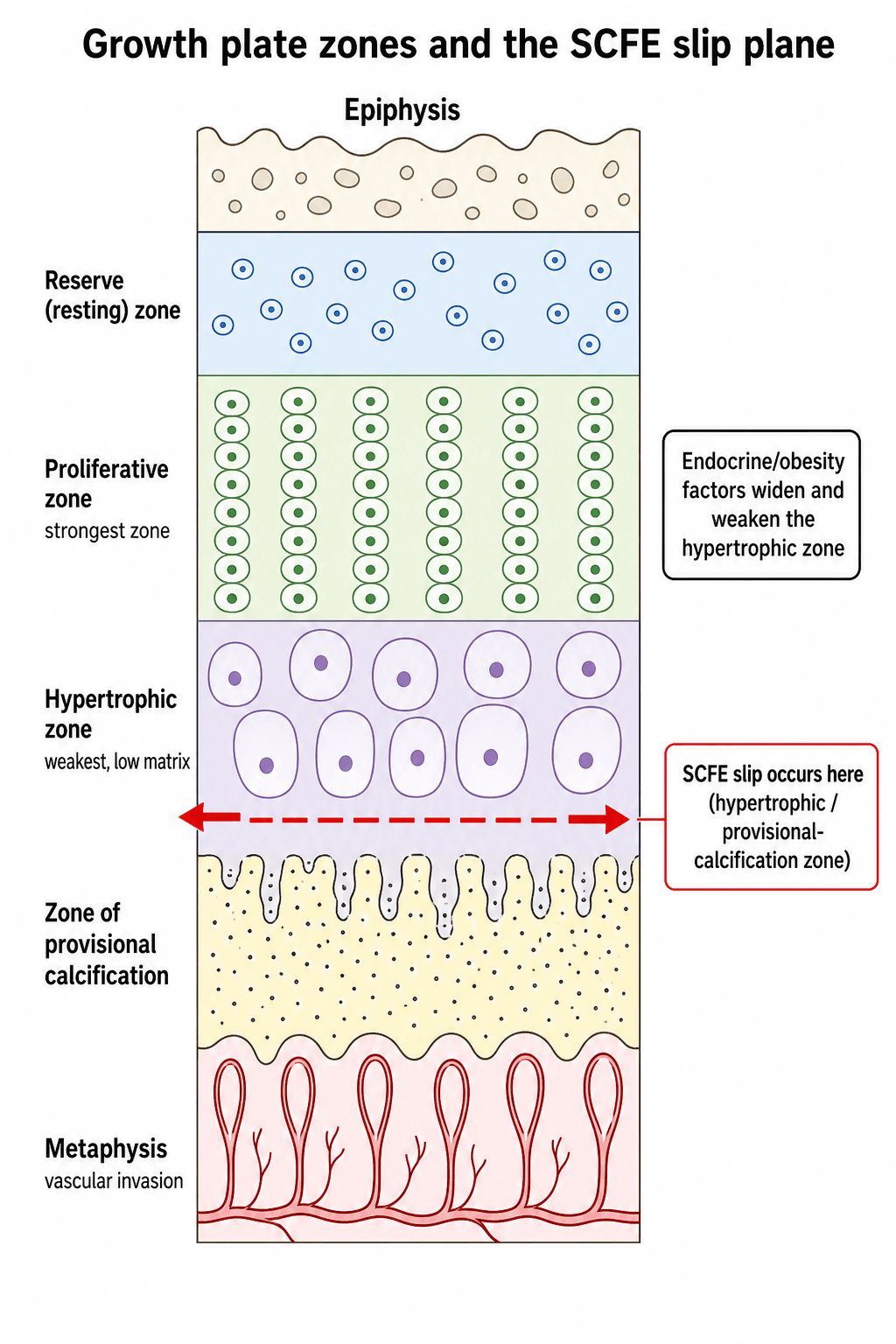
Where the slip happens. The growth plate is layered — from the epiphysis downward into the reserve (resting) zone, the proliferative zone, the hypertrophic zone, and the zone of provisional calcification — before it meets the metaphyseal primary spongiosa. The hypertrophic zone is the mechanically weakest layer, and in SCFE it is abnormally widened and disorganised under combined hormonal and mechanical influence. The adolescent physis is especially vulnerable because, around the growth spurt, it becomes more obliquely/vertically oriented (raising the shear across it), the reinforcing perichondrial ring thins, and sex-hormone / growth-hormone shifts plus excess body weight further reduce its resistance to shear — which is exactly why obesity and endocrinopathy are such strong risk factors. The slip therefore propagates as a cleavage through the hypertrophic zone and zone of provisional calcification: the epiphysis stays seated in the acetabulum while the metaphysis and shaft displace anteriorly and rotate externally. Recognising this cleavage plane explains both why the slip occurs where it does and why a single screw across the physis arrests it.
Why avascular necrosis is the dominant fear. The femoral head is fed by the retinacular vessels (terminal branches of the medial femoral circumflex artery, MFCA), which run along the posterosuperior femoral neck and are held in a periosteal sleeve that stays attached to the epiphysis. When the slip occurs, those vessels are stretched and, in an unstable slip, may already be torn or thrombosed — which is why unstable slips carry such a high AVN rate regardless of treatment.
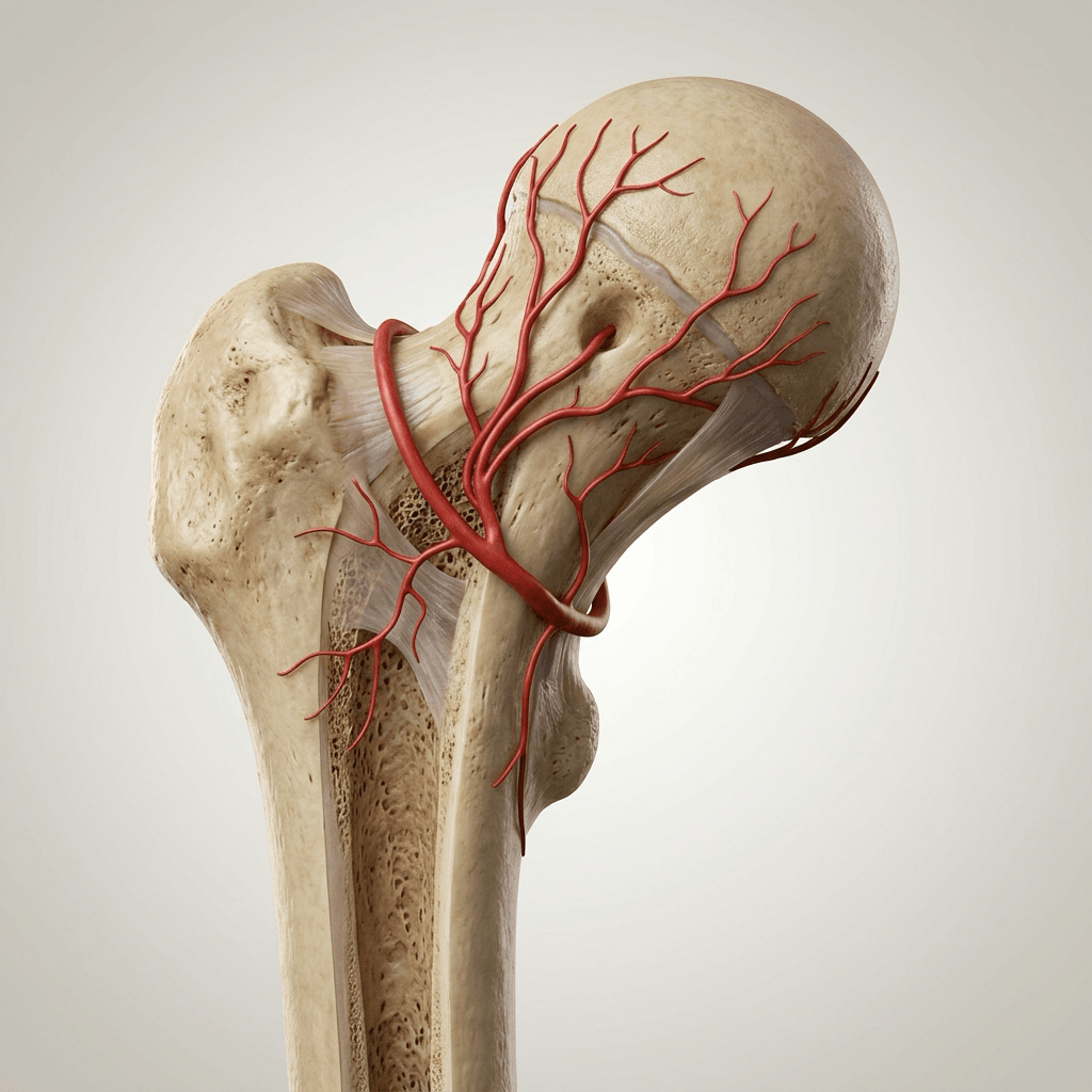
Any forceful reduction — open or closed — or aggressive dissection along the posterosuperior neck risks tearing the retinacular vessels and causing avascular necrosis. This is the single reason in situ fixation without reduction is the rule. The posterior periosteum and its vessels are the structures you are protecting.
Classification
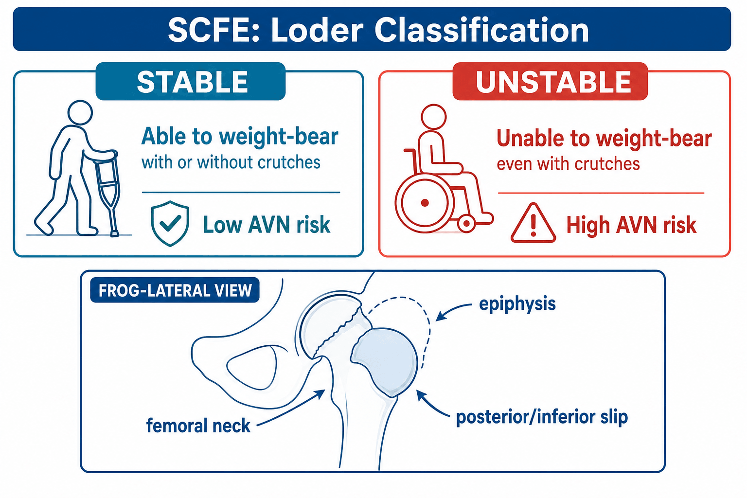
Three classifications are used, but they are not equal: stability (Loder) predicts the outcome, while Southwick grades the deformity and the temporal labels describe the history.
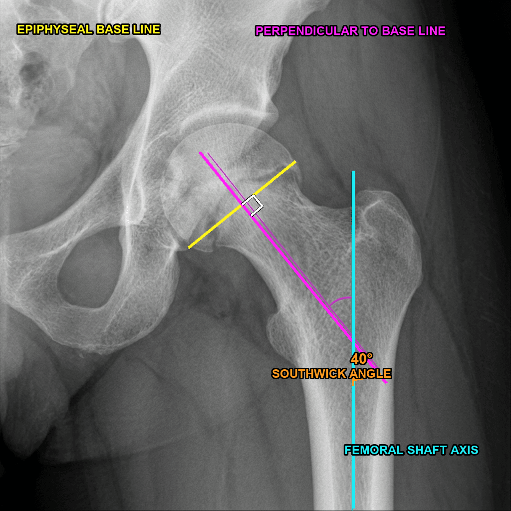
Southwick (severity) is the lateral epiphyseal–shaft (head–shaft) angle, measured ideally on the frog-leg lateral of both hips. How to measure it:
- Mark the anterior and posterior tips of the capital epiphysis at the physis and join them — the epiphyseal base line.
- Draw a line perpendicular to that base line.
- Draw the femoral shaft axis.
- The angle between the perpendicular and the shaft axis is the head–shaft angle.
- Subtract the contralateral (normal) hip's head–shaft angle — the difference is the Southwick slip angle. If both hips are slipped, use the normal reference of ~12°.
- Southwick slip angle
- Less than 30°
- Clinical
- Minimal deformity
- Southwick slip angle
- 30–50°
- Clinical
- Some deformity
- Southwick slip angle
- Greater than 50°
- Clinical
- Significant deformity, cam-FAI risk
Severe slips drive cam-type femoroacetabular impingement and early osteoarthritis, and may need later deformity correction.
Clinical assessment
- Groin, thigh, or knee pain (referred — the classic trap)
- Limp (antalgic or abductor)
- Can the child weight-bear? — the critical question for stability
- Endocrine/renal history; family history of SCFE
- Obligate external rotation with hip flexion (the hallmark)
- Loss of internal rotation
- Apparent limb shortening if a severe slip
- Trendelenburg gait; always examine the other hip
The hallmark sign is obligate external rotation (the Drehmann sign): as you flex the hip it is forced into external rotation, because the anteriorly-displaced metaphysis drives against the anterior acetabulum. It is, in effect, the slip made visible on examination.
Knee or thigh pain in an adolescent = X-ray the hip. Referred pain is common in SCFE, and a child who is "limping with knee pain" but has a normal knee examination must have hip radiographs. Missing a SCFE presenting as knee pain is a classic, avoidable, medicolegal pitfall.
Imaging
Get an AP pelvis and a frog-leg (or cross-table) lateral — the lateral is essential because a mild slip can look normal on the AP. The frog-lateral shows the posterior slip and allows the Southwick angle to be measured.
Learn the early AP signs, because they catch the slip before the displacement is obvious:
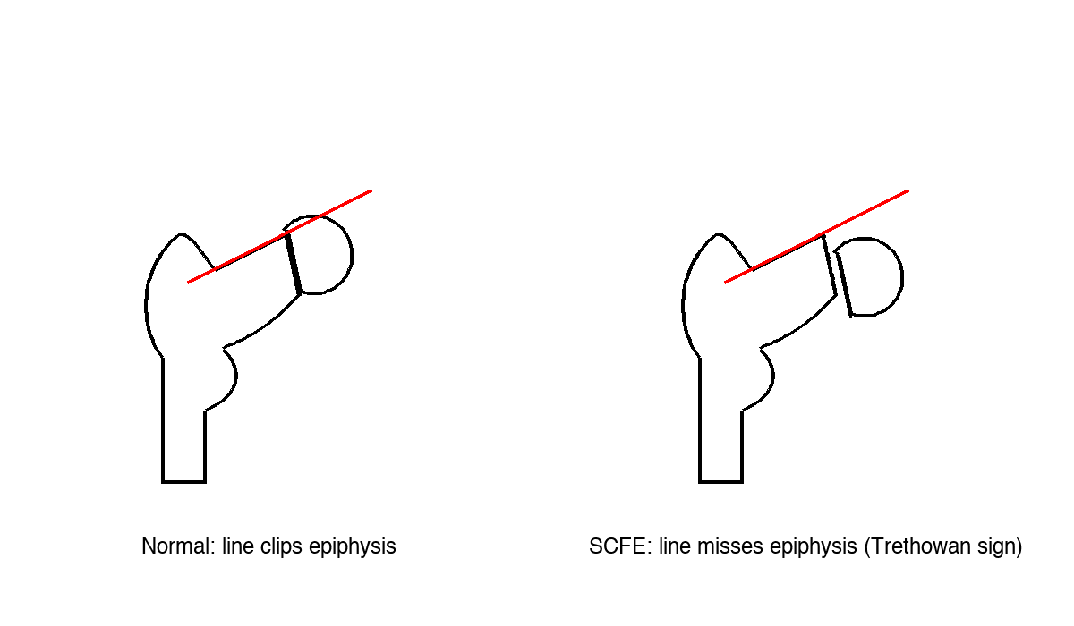
- Klein's line / Trethowan sign — a line along the superior border of the femoral neck should clip the lateral epiphysis; if it does not (Trethowan sign), the epiphysis has slipped. The modified Klein's line (less of the epiphysis projecting lateral to the line than on the normal side) improves sensitivity for subtle slips.
- Steel's metaphyseal blanch sign — a crescent of increased density over the proximal metaphysis where the posteriorly-displaced epiphysis overlaps it; an early sign on the AP.
- Physeal widening and irregularity — the "pre-slip": a widened, fuzzy physis before any displacement.
- Loss of Capener's sign — normally the posteromedial neck overlaps the posterior acetabular wall (a triangular overlap); this triangle is lost as the epiphysis slips.
- Key finding
- Klein's line fails to intersect the epiphysis; physeal widening
- Use
- Detect the slip; compare both hips
- Key finding
- Posterior/inferior slip of the epiphysis
- Use
- Confirm the slip; measure the Southwick angle
- Key finding
- Same findings without moving a painful hip
- Use
- Alternative when the frog-leg is too painful (unstable slip)
MRI is not needed in a classic case with positive films, but it can detect a pre-slip (physeal widening and oedema before displacement) and is sometimes used to assess epiphyseal perfusion in an unstable slip — though it must never delay urgent surgery.
Differential diagnosis
- Typical patient
- 10–16 yr, often obese, growth spurt
- Discriminating features
- Obligate external rotation on flexion; knee/thigh pain
- Key investigation
- AP + frog-leg lateral (Klein's line, Southwick)
- Typical patient
- 4–9 yr, often small/thin
- Discriminating features
- Insidious limp, younger child, lateral pillar changes
- Key investigation
- AP + frog-leg; MRI early
- Typical patient
- Any age, systemic upset
- Discriminating features
- Fever, refusal to bear weight, raised CRP/ESR/WCC, pain on micro-movement
- Key investigation
- Joint aspiration; ultrasound; markers
- Typical patient
- 3–8 yr, post-viral
- Discriminating features
- Well child, settles in days, mild markers
- Key investigation
- Diagnosis of exclusion after sepsis ruled out
- Typical patient
- Athletic adolescent, RED-S
- Discriminating features
- Activity-related groin pain, normal early X-ray
- Key investigation
- MRI
Two cannot-miss traps sit on either side of SCFE: a febrile child who refuses to weight-bear is septic arthritis until proven otherwise (aspirate, do not just X-ray), and a well adolescent with isolated knee pain may have a SCFE (always image the hip).
Management
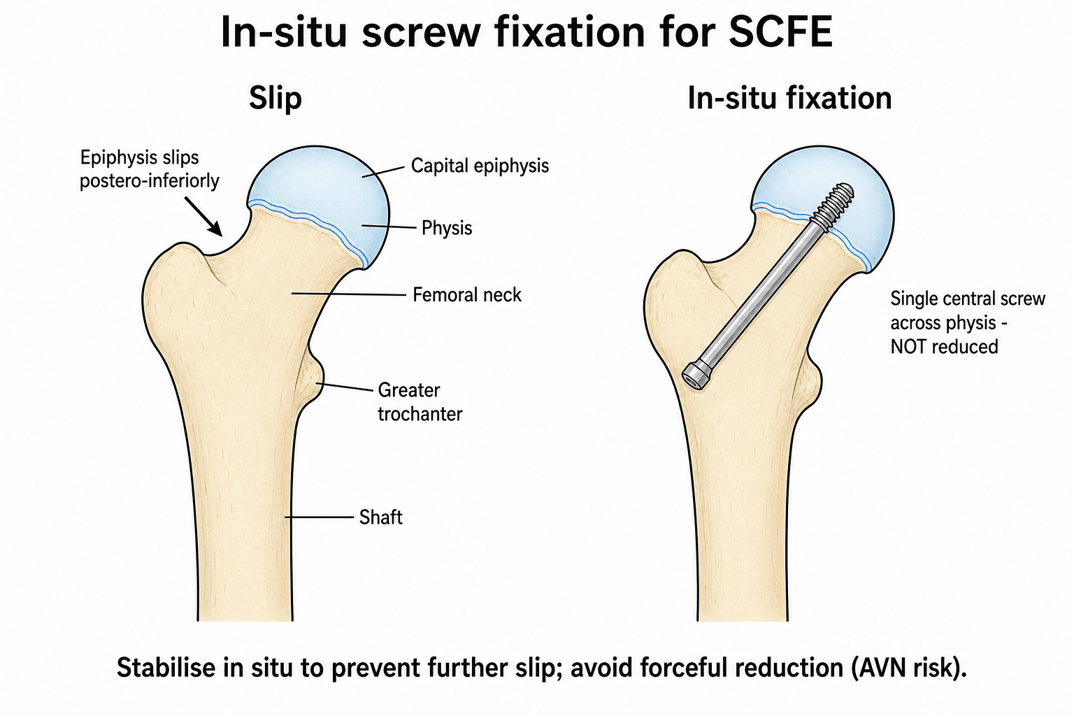
In situ fixation — do NOT reduce. This is the single most important principle in SCFE. Any attempt at reduction, open or closed, dramatically increases the AVN risk (up to 100% in some series). Even a severe slip is pinned in situ and the residual deformity addressed later; that is safer than realigning the head acutely.
The pathway is the same in principle for every slip — confirm stability, fix in situ, then decide about the other hip — but the urgency differs sharply with stability.
- 1Offload & classifyMake the child non-weight-bearing immediately and classify stability (Loder): can they weight-bear or not? This sets both urgency and prognosis.
- 2Fix in situSingle central cannulated screw, perpendicular to the physis, aimed at the centre of the epiphysis on both views — never a forced reduction. Stable: semi-urgent (days). Unstable: urgent (within ~24 h), gentle positioning only, no traction.
- 3Decide on the other hipCounsel about the 20–40% bilateral risk and consider prophylactic fixation — most strongly when the child is young (under ~13), or has an endocrine/renal cause or unreliable follow-up.
- Stable SCFE
- Able to walk (± crutches)
- Unstable SCFE
- Unable to walk even with crutches
- Stable SCFE
- Less than 1%
- Unstable SCFE
- ~47%
- Stable SCFE
- Semi-urgent (days)
- Unstable SCFE
- Emergency (within ~24 h)
- Stable SCFE
- In situ single screw
- Unstable SCFE
- In situ single screw, gentle positioning only
- Stable SCFE
- Excellent
- Unstable SCFE
- Guarded (high AVN risk)
In situ single-screw fixation. Supine on a fracture table with fluoroscopy. Enter the anterolateral femur (below the vastus lateralis ridge) so the screw lines up with the centre of the epiphysis on the lateral view. Aim for the centre of the epiphysis on both AP and lateral, perpendicular to the physis, with at least five threads crossing into the epiphysis. Confirm no joint penetration on a frog-lateral (tip more than ~5 mm from the articular surface). A single screw is sufficient for almost all slips — extra screws add AVN risk without adding stability.
Complications
- Cause
- Reduction attempt; unstable slip vascular insult
- Prevention / treatment
- In situ fixation, no reduction
- Cause
- Screw penetration of the joint
- Prevention / treatment
- Confirm no penetration on live fluoroscopy
- Cause
- Inadequate fixation or missed diagnosis
- Prevention / treatment
- Secure central screw placement
- Cause
- Residual metaphyseal prominence
- Prevention / treatment
- Later osteochondroplasty or osteotomy
- Cause
- Natural history (20–40%)
- Prevention / treatment
- Prophylactic pinning if high-risk
- Cause
- Screw prominence / backout
- Prevention / treatment
- Bury the head; confirm fixation
The numbers worth carrying: AVN is under 1% in stable slips but around 47%unstable in unstable slips — and rises steeply with any reduction attempt. AVN, when it occurs, is the principal driver of a poor long-term hip; cam-FAI from residual deformity is the slower, later cause of osteoarthritis even in mild slips.
Chondrolysis deserves emphasis as a distinct complication. It is acute loss of articular cartilage, defined radiographically as joint-space narrowing to under ~3 mm (or under 50% of the contralateral hip), and it presents with progressive pain and a globally stiff, restricted hip. The classic and avoidable cause is persistent intra-articular screw penetration — so live fluoroscopy through a full arc (the "approach-withdrawal" technique) to exclude penetration is mandatory. Crucially, however, chondrolysis can also occur idiopathically — without any pin penetration — and is associated with severe slips and prolonged spica immobilisation. Management is to confirm there is no retained intra-articular hardware, then NSAIDs, analgesia and physiotherapy to maintain motion; it is often partly self-limiting but can leave permanent stiffness.
Guidelines, registries & global practice
- Overall incidence ~10–11 per 100,000 children aged 9–16 (US data); regional rates vary widely
- Boys more than girls (~13 vs 8 per 100,000); peak at the growth spurt
- Much higher in African and Pacific Islander ancestry; lowest in East Asian populations
- Rising in parallel with childhood obesity; bilateral disease in ~18–50% across series
- No dedicated SCFE registry; evidence comes from national paediatric cohorts (Swedish, Norwegian) and the US KID database
- National cohorts identify younger chronological age as the dominant contralateral-slip predictor
- Long-term cohorts confirm cam-FAI and early OA as the principal late burden, even after mild slips
- Modified-Dunn durability data come from single expert centres, not population registries
- Common ground (AAOS / BOA-BSCOS / AO / EFORT)
- AP + frog-leg (or cross-table) lateral; treat adolescent knee/thigh pain as a hip until excluded
- Where practice genuinely varies
- Threshold for MRI to detect a pre-slip or assess perfusion
- Common ground (AAOS / BOA-BSCOS / AO / EFORT)
- In situ fixation for stable and most unstable slips; avoid forced reduction
- Where practice genuinely varies
- Single vs two screws for unstable slips; screw vs smooth pins in the very young
- Common ground (AAOS / BOA-BSCOS / AO / EFORT)
- Urgent stabilisation; non-weight-bearing until theatre
- Where practice genuinely varies
- Emergency under 24 h vs planned-urgent after synovitis settles; capsular decompression debated
- Common ground (AAOS / BOA-BSCOS / AO / EFORT)
- Address symptomatic cam-FAI; protect the MFCA
- Where practice genuinely varies
- Modified Dunn (capable centres) vs Imhauser osteotomy vs osteochondroplasty
- Common ground (AAOS / BOA-BSCOS / AO / EFORT)
- Counsel on bilateral risk; monitor to maturity
- Where practice genuinely varies
- Routine prophylaxis under age 13 vs selective vs watchful waiting
Special situations & controversies
Suspect an endocrine cause if the slip is bilateral, the child is younger than usual, short, or has delayed puberty. Screen for hypothyroidism, growth-hormone deficiency, hypogonadism and renal osteodystrophy — and lower the threshold for prophylactic contralateral fixation.
The classical doctrine is no forced reduction. Modified-Dunn series report anatomical realignment with low AVN, but only in expert centres; elsewhere AVN climbs steeply, and there is no randomised comparison. Outside high-volume units, in situ pinning is the safe default.
Some argue urgent decompression of the tense haemarthrosis in an unstable slip relieves tamponade on the retinacular vessels and lowers AVN. Evidence is conflicting and it is not universally adopted.
A chronic healed slip may present years later with cam-FAI and early osteoarthritis. Correct the mechanics with an osteotomy or osteochondroplasty — but avoid osteotomy through the old slip site, which carries a high AVN risk even years on.
Exam & revision
Everything below condenses SCFE for revision and viva practice — the high-yield points, the memory hooks, worked vivas, rapid-fire MCQ points, and a one-screen cheat sheet.
- Stability (Loder) predicts AVN — can the child weight-bear? Stable under 1%, unstable ~47%.
- In situ fixation, never a forced reduction — reduction is the route to AVN.
- Single central screw, perpendicular to the physis — extra screws add AVN risk, not stability.
- Unstable = emergency (~24 h); stable = semi-urgent (days) — and non-weight-bearing until theatre either way.
- Knee/thigh pain in an adolescent = hip X-ray — and obligate external rotation on flexion is the hallmark sign.
- 20–40% bilateral — consider prophylactic pinning when young (under ~13), endocrine/renal, or unreliable follow-up.
FAT HIPSCFE risk factors
Hook:FAT HIP — the fat hip is at risk of slipping.
SCREWIn situ fixation principles
Hook:Get the SCREW right — single, central, perpendicular.
Viva practice
Practise clinical reasoning and management decisions out loud
“A 13-year-old obese boy presents with 3 weeks of left groin and thigh pain. He is able to walk with a limp. Examination shows obligate external rotation with hip flexion and loss of internal rotation. X-rays show a SCFE with Southwick angle 35 degrees. How would you manage this?”
“A 12-year-old girl presents with sudden onset left hip pain after a minor fall. She is unable to weight-bear. The leg is held in external rotation and she screams with any hip movement. X-rays show a significantly displaced SCFE. How would you manage this?”
“A 10-year-old boy with known hypothyroidism presents with new right hip pain. He had a left SCFE pinned 6 months ago. X-rays show a new right SCFE. How would you manage this, and what would you do differently if asked at the time of the first SCFE?”
Key facts
- Peak age 10–16 years; obesity the main risk factor
- 20–40% bilateral risk
- Knee/thigh pain in an adolescent = X-ray the hip
- Hallmark sign: obligate external rotation on flexion
Loder classification
- Stable = can weight-bear → under 1% AVN
- Unstable = cannot weight-bear → ~47% AVN
- Stability predicts outcome better than symptom duration
Treatment principles
- In situ fixation — do NOT reduce
- Single screw, central, perpendicular to the physis
- Unstable = emergency (~24 h); stable = semi-urgent (days)
- Consider contralateral prophylaxis if young/endocrine/renal
Radiographic signs
- Klein's line fails to intersect the epiphysis (AP)
- Posterior slip on the frog-leg lateral
- Southwick angle grades severity
- Physeal widening — an early sign
Evidence
Loder et al. - Landmark Stability Classification
- 55 hips presenting acutely (symptoms under 3 weeks) reclassified by physeal stability
- 30 unstable (cannot weight-bear even with crutches), 25 stable (can weight-bear)
- AVN developed in 14 of 30 unstable hips (47%) and 0 of 25 stable hips
- No demonstrable association between early reduction and AVN in this series
Aronsson & Loder - Treatment of the Unstable Slip
- Treatment priorities: avoid AVN, avoid chondrolysis, prevent further slip, then correct deformity
- Manipulative reduction and acute corrective osteotomy NOT recommended (high AVN/chondrolysis)
- Recommends preoperative bed rest to settle synovitis, then stabilisation with a single central screw
- Careful positioning on the fracture table may give incidental reduction - no active manipulation
Lehmann et al. - Epidemiology Update
- Overall US incidence 10.8 per 100,000 children aged 9-16 years (Kids' Inpatient Database)
- Incidence 3.94x higher in Black and 2.53x higher in Hispanic children vs White children
- Higher in boys (13.4/100,000) than girls (8.1/100,000)
- Seasonal and latitude variation suggests environmental contribution
Tannast/Ziebarth et al. - Modified Dunn (Bernese)
- Modified Dunn via surgical hip dislocation with a retinacular soft-tissue flap protecting the MFCA
- AVN rate at the inventor institution approximately 2% - far lower than historical open reduction
- AVN occurred only where no head perfusion was evident before reduction
- Other centres report AVN up to 24% during their early learning curve
Ziebarth et al. - Modified Dunn 10-Year Outcomes
- 43 hips (mild to severe slips) treated with modified Dunn, 98% available at minimum 10 years
- Cumulative survivorship 93% at 10 years (95% CI 85-100%)
- No hips showed AVN on plain radiographs at follow-up
- Secondary impingement persisted in some hips; 14% needed further surgery for impingement
Lindell et al. - Prophylactic Fixation Algorithm
- National cohort of 379 children with SCFE (2007-2013)
- Chronological age was the only independent predictor of a subsequent contralateral slip
- Using age under 13 years as the threshold: sensitivity 88%, specificity 51% for preventing contralateral slip
- Triradiate cartilage assessment had poor inter-observer agreement and was unreliable
Ziebarth/Leunig et al. - Cartilage Damage & Perfusion
- 119 SCFE hips assessed at open surgery; acetabular cartilage damage in 97 of 109 (89%)
- Cam impingement from the slip - not slip angle alone - drives acetabular cartilage damage
- In disconnected (unstable) epiphyses, perfusion loss increased with longer time to surgery
- Posterior callus resection improved laser-Doppler epiphyseal perfusion
Loder - Controversies in SCFE
- Frames the four enduring controversies: unstable slip management, role of osteotomy, contralateral prophylaxis, fixation in the very young
- Reduction of the unstable slip remains contentious because of AVN risk
- Decision analysis is needed to weigh prophylactic fixation against expectant follow-up
- Implant choice in young children must allow continued growth