90% Posterior | Reduce within 6 Hours | Sciatic Nerve at Risk
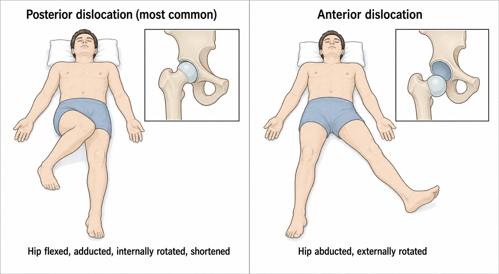
- Posterior dislocation is most common (90%) - mechanism is dashboard injury (knee hitting dashboard = flexed hip)
- Sciatic nerve at risk in posterior dislocation (10-20%) - peroneal division most vulnerable
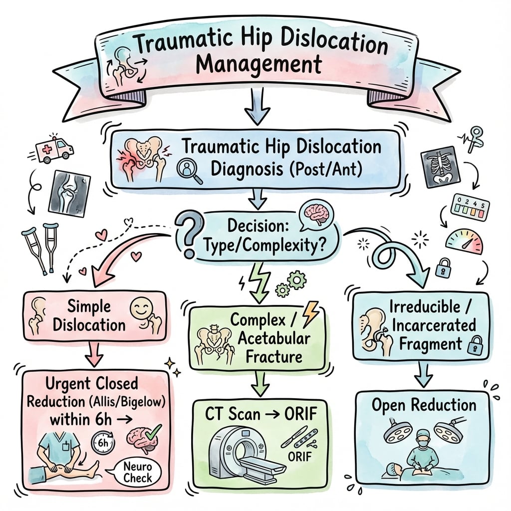
- 6-hour golden window - AVN risk increases significantly if reduction delayed beyond 6 hours
- Associated fractures common - femoral head (Pipkin), acetabulum (posterior wall), femoral neck
- CT post-reduction mandatory to assess concentric reduction and associated fractures
- “Posterior: leg shortened, adducted, internally rotated. Anterior: externally rotated, abducted
- “Sciatic nerve palsy: most recover by 2 years, explore if no recovery by 6 months
- “Pipkin I and II = different - Pipkin I below fovea (ORIF), Pipkin II above fovea (excise if small)
- “Thompson-Epstein classification for posterior; Epstein classification for anterior
6-hour golden window for reduction. AVN risk increases from 5% (under 6h) to 40%+ (over 12h). This is a true orthopaedic emergency. Reduce as soon as possible.
10-20% sciatic nerve injury in posterior dislocation. Peroneal division most vulnerable. Document nerve function BEFORE and AFTER reduction. Most recover by 2 years.
High-energy mechanism - look for associated injuries: femoral head fracture (Pipkin), acetabular fracture (posterior wall), femoral neck fracture (15%), knee injuries (PCL, patella).
CT mandatory after reduction. Assess for: concentric reduction, incarcerated fragments, femoral head impaction, acetabular fracture, femoral neck fracture.
- Key Finding
- No fracture on X-ray
- Treatment
- Urgent closed reduction, CT after
- Pearl
- 6-hour window - treat as emergency
- Key Finding
- Posterior wall on CT
- Treatment
- Reduce, CT assess wall size, ORIF wall
- Pearl
- Wall over 40% = unstable, needs fixation
- Key Finding
- Femoral head fragment
- Treatment
- Reduce, Pipkin classification guides Rx
- Pearl
- Pipkin I (below fovea) = consider ORIF
- Key Finding
- Femoral neck visible
- Treatment
- Cannot reduce closed - open reduction
- Pearl
- Devastating injury - high AVN risk
- Key Finding
- Leg externally rotated, abducted
- Treatment
- Closed reduction
- Pearl
- Less common (10%), check femoral vessels
SAIDPosterior Hip Dislocation Position
Hook:The patient SAID 'my knee hit the dashboard' - leg is Short, Adducted, Internally rotated, from Dashboard injury!
FABERAnterior Hip Dislocation Position
Hook:Anterior dislocation puts the hip in forced FABER position!
6 AVN6-Hour Rule Importance
Hook:After 6 hours, AVN risk climbs - '6 AVN' reminds you time is hip!
PERONEAL PRONESciatic Nerve in Posterior Dislocation
Hook:Peroneal is prone to injury when the hip goes posterior!
Overview and Epidemiology
Hip dislocation is a true orthopaedic emergency with time-sensitive management. The examiner will test your understanding of the 6-hour rule, sciatic nerve considerations, and associated injuries. Getting the basics wrong shows poor emergency management understanding.
- Young adults (MVA, high-energy trauma)
- Native hips - dislocation rare in healthy hip
- Male predominance (2:1) - high-risk activities
- Prosthetic hips - much more common (different topic)
- MVA most common (dashboard injury)
- High-energy sports (rugby, American football, skiing)
- Industrial accidents
- Fall from height
- Force vector determines direction
Anatomy and Mechanism
The hip is inherently stable - a ball-and-socket joint requiring significant force to dislocate. Stability comes from: bony congruity, acetabular labrum, strong capsular ligaments (iliofemoral, pubofemoral, ischiofemoral), and surrounding muscles.
Femoral Head Blood Supply
- Source
- Profunda femoris
- Contribution
- 80-90% of head
- Clinical Relevance
- Damaged in dislocation - AVN risk
- Source
- Profunda femoris
- Contribution
- Minimal
- Clinical Relevance
- Minor contribution
- Source
- Obturator artery
- Contribution
- 10-20% (variable)
- Clinical Relevance
- More important in children
- Source
- Metaphyseal
- Contribution
- Variable
- Clinical Relevance
- May be compromised
Dislocation stretches/tears the retinacular vessels (from MFCA) that supply the femoral head. Prolonged dislocation = prolonged ischemia. AVN risk doubles after 6 hours and approaches 50% after 12-24 hours.
Classification Systems
Thompson-Epstein Classification (Posterior)
- Description
- Simple dislocation, no/minimal fracture
- Treatment
- Closed reduction, conservative
- Description
- Posterior wall rim fracture (small)
- Treatment
- Closed reduction, assess stability
- Description
- Large posterior wall fracture (unstable)
- Treatment
- ORIF posterior wall
- Description
- With acetabular floor fracture
- Treatment
- ORIF acetabulum
- Description
- With femoral head fracture (Pipkin)
- Treatment
- Pipkin classification guides treatment
In practice, think: Simple dislocation (reduce urgently) vs Complex (with fracture - reduce, then definitive fix). CT post-reduction determines if wall/head fixation needed.
Clinical Assessment
- Mechanism: MVA, dashboard, sport - force vector matters
- Time of injury: 6-hour rule critical
- Associated injuries: polytrauma? knee injury?
- Medical history: native vs prosthetic hip
- Anticoagulation: bleeding risk assessment
- ATLS first in polytrauma
- Obvious deformity - position diagnostic
- Leg length discrepancy
- Neurovascular status: sciatic (peroneal), femoral
- Knee examination: PCL, patella (dashboard)
- Log roll: pelvic stability
- Posterior
- Shortened, adducted, IR (SAID)
- Anterior
- Abducted, externally rotated
- Posterior
- Flexed
- Anterior
- May be extended
- Posterior
- Not anteriorly (empty)
- Anterior
- Palpable in groin (bulge)
- Posterior
- Sciatic (peroneal)
- Anterior
- Femoral, lateral cutaneous
- Posterior
- 90%
- Anterior
- 10%
ATLS takes priority. Hip dislocation often occurs in polytrauma setting. Complete primary and secondary survey. Missed ipsilateral femoral neck fracture is a disaster - do not attempt closed reduction if neck fracture suspected.
- Discriminating Features
- Shortened, adducted, internally rotated (SAID); empty acetabulum
- Key Imaging
- AP pelvis: head superolateral, smaller apparent head
- Why It Matters
- Time-critical reduction to limit AVN
- Discriminating Features
- Abducted, externally rotated; head palpable in groin
- Key Imaging
- AP pelvis: head infero/anteromedial, larger apparent head
- Why It Matters
- Different reduction vector; femoral vessel proximity
- Discriminating Features
- Shortened, externally rotated; unable to straight-leg raise
- Key Imaging
- AP/lateral hip; CT if occult
- Why It Matters
- Closed reduction of a coexisting dislocation can displace/comminute the neck - catastrophic
- Discriminating Features
- Shortened, externally rotated; elderly low-energy
- Key Imaging
- AP hip/pelvis radiograph
- Why It Matters
- No dislocation; different fixation pathway
- Discriminating Features
- Pain, reduced weight-bearing; head may be central/subluxed
- Key Imaging
- Judet views + CT
- Why It Matters
- May be central fracture-dislocation; staged reconstruction
- Discriminating Features
- Surgical scar, known arthroplasty, low-energy mechanism
- Key Imaging
- Radiograph shows implant
- Why It Matters
- Prosthetic instability has a separate work-up and reduction risk profile
The single most dangerous differential is a coexisting ipsilateral femoral neck fracture. If the neck is fractured, closed reduction of the dislocation can convert a salvageable injury into a displaced neck fracture with devastating AVN risk. Scrutinise the neck on the pre-reduction film, and if there is any doubt obtain CT or proceed to open reduction.
Investigations
Imaging Protocol
Diagnosis usually obvious. Look for associated fractures: posterior wall, femoral head (shenton's arc), femoral neck. Do not delay reduction for extensive imaging.
Judet views if stable. Do not delay reduction - X-ray after if unstable patient.
Assess: concentric reduction, incarcerated fragments, femoral head impaction, posterior wall size, occult femoral neck fracture. This dictates further treatment.
Suspected labral pathology, soft tissue incarceration, early AVN assessment. Not urgent.
X-ray Signs of Hip Dislocation
- Head superolateral to acetabulum
- Smaller apparent head size (further from plate)
- Shenton's line disrupted
- Look for posterior wall fragment
- Internal rotation of lesser trochanter
- Head inferomedial (obturator) or anteromedial (pubic)
- Larger apparent head size (closer to plate)
- Shenton's line disrupted
- External rotation - lesser trochanter prominent
- Head may overlap obturator foramen

Management Algorithm
Reduce within 6 hours! Every hour of dislocation increases AVN risk. Under 6h = 5% AVN. 6-12h = 15-20% AVN. Over 12h = 30-50% AVN. This is non-negotiable - prioritize reduction.
Closed Reduction Technique - Comprehensive Guide
Pre-Procedure Preparation
- Time of injury - 6-hour rule critical
- Sciatic nerve exam - document BEFORE reduction
- Peroneal: dorsiflexion, toe extension, dorsal foot sensation
- Tibial: plantarflexion, plantar sensation
- Informed consent - AVN, nerve injury, need for open procedure
- General anaesthesia or deep sedation - muscle relaxation essential
- Theatre or ED resuscitation bay (if stable)
- Fluoroscopy available - confirm reduction
- Assistant to stabilize pelvis - this is crucial
- Padded table - patient supine
- Backup plan for open reduction if needed
Allis Maneuver (Preferred Technique)
Step-by-Step Allis Maneuver
Supine on firm table. Assistant positioned at patient's pelvis with hands pressing down on ASIS bilaterally. This counterforce is essential - without it, you'll pull the whole patient toward you.
Stand at the side of the table, facing the patient's head. Flex the hip to 90° and knee to 90°. This relaxes the iliofemoral ligament (strongest ligament in body) and positions the head at the acetabular opening.
Grasp the proximal tibia/knee region. Apply steady, in-line axial traction pulling the femur toward the ceiling (with hip at 90°). This is NOT a jerking motion - steady sustained force.
- Some surgeons place knee in crook of elbow for leverage
- Full body weight can be used - stand on table if needed
While maintaining traction, apply gentle internal and external rotation to the leg. This helps disengage the femoral head from behind the acetabulum and guides it over the rim.
- Listen/feel for a satisfying clunk of reduction
- Do NOT use excessive force or rotation
Once reduced, leg should return to normal length and alignment. Test ROM carefully - hip should move through flexion, extension, rotation smoothly. Obtain fluoroscopy or X-ray immediately.
For strong patients or difficult reductions, stand on the table for better leverage. Place the patient's knee in the crook of your elbow, hold the tibia, and use your body weight to provide steady axial traction. This is the "Captain Morgan" technique variation.
Alternative: Stimson Maneuver
- Patient prone on table with injured leg hanging off
- Hip flexed over edge of table (90°)
- Knee flexed to 90°
- Gravity provides traction on the hanging limb
- Assistant stabilizes pelvis
- Surgeon applies downward pressure on calf with gentle rotation
- Useful when Allis fails, but requires rolling patient prone
Advantages:
- Gravity provides continuous traction
- Less physical effort for surgeon
- Muscle relaxation with gravity
Disadvantages:
- Requires rolling polytrauma patient prone
- Cannot visualize patient face (airway)
- Not suitable for unstable patients
Critical Technical Points
- Document sciatic nerve function before AND after
- Ensure adequate sedation/relaxation - fighting muscles = failure
- Use steady sustained traction - not jerky movements
- Have assistant stabilize pelvis firmly
- Confirm reduction with imaging immediately
- Get CT post-reduction - mandatory
- Don't use excessive force - risk of iatrogenic fracture
- Don't attempt more than 2-3 times - go to open if failing
- Don't reduce if femoral neck fracture suspected - catastrophic
- Don't forget post-reduction CT - miss fragments or wall
- Don't delay for imaging - time is hip, reduce first
- Don't wait for specialist if unavailable - reduce within 6h
Troubleshooting Failed Reduction
- Likely Cause
- Incarcerated fragment (labrum, osteochondral)
- Solution
- Proceed to open reduction - cannot force closed
- Likely Cause
- Posterior wall fracture (unstable)
- Solution
- Maintain reduction with traction, CT, plan ORIF wall
- Likely Cause
- Inadequate muscle relaxation
- Solution
- Increase sedation/paralysis, ensure full GA
- Likely Cause
- Not actually reduced, head still out
- Solution
- Obtain fluoro immediately, reattempt or open
- Likely Cause
- Incarcerated soft tissue or fragment
- Solution
- Urgent operative removal of incarcerated tissue
Stop closed reduction if: (1) X-ray shows femoral neck fracture - closed forces will displace it, (2) More than 2-3 gentle attempts fail - something is blocking reduction, (3) Patient becomes hemodynamically unstable. Proceed to open reduction urgently - still need to meet 6-hour window.

A traumatic hip dislocation that presents late — weeks to months unreduced is a recognised problem, especially where access to acute care is limited, and it behaves very differently from the acute injury. The femoral head becomes fixed in soft-tissue scar with progressive cartilage loss and capsular contracture, so simple closed reduction is almost never possible and must not be forced (high risk of fracturing the femoral neck or shaft). Management is staged and age/cartilage-dependent: a period of preliminary skeletal traction to gradually bring the head down, then open reduction (capsulotomy, clearance of the acetabular scar and fibrofatty tissue, and sometimes a femoral shortening osteotomy to allow tension-free relocation and protect the sciatic nerve). In a young patient with preserved cartilage the aim is to relocate and preserve the joint, accepting a high AVN/arthrosis risk; where the head or cartilage is destroyed, arthrodesis or arthroplasty is selected by age and demand. The honest viva point: outcomes are guarded and the patient must be counselled that AVN and post-traumatic arthritis are likely.
Surgical Technique
Closed Reduction Technique
- Adequate anesthesia and muscle relaxation (GA preferred)
- Fluoroscopy available
- Assistant required for counter-traction
- Patient supine, assistant stabilizes pelvis
- Hip flexed to 90 degrees, knee flexed
- Inline traction along femoral axis
- Gentle internal rotation while maintaining traction
- Feel for "clunk" as head relocates
- Assess stability through range of motion
- External rotation, extension, and traction
- Less common, often require open reduction
- Fluoroscopy to confirm concentric reduction
- Full ROM under fluoro to assess stability
- CT scan to exclude loose fragments or fracture
Emergency reduction within 6 hours is critical to minimize AVN risk.
Complications
- Incidence
- 10-40%
- Risk Factors
- Delayed reduction over 6h, repeated attempts
- Management
- Surveillance, THA if symptomatic
- Incidence
- 10-20%
- Risk Factors
- Posterior dislocation, bone fragments
- Management
- Observe, most recover by 2 years
- Incidence
- 20-40%
- Risk Factors
- Cartilage damage, malreduction, AVN
- Management
- Activity modification, THA long-term
- Incidence
- Rare in native hip
- Risk Factors
- Posterior wall deficiency, malreduction
- Management
- Revision ORIF or THA
- Incidence
- 3-5%
- Risk Factors
- Delayed surgery, brain injury
- Management
- Prophylaxis: indomethacin or RT
- Incidence
- Variable
- Risk Factors
- AVN, untreated Pipkin
- Management
- THA
Avascular Necrosis
AVN may take 6-24 months to become apparent on X-ray. MRI can detect earlier (marrow changes). Follow with X-rays at 6 weeks, 3 months, 6 months, 1 year, 2 years. Treat symptomatic AVN with THA.
Postoperative Care and Rehabilitation
Rehabilitation Protocol
Toe-touch or protected weight bearing depending on stability. Bed rest initially. Thromboprophylaxis. ROM exercises.
If stable and no fracture: progress to full weight bearing. If ORIF: protected until fracture healing assessed on X-ray.
Full weight bearing. Progressive strengthening. Gait training. Hip abductor focus.
Return to sport/activity once strength restored. Long-term surveillance for AVN (X-rays).
Simple dislocation (no fracture, concentric reduction): May weight bear as tolerated early. With posterior wall ORIF: Protected until wall healed (6-12 weeks). Pipkin ORIF: Depends on fragment size and fixation.
Outcomes and Prognosis
Prognostic Factors
- Good Outcome Rate
- 70-80%
- Key Factors
- Early reduction, no fracture
- Complications
- AVN 5-10%, OA 15-20%
- Good Outcome Rate
- 50-70%
- Key Factors
- Quality of reduction, wall stability
- Complications
- AVN 10-20%, OA 20-40%
- Good Outcome Rate
- 60-70%
- Key Factors
- Fragment size, excision vs ORIF
- Complications
- AVN 10-20%
- Good Outcome Rate
- 40-60%
- Key Factors
- Articular involvement, reduction quality
- Complications
- AVN 20-30%, OA 30-50%
- Good Outcome Rate
- 20-40%
- Key Factors
- Age, dual blood supply injury
- Complications
- AVN 40-60%, often THA
Factors Affecting Outcome
Reduction within 6 hours, Simple dislocation without fracture, Concentric stable reduction achieved, Young patient without comorbidities, No sciatic nerve injury
Delayed reduction over 6-12 hours, Associated acetabular or femoral head fracture, Non-concentric or unstable reduction, Sciatic nerve injury (peroneal component), Repeated closed reduction attempts
Long-term Outcomes
Most patients with simple posterior dislocations reduced within 6 hours have good functional outcomes. The main determinants of long-term outcome are development of AVN and post-traumatic osteoarthritis. Younger patients may tolerate mild degenerative change better than older patients. THA remains the salvage procedure for severe AVN or debilitating arthritis.
Guidelines, Registries & Global Practice
- Traumatic native-hip dislocation is uncommon and high-energy worldwide; the great majority (~85-90%) are posterior
- Dominant mechanism globally is road traffic collision (dashboard/front-seat passenger); motorcycle and pedestrian trauma predominate in low- and middle-income settings
- Geographic remoteness and retrieval logistics — not injury energy alone — drive time-to-reduction and therefore AVN risk; rural and low/middle-income settings see the worst delays despite road-safety legislation
- Sport (rugby codes, American football, skiing, soccer) is a recognised cause in young athletes
- In children, posterior dislocation predominates and AVN remains the key long-term adverse event (posterior ~86%, AVN ~15% of associated pathologies in a paediatric systematic review, Baumann 2023, PMID 37947036)
- Most fragility-type hip "dislocations" in the elderly are in fact prosthetic instability - a separate clinical entity
- Where reduction happens varies: many systems reduce in the emergency department under procedural sedation; others mandate theatre/GA - both are defensible if muscle relaxation is adequate
- Time-to-reduction targets differ but the universal principle is "as soon as safely possible"; the historical 6-hour figure is a teaching anchor, not a regulatory threshold
- Stability assessment of intermediate posterior wall fractures varies between CT-only and examination under anaesthesia (EUA), with trauma units increasingly favouring EUA (Moed, PMID 19104298)
- Approach for femoral head fractures varies between anterior (Smith-Petersen), surgical hip dislocation (Ganz) and posterior (Kocher-Langenbeck), with a trend toward vascular-protective approaches
- Position on Hip Dislocation
- Emergent reduction of the dislocated hip; mandatory post-reduction CT; EUA or CT to decide posterior wall stability; ORIF for unstable walls
- Evidence Basis
- Expert consensus + retrospective series (Moed, Grimshaw)
- Position on Hip Dislocation
- Polytrauma hips managed in the ATLS framework within trauma networks; urgent reduction and senior-led decision-making; combined ortho-plastic and pelvic/acetabular referral pathways for associated fractures
- Evidence Basis
- BOAST standards for pelvic/acetabular and open fractures (principle-level)
- Position on Hip Dislocation
- Reduction priority, careful classification (Thompson-Epstein, Pipkin/Brumback), MFCA-protective approaches, anatomic articular reconstruction of wall/head fractures
- Evidence Basis
- AO surgical principles and technique teaching
- Position on Hip Dislocation
- Reinforces prompt reduction, mandatory cross-sectional imaging, and referral of complex fracture-dislocations to specialist acetabular units
- Evidence Basis
- European educational consensus
There is no high-level (NICE-style) clinical guideline specific to traumatic native hip dislocation because the injury is rare and randomised data are lacking. Practice rests on retrospective cohorts and meta-analysis (Hougaard 1986, Kellam & Ostrum 2016) plus expert consensus. In a viva, state this honestly: management is principle-driven (rapid reduction, mandatory post-reduction CT, stability-guided fixation), not protocol-driven.
- There is no dedicated traumatic-dislocation registry; long-term salvage data come from arthroplasty registries
- The AOANJRR, NJR, Nordic and other national joint-replacement registries capture THA performed for post-traumatic osteoarthritis and post-dislocation AVN, which is the common end-point of failed hips
- Post-traumatic / AVN indications are a recognised subgroup with their own revision profile, informing counselling about the possibility of eventual hip replacement
Key documentation: (1) Time of injury AND time of reduction - the 6-hour rule, (2) Sciatic nerve exam before AND after reduction, (3) Consent including AVN and nerve risks, (4) Post-reduction CT performed and reviewed, (5) Follow-up plan for AVN surveillance. Delayed reduction and missed sciatic nerve injury are litigation risks.
MCQ Practice Points
Q: What is the approximate AVN rate if a posterior hip dislocation is reduced after 12 hours? A: 40-50%. The 6-hour rule: under 6h = 5% AVN, 6-12h = 20%, over 12h = 40%+. Time to reduction is the most important modifiable factor.
Q: Which division of the sciatic nerve is most commonly injured in posterior hip dislocation? A: Peroneal (common peroneal) division. It is more lateral and tethered, making it more vulnerable. Clinically presents as foot drop and dorsal foot numbness.
Q: What is the classic leg position in posterior hip dislocation? A: Shortened, Adducted, Internally Rotated (SAID). The leg appears shorter, is pulled toward midline, with the foot pointing inward. This is the dashboard injury position.
Q: At what percentage of posterior wall involvement does the hip become unstable post-reduction? A: Over 40% posterior wall involvement. This threshold indicates the need for ORIF to prevent redislocation. Assess on CT post-reduction.
Q: What distinguishes Pipkin I from Pipkin II femoral head fractures? A: Pipkin I = below the fovea (infrafoveal), spares weight-bearing surface. Pipkin II = above the fovea (suprafoveal), involves weight-bearing surface and has worse prognosis.
Q: What mandatory imaging is required after closed reduction of a hip dislocation? A: CT scan. Essential to assess: concentric reduction (no incarcerated fragments), posterior wall integrity, femoral head fracture, occult femoral neck fracture, and loose bodies.
Exam Viva Scenarios
Practise clinical reasoning and management decisions out loud
“A 25-year-old male is brought to ED after an MVA. He was an unrestrained front passenger. His left leg is shortened, adducted, and internally rotated. He has no other obvious injuries and is haemodynamically stable. X-ray shows a posterior hip dislocation with no obvious fracture. It is 2 hours since the accident. How would you manage this patient?”
“A 30-year-old female presents after a motorcycle accident with a posterior hip dislocation. You perform closed reduction successfully at 4 hours post-injury. Post-reduction CT shows concentric reduction but a displaced posterior wall fragment involving approximately 50% of the articular surface. She has normal sciatic nerve function. What is your further management?”
“A 35-year-old presents after an MVA with a posterior hip dislocation. You reduce the hip successfully at 5 hours. CT post-reduction shows a femoral head fragment (Pipkin type II - above the fovea involving 30% of the weight-bearing surface). The acetabulum appears intact. There is no sciatic nerve injury. How would you manage this?”
Key Facts
- Posterior 90%, Anterior 10%
- 6-hour rule: reduce early to minimize AVN
- SAID position: Shortened, Adducted, IR (posterior)
- Dashboard injury = posterior dislocation
Sciatic Nerve
- 10-20% injury rate in posterior dislocation
- Peroneal division most vulnerable
- Document BEFORE and AFTER reduction
- 90% recover by 2 years, explore at 6 months if not
Thompson-Epstein
- Type I-II: Simple or small wall fragment
- Type III: Large posterior wall (over 40%) - ORIF
- Type IV: Acetabular floor fracture
- Type V: Femoral head fracture (Pipkin)
Pipkin Classification
- Pipkin I: Below fovea - better prognosis
- Pipkin II: Above fovea - weight-bearing involved
- Pipkin III: With femoral neck - disaster
- Pipkin IV: With acetabular fracture
Post-Reduction CT
- MANDATORY after every reduction
- Check: concentric reduction, wall, fragments
- Non-concentric = incarcerated tissue/bone
- Over 40% wall = needs ORIF
AVN Rates
- Under 6 hours: 5% AVN
- 6-12 hours: 20% AVN
- Over 12 hours: 40%+ AVN
- Surveillance X-rays for 2 years
Evidence Base and Key Trials
Timing and AVN - Hougaard & Thomsen (origin of the time-to-reduction principle)
- 98 adults with 100 traumatic posterior dislocations, minimum 5-year follow-up
- Avascular necrosis in only 4.8% of hips reduced within 6 hours
- Avascular necrosis in 52.9% of hips reduced more than 6 hours after injury
- Higher AVN incidence with Stewart-Milford grade III and IV dislocations; no benefit demonstrated from skeletal traction or non-weight-bearing
AVN & Post-Traumatic OA Meta-Analysis - Kellam & Ostrum
- Systematic review and meta-analysis of AVN and post-traumatic arthritis after traumatic hip dislocation
- Posterior dislocation AVN event rate 10.6-43.0%; PTA event rate 19.4-58.6%
- Odds ratio of AVN 5.6 for reduction after 12 hours versus before 12 hours
- Injury severity (higher Thompson-Epstein grade) correlates with more AVN and PTA
Posterior Wall Stability on CT - Moed
- Walls involving less than 20% are generally stable; greater than 40-50% are generally unstable, leaving an indeterminate middle zone
- Static 2D CT measurement was an unreliable predictor of instability for fragments around 20-40%
- Dynamic fluoroscopic stress examination under general anaesthesia is the recommended gold-standard test of stability
- Small CT wall size does not reliably exclude instability
Non-Operative Posterior Wall after Stable EUA - Grimshaw & Moed
- 21 posterior wall fractures shown to be stable on dynamic stress fluoroscopy under anaesthesia, treated non-operatively
- At minimum 2-year follow-up, modified Merle d'Aubigne scores were good to very good in all
- All radiographically assessed hips had a congruent joint with no post-traumatic arthritis
- Stable EUA reliably predicts a congruent joint and good early outcome without surgery
Femoral Head (Pipkin) Fracture Outcomes & Approach - Stannard
- 26 femoral head fractures associated with hip dislocation - often poor functional outcome
- Kocher-Langenbeck posterior approach carried a 3.2x higher odds of AVN versus the Smith-Petersen anterior approach
- Brumback classification differentiated fracture types better than Pipkin in this series
- 3-mm cannulated screws with washers gave poor results and are contraindicated for head fixation
Vascular-Safe Surgical Dislocation - Ganz Technique
- Trochanteric flip osteotomy with anterior dislocation through a posterior approach, based on detailed MFCA anatomy
- External rotators are not divided and the obturator externus protects the medial femoral circumflex artery
- 213 hips over 7 years with intra-operative confirmation of head perfusion - no subsequent AVN reported in the series
- Gives full access to the femoral head and acetabulum for fixation of head fractures and loose bodies
HO Prophylaxis Caution - Indomethacin & Long-Bone Nonunion (Burd RCT)
- Patients with acetabular fractures randomised to indomethacin or radiation for HO prophylaxis
- Among 112 with a concomitant long-bone fracture, nonunion occurred in 26% of the indomethacin group versus 7% without indomethacin (p=0.004)
- Radiation therapy did not carry the same nonunion penalty
- Companion RCT (Moore 1998, JBJS Br, PMID 9546456) showed indomethacin and single-dose ~800 cGy radiation were equally effective at preventing HO