Osteochondrosis | Second Metatarsal Head | Females
- Second metatarsal head most commonly affected (68%)
- Adolescent females predominantly affected
- Osteonecrosis/infraction of metatarsal head
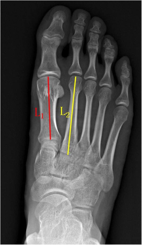
- Long second metatarsal may be risk factor (Morton's foot)
- Dorsal wedge osteotomy can reposition healthy cartilage
- “Smillie classification guides treatment
- “X-ray shows flattening, sclerosis, fragmentation
- “Conservative treatment effective early stages
- “Debridement, osteotomy, or arthroplasty for advanced
Overview and Epidemiology
Freiberg disease is an osteochondrosis of the metatarsal head, most commonly the second: an osteonecrosis that goes on to collapse and articular damage. The alternative name, Freiberg infraction, refers to the subchondral insufficiency fracture with which the process begins.
Who. An adolescent female. Females outnumber males 4:1, the peak age is 13-18 years, and the disease is bilateral in approximately 10% of cases.
Where. The second metatarsal in 68% of cases and the third in 27%.
Pathophysiology
The cause is debated and probably multifactorial, combining vascular insufficiency, repetitive microtrauma with a subchondral stress fracture, and a biomechanical or genetic predisposition.
The vascular argument. The developing metatarsal head epiphysis has a relatively tenuous blood supply and is vulnerable to ischaemia during adolescent growth.
The mechanical argument. A long second metatarsal (Morton foot) increases peak pressure on the second head, a 20-30% increase in loading, and may be a risk factor. High heels and tight footwear concentrate forefoot load, and tight footwear compresses the forefoot and restricts blood flow. The disease may be related to increased activity: repetitive activity such as ballet, especially en pointe, supplies the microtrauma.
The final common pathway. Whichever starts it, a subchondral insufficiency fracture leads to osteonecrosis, collapse and progressive articular destruction, which is the sequence Smillie staged.
Smillie Classification
Smillie's five stages follow the head from an ischaemic fissure to an arthritic joint, and each stage maps to a treatment.
- Pathology
- Ischaemia with an epiphyseal fissure fracture; articular cartilage intact
- Radiographic Findings
- Often normal (MRI sensitive)
- Treatment
- Conservative: offloading, stiff shoe, metatarsal pad
- Pathology
- Resorption of subchondral bone; the cartilage sinks
- Radiographic Findings
- Subtle sclerosis, altered density
- Treatment
- Conservative: activity modification, orthotics
- Pathology
- Central depression of the head; the dorsal rim and the plantar fragment project
- Radiographic Findings
- Flattening, dorsal rim projection (the 'rim sign')
- Treatment
- Surgical: dorsal closing wedge osteotomy (Gauthier)
- Pathology
- Central fragment becomes a loose body; the peripheral rim may fragment
- Radiographic Findings
- Loose bodies visible, fragmented head
- Treatment
- Surgical: debridement, loose body removal
- Pathology
- Flattening and widening of the head with secondary arthrosis
- Radiographic Findings
- Widened head, OA changes, joint destruction
- Treatment
- Salvage: arthroplasty or MTP arthrodesis
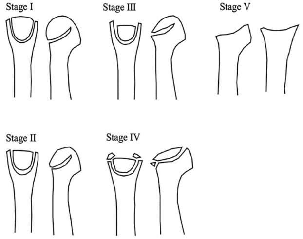
What actually does the guiding is the plantar articular surface. Freiberg destroys the head from dorsal to plantar, so through the earlier stages the plantar cartilage and its subchondral bone remain intact while the dorsal surface collapses. That single fact is what makes the classification operative rather than descriptive.
Stages 1-3: plantar surface intact. There is healthy cartilage available to be brought into load, which is exactly what the Gauthier dorsal closing wedge osteotomy does: it excises the damaged dorsal segment and rotates the preserved plantar cartilage up into the weight-bearing position. Stage 3 is the last stage at which that cartilage is reliably present, hence "good for Smillie 3".
Stage 4: the plantar hinge fails. The central fragment separates as a loose body and the peripheral rim fragments, so there is no longer an intact plantar surface to rotate up. Treatment shifts towards debridement and loose body removal, or to procedures that restore or resurface the head rather than reorient it.
Stage 5: established arthrosis. Reorientation is no longer the question; this is salvage.
Not "how collapsed is the dorsal surface" but "is there still a plantar surface worth rotating".
Clinical Presentation
History. An adolescent female with forefoot pain at the affected metatarsal head, worse with activity and weight-bearing. The onset may be insidious, and symptoms are sometimes minimal at first.
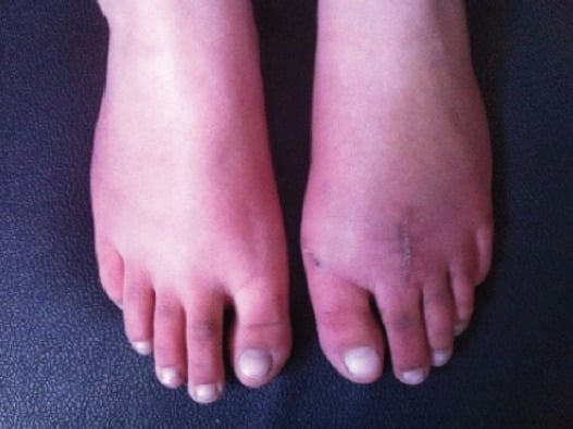
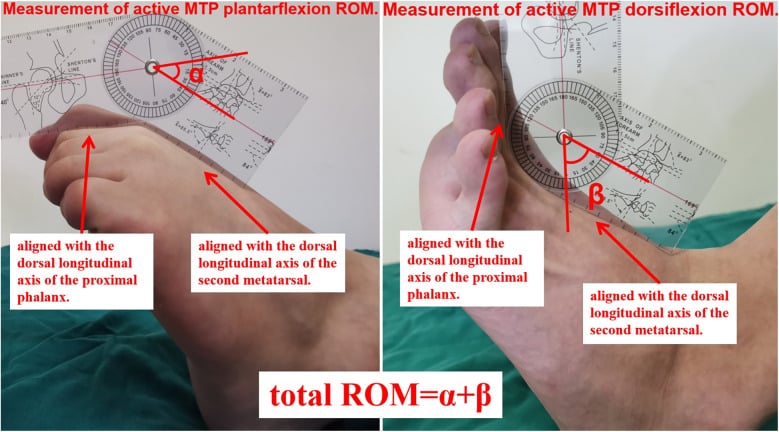
Examination. There may be swelling over the affected MTP joint, and the metatarsal head is tender dorsally and plantarly. MTP motion is reduced, dorsiflexion most of all because it loads the damaged articular surface, and the gait is modified to offload the forefoot. The signs are subtle: mild shortening of the involved toe and a broadened head are easier to see against the other foot than in isolation.
Investigations
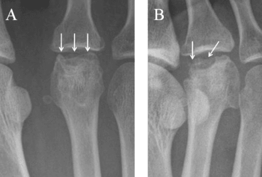
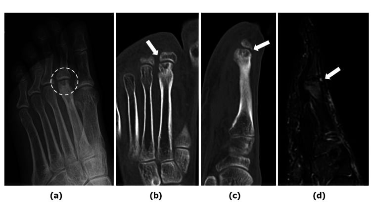
Radiographs. Anteroposterior, oblique and lateral views. Early disease may look normal or show subtle flattening; as it progresses the head becomes sclerotic and flattened, then centrally collapsed, then accompanied by loose bodies. More than one view matters, because head changes subtle on a single projection show on another: the oblique shows a flattened head and subchondral cysts that an AP view can hide.
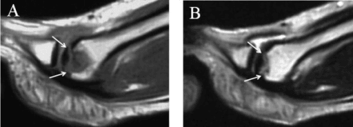
MRI. Sensitive for early disease before any radiographic change: it shows bone marrow oedema and subchondral change. It also shows how much of the head is still viable and the state of the plantar cartilage, which is what decides whether a joint-preserving osteotomy is possible; the sagittal plane shows the dorsal collapse and the plantar cartilage together.
CT. Detail of the collapse pattern for surgical planning: it quantifies the deformity.
Differential diagnosis. Forefoot pain has several other causes, and marrow oedema on MRI is not specific to Freiberg:
- Typical Patient / Site
- Adolescent female; 2nd (then 3rd) MT head
- Key Distinguishing Feature
- Tender, stiff MTP head; pain on dorsiflexion
- Imaging Clue
- Head flattening/collapse; MRI marrow oedema before X-ray change
- Typical Patient / Site
- Athlete/runner; MT shaft (often 2nd/3rd)
- Key Distinguishing Feature
- Tenderness over the shaft, not the head
- Imaging Clue
- Periosteal reaction or linear shaft signal; not focal head
- Typical Patient / Site
- Adults; 3rd–4th web space
- Key Distinguishing Feature
- Burning, radiating toe pain; Mulder click
- Imaging Clue
- No bone change; MRI/US shows interdigital mass
- Typical Patient / Site
- Adults; 2nd MTP
- Key Distinguishing Feature
- Positive drawer; toe deviation/crossover
- Imaging Clue
- Plantar plate disruption on MRI; head preserved
- Typical Patient / Site
- Any age; often multiple joints
- Key Distinguishing Feature
- Effusion, multiple-joint involvement, raised CRP/ESR
- Imaging Clue
- Erosions/effusion; bone marrow oedema can mimic Freiberg
- Typical Patient / Site
- Acute; any MTP
- Key Distinguishing Feature
- Fever, hot swollen joint, rapid onset
- Imaging Clue
- Joint effusion/abscess on MRI; raised inflammatory markers
Management
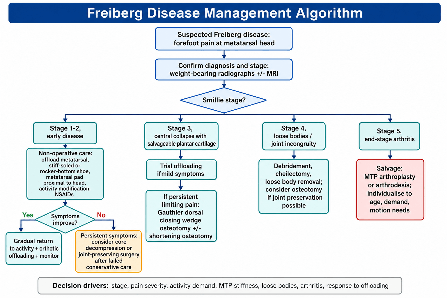
Who. Early disease (Smillie 1-2), symptoms that are not severe, and the patient who prefers it.
What. Offload the affected metatarsal: a stiff-soled or rocker-bottom shoe, a metatarsal pad placed proximal to the head, activity modification, and NSAIDs for pain.
What to expect. It may halt progression if the disease is caught early; the higher the stage, the less likely a response. Where symptoms settle, the patient returns gradually; core decompression or a joint-preserving procedure is an escalation only after failed conservative care.
Joint-Preserving Surgery
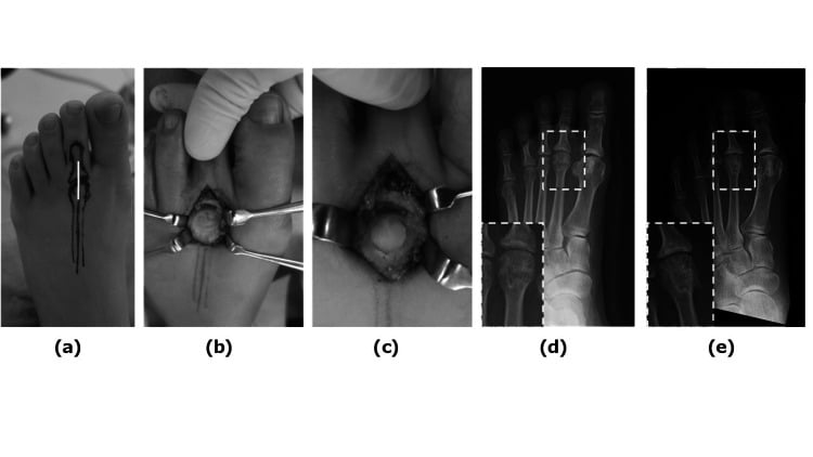
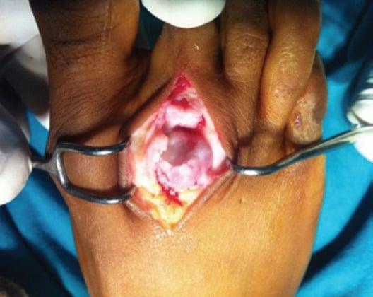
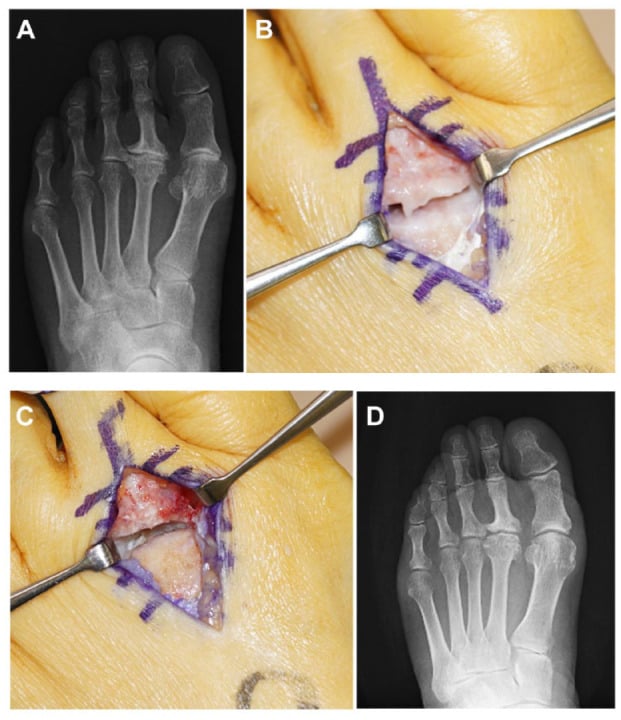
The operation. Plan the osteotomy line at the proximal margin of the necrotic segment, excise the lesion as a dorsal wedge, close the wedge so that the intact plantar articular surface rotates up to face the phalanx, and fix it rigidly, so that the toe regains a functional arc. The point is to bring healthy plantar cartilage into the load-bearing position rather than to resect the head. It shortens the ray far less than excising the head does: a mean of 2.2 mm against 9.8 mm in the Özkul comparison. Union of the osteotomy and a maintained arc of motion together define a good result.
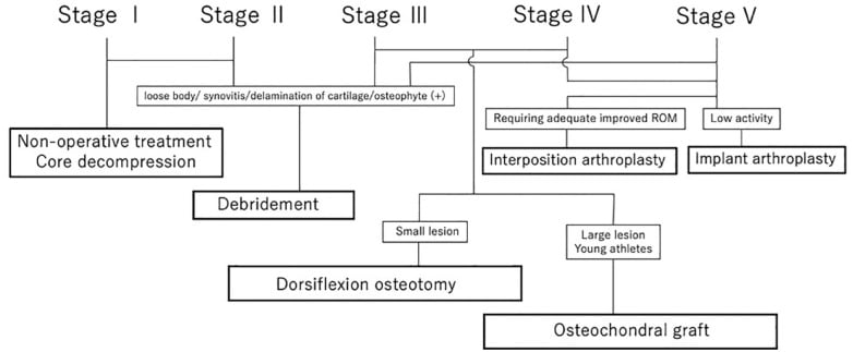
Which stage. Gauthier's osteotomy is the operation for Smillie 3, the last stage at which the plantar cartilage is reliably present. The published series have applied a dorsal wedge more widely, from stage II to V in Lee's series and to stage IV-V heads in Özkul's, and the algorithm adds an osteotomy at stage 4 where joint preservation is still possible. What decides is whether the imaging still shows a plantar surface worth rotating.
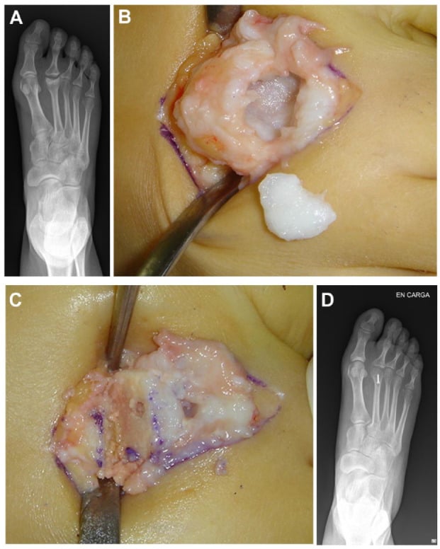
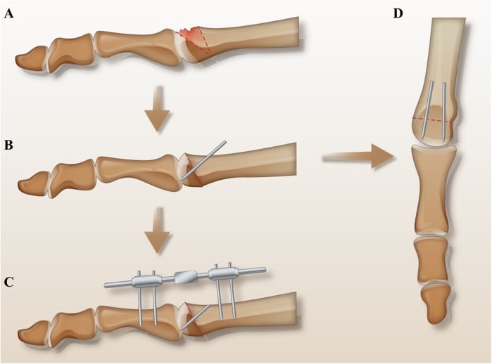
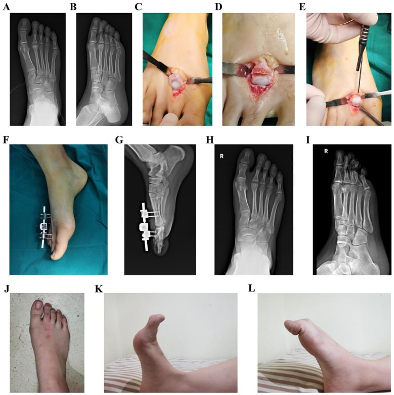
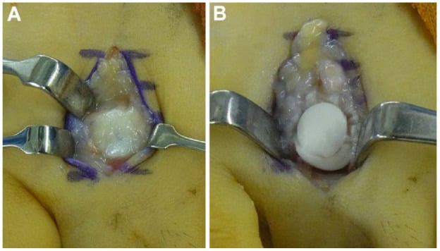
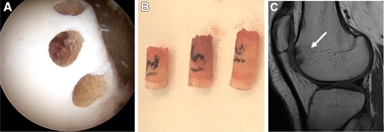
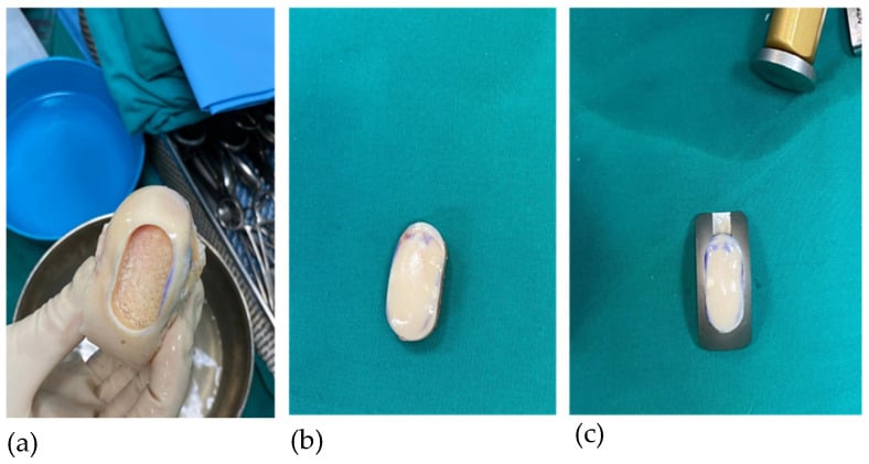
The Gauthier osteotomy reorients the patient's own cartilage, head restoration elevates the patient's own bone beneath it, and an osteochondral autograft brings in new cartilage and bone. None of them suits an end-stage head.
Complications
Of the disease. Left untreated, the joint progresses through:
- Collapse and secondary osteoarthritis of the MTP joint (Smillie 5)
- Loose bodies and mechanical locking
- Stiffness and dorsal impingement limiting push-off
- Transfer metatarsalgia to adjacent rays from altered loading
Of treatment. Each operation has its own signature:
- After osteotomy: shortening, transfer metatarsalgia, stiffness, nonunion or malunion, recurrence
- After head resection: floating or dorsiflexed toe, instability, marked shortening, transfer metatarsalgia
- After arthrodesis: nonunion (10-15 percent), malunion, hardware irritation, loss of MTP motion
- After silicone implant: implant fracture or migration and silicone synovitis (high mid-term failure, especially in the young)
Guidelines, Registries & Global Practice
Freiberg disease is too uncommon to feature in formal national arthroplasty registries or major society guidelines; management is driven by case series and consensus rather than guideline statements. The principles below are consistent in practice worldwide.
Global Epidemiology
- Fourth most common osteochondrosis (after Köhler, Panner and Sever disease).
- Strong female predominance and adolescent onset, though adults present later; second metatarsal head is most often affected, then the third.
- Long second metatarsal (Morton foot) and high-impact forefoot loading (dance, running) are recurrent risk factors across populations.
Consensus Practice (Side by Side)
- Widely Agreed Position
- Conservative first: offloading, stiff/rocker sole, metatarsal pad, activity modification
- Where Practice Varies
- Threshold and enthusiasm for early core decompression
- Widely Agreed Position
- Joint-preserving surgery preferred (dorsal wedge, shortening or head restoration)
- Where Practice Varies
- Choice of specific osteotomy; availability of restoration/osteochondral techniques
- Widely Agreed Position
- Salvage: arthrodesis or resection; avoid silicone implants in young patients
- Where Practice Varies
- Resection vs arthrodesis preference by region and surgeon
- Widely Agreed Position
- MRI for early/radiograph-negative disease; plain films stage collapse
- Where Practice Varies
- Availability of MRI in limited-resource settings
High- vs Limited-Resource Variation
- Well-resourced settings: early MRI, arthroscopic debridement and head-restoration/osteochondral techniques are increasingly used.
- Limited-resource settings: diagnosis relies more on plain radiographs and clinical staging; prolonged conservative care and simpler open procedures (debridement, dorsal wedge osteotomy, head resection) predominate. Outcomes for these established operations remain good, which is reassuring where advanced techniques are unavailable.
Controversies & Areas of Uncertainty
The entire surgical literature for Freiberg disease is Level III-IV, small retrospective series and one small randomised trial. There are no large randomised comparisons, so most "best operation" claims rest on low-grade evidence and surgeon preference.
Aetiology. None of the three mechanisms in the pathophysiology section is proven, and the likeliest answer is that all of them contribute.
Core decompression. Extrapolated from femoral-head AVN. For Smillie 1-2 it has only small case series, no controlled comparison with conservative care, and a real risk of iatrogenic fracture, so many surgeons reserve it for failed conservative treatment.
Which osteotomy. Dorsal closing wedge (Gauthier), shortening (Weil) and head restoration all report good results. The comparative data (Mutlu, Özkul 2016) favour preservation or restoration over excision, but the series are single-centre.
Salvage in the young. For end-stage disease, arthrodesis gives the most reliable pain relief but sacrifices motion; resection preserves motion but risks transfer metatarsalgia and a floating toe; silicone implants have high mid-term failure and are generally avoided in young patients.
State the principle, then the evidence grade: "I would preserve the joint where possible because comparative series favour osteotomy or restoration over excision, but I would acknowledge the evidence is Level III at best and decisions are individualised to stage, age and demand."
MCQ Practice Points
Q: Which metatarsal head is most commonly affected in Freiberg disease? A: Second metatarsal (68%). Third metatarsal is second most common (27%).
Q: What is the typical patient with Freiberg disease? A: Adolescent female (4:1 F:M ratio). Peak age 13-18 years. May be associated with long second metatarsal.
Q: Radiographs are normal but you suspect early Freiberg disease - what next? A: MRI. Stage 1 is radiographically occult; MRI shows bone marrow oedema and subchondral change before plain films flatten.
Exam Viva Scenarios
Practise clinical reasoning and management decisions out loud
“A 15-year-old girl has pain at her second toe. X-ray shows flattening and sclerosis of the second metatarsal head. What is the diagnosis and management?”
“You are seeing a 16-year-old competitive ballet dancer in your clinic who has developed insidious onset pain in her right forefoot over the past 3 months. She describes the pain as deep and aching, localized to the base of her second toe, particularly painful when she is en pointe (on her toes) during dance rehearsals. The pain initially only occurred during intense training sessions but has now progressed to being present with daily activities like walking. She is highly motivated to continue her dance career and is concerned this injury may jeopardize her upcoming performance season. She has tried 2 weeks of rest, ice, and NSAIDs prescribed by her GP with minimal improvement. On examination, she has a visibly longer second metatarsal compared to her first (Morton's foot type - second metatarsal protrudes beyond first by approximately 1cm when comparing toe lengths). There is mild swelling over the second MTP joint with tenderness on palpation both dorsally and plantarly over the metatarsal head. Active and passive range of motion of the second MTP joint is reduced compared to the contralateral side - dorsiflexion is particularly limited (20° vs 40° on left) and painful at end range. There is no instability or crepitus. Her gait shows subtle offloading of the forefoot on the right side. You order plain radiographs (AP, oblique, lateral views of the foot) which are reported as: 'No fracture. No dislocation. Second metatarsal is longer than first. Metatarsal heads appear normal. No sclerosis or collapse identified. Joint spaces preserved. No loose bodies.' Concerned about early Freiberg disease given the clinical presentation and risk factors, you order an MRI which reports: 'Abnormal bone marrow signal in the second metatarsal head with diffuse bone marrow edema (high T2 signal, low T1 signal). Subchondral T2 hyperintensity suggesting early insufficiency/microfracture. No definite collapse or fragmentation. Articular cartilage appears intact. Mild synovitis of second MTP joint. No loose bodies. Findings consistent with early Freiberg disease (Smillie Stage 1-2).' The patient and her parents ask whether this can heal with rest alone or if she needs surgery now. They are particularly concerned about: (1) Will this progress if she continues dancing? (2) Should she have surgery now to prevent worsening? (3) What is the natural history if treated conservatively? (4) What are the risks of core decompression surgery? How do you counsel them and what is your management plan?”
“You are seeing a 24-year-old administrative assistant in your reconstructive foot and ankle clinic, referred from another surgeon for a second opinion. She has a long history of right second toe pain dating back to age 17 when she was first diagnosed with Freiberg disease. At that time, she underwent 'cleaning out of the joint' (operative report describes debridement and cheilectomy - removal of loose bodies, excision of dorsal osteophytes, debridement of damaged cartilage). She had good pain relief for approximately 2 years post-operatively but over the past 3 years her symptoms have gradually worsened. She now has constant dull aching pain in her right second toe, significantly worse with any prolonged walking or standing. She describes the toe as feeling 'stiff' and 'stuck'. She has tried conservative measures including custom orthotics with metatarsal pad, stiff-soled rocker-bottom shoes, multiple courses of physiotherapy, and regular NSAIDs. She has modified her work duties to minimize standing (now primarily desk-based) but still has significant pain even with activities of daily living like grocery shopping. The pain is impacting her quality of life - she avoids social activities involving walking, cannot exercise for fitness, and is concerned about her future. She is very motivated for surgical treatment if it can improve her pain and function. On examination, there is a well-healed dorsal scar over the second MTP joint from her previous surgery. The second toe appears slightly dorsiflexed and 'stuck up' compared to the other toes. There is no swelling or erythema. Palpation reveals significant tenderness over the second metatarsal head both dorsally and plantarly. Range of motion testing of the second MTP joint shows severe restriction - only 5° of plantarflexion and 10° of dorsiflexion (normal approximately 30-40° each direction), passive motion is blocked by hard endpoint (bony block, not soft tissue), attempting to move the joint causes significant pain. There is no instability. The adjacent third and fourth MTP joints have normal range of motion and are non-tender, suggesting no significant transfer metatarsalgia yet. Her gait shows antalgic offloading of the right forefoot with shortened stance phase on the right. You review her radiographs (AP, oblique, lateral) which show: 'Second metatarsal head markedly flattened and widened. Severe osteoarthritis with joint space narrowing (less than 1mm), large dorsal and plantar osteophytes, subchondral sclerosis and cysts. Dorsal subluxation of proximal phalanx. No hardware present. Findings consistent with advanced Freiberg disease (Smillie Stage 5).' You review her previous operative report which documents removal of 3 loose bodies, debridement of approximately 60% of metatarsal head articular cartilage (described as 'extensively damaged'), and excision of dorsal osteophytes. No osteotomy was performed. She asks: 'Can you fix this? I'm only 24 and I can't live the rest of my life with this pain. My last surgeon said there's nothing more he can do. What are my options?' How do you counsel her and what is your surgical recommendation?”
Key Facts
- Second metatarsal head (68%)
- Adolescent females (4:1)
- Osteonecrosis mechanism
- Smillie classification (1-5)
Smillie Stages
- 1: Fissure fracture
- 2: Absorption
- 3: Central depression
- 4: Loose bodies
- 5: Arthritis
Treatment
- Conservative: Offload, stiff shoe, metatarsal pad
- Debridement for loose bodies
- Dorsal wedge osteotomy for Stage 3
- Arthroplasty for end-stage
Evidence Base
Smillie (classic)
- Original five-stage radiographic classification of Freiberg disease
- Proposed an ischaemic / subchondral fracture aetiology
- Linked progressive collapse to articular destruction
- Remains the most widely used staging system worldwide
Gauthier & Elbaz (classic)
- Described the dorsal closing-wedge osteotomy of the metatarsal head
- Excises damaged dorsal cartilage and rotates intact plantar cartilage into load
- Joint-preserving alternative to head excision
- Conceptual basis for most modern preservation procedures
Talusan et al. (narrative review)
- Second and third metatarsals most commonly affected; female predominance
- Multifactorial aetiology: vascular compromise, genetic predisposition, altered biomechanics
- Early radiographs are often normal; MRI/bone scan detect disease earlier
- Non-operative and operative outcomes both reported as good to excellent