Posterosuperior Calcaneal Prominence | Conservative First 3-6 Months | Surgery for Refractory Cases
- Haglund's Triad: posterosuperior calcaneal prominence, retrocalcaneal bursitis, insertional Achilles tendinopathy
- Parallel Pitch Lines - radiographic assessment on lateral X-ray to quantify posterior calcaneal prominence
- Conservative management (heel lifts, eccentric exercises, orthotics) successful in 70-80% of cases
- Central tendon-splitting approach - preserves insertional Achilles fibres, allows direct access to bursa and bone
- Complication risk: Achilles rupture (2-5%), wound healing problems, sural nerve injury
- “Two-incision technique vs central tendon-splitting - know indications for each approach
- “Parallel pitch lines: posterosuperior angle greater than 75 degrees indicates prominent tuberosity
- “Avoid complete Achilles detachment - increases rupture risk and requires prolonged immobilisation
- “Retrocalcaneal exostosis may recur if inadequate resection or continued mechanical irritation
70-80% respond to non-operative treatment. Demonstrate systematic conservative algorithm: activity modification, heel lifts, eccentric Achilles strengthening, orthotics with open heel counter. Minimum 3-6 months before considering surgery.
Radiographic diagnosis uses parallel pitch lines on lateral radiograph. Three lines assess prominence: posterior calcaneal line, superior calcaneal line, parallel pitch line. Posterosuperior angle greater than 75 degrees indicates significant deformity.
Preserves Achilles insertion while allowing access to retrocalcaneal bursa and posterosuperior prominence. Split tendon longitudinally in midline, reflect medially and laterally. Avoids complete detachment and reattachment complications.
Most feared complication (2-5% incidence). Higher risk with complete Achilles detachment, inadequate tissue quality, over-aggressive debridement. Protect repair with prolonged immobilisation and gradual weight-bearing progression.
- Primary Pathology
- Retrocalcaneal bursitis alone
- Treatment Approach
- NSAIDs, ice, activity modification, heel lift
- Key Pearl
- Usually resolves within 4-6 weeks with rest
- Primary Pathology
- Haglund's with insertional tendinopathy
- Treatment Approach
- Eccentric exercises, orthotics, consider ESWT
- Key Pearl
- 3-6 month trial essential before surgery
- Primary Pathology
- Refractory Haglund's triad
- Treatment Approach
- Surgical: ostectomy plus bursa excision plus tendon debridement
- Key Pearl
- 80-90% good outcomes with appropriate patient selection
- Primary Pathology
- Insertional calcific tendinopathy
- Treatment Approach
- Surgical debridement with suture anchor repair
- Key Pearl
- May require Achilles detachment and reattachment
Overview and Epidemiology
Definition
Haglund's deformity is a bony enlargement of the posterosuperior aspect of the calcaneus that causes posterior heel pain through mechanical irritation of the retrocalcaneal bursa and Achilles tendon insertion. First described by Patrick Haglund in 1928, the condition is colloquially known as "pump bump" due to its association with rigid-heeled footwear.
Epidemiology
- Anatomic prominence present in 20-30% of general population
- Symptomatic in subset of those with prominence
- Peak age: 20-40 years (active working and athletic population)
- Gender: Female predominance (1.5-2:1 ratio)
- Runners and endurance athletes
- Ice skaters and figure skaters (rigid boot compression)
- Women wearing high-heeled dress shoes ("pump bump")
- Military personnel and police (occupational footwear)
- Workers in hospitality industry (dress shoe requirements)
- Self-limiting in many cases with activity modification
- 70-80% respond to conservative management over 3-6 months
- Surgical intervention required in 20-30% of symptomatic cases
- Bilateral involvement in 60% of symptomatic patients
Aetiology
Haglund's deformity develops through a combination of intrinsic and extrinsic factors:
- Prominent posterosuperior calcaneal tuberosity (anatomic variant)
- High-arched cavus foot deformity (increased posterior heel pitch)
- Pes planus with valgus hindfoot (altered Achilles pull vector)
- Tight Achilles tendon (gastrocnemius-soleus contracture)
- Inflammatory arthropathy (predisposes to bursal inflammation)
- Rigid-backed footwear causing repeated heel counter friction
- Sudden increase in training intensity or running mileage
- Hill running and stair climbing (increased Achilles tension)
- Occupational footwear requirements
- Ice skating and figure skating activities
Anatomy and Pathophysiology
Relevant Anatomy
Posterior Heel Structures
The posterosuperior aspect of the calcaneus is a critical zone with multiple overlapping structures that contribute to Haglund's pathology:
- Posterosuperior calcaneal tuberosity - anatomic insertion site for Achilles tendon
- Lateral process - prominence often enlarged in Haglund's deformity
- Bursal projection - superior ridge that defines retrocalcaneal bursa location
- Medial tubercle - attachment for superficial Achilles fibres
- Achilles tendon insertion - crescent-shaped footprint approximately 2cm wide
- Retrocalcaneal bursa - located between posterosuperior calcaneus and anterior Achilles surface
- Superficial calcaneal bursa - between skin and posterior Achilles surface (adventitial bursa)
- Kager's fat pad - triangular radiolucent area anterior to Achilles on lateral X-ray
Neurovascular Structures at Risk
- Courses along lateral border of Achilles tendon
- Average distance 18-20mm from lateral border at insertion level
- At risk with lateral incision approach
- Injury causes lateral foot numbness and painful neuroma
- Proximal: muscular branches from posterior tibial and peroneal arteries
- Mid-portion: watershed area with poorest blood supply (2-6cm proximal to insertion)
- Distal insertion: branches from posterior tibial and peroneal arteries via periosteal vessels
- Surgical dissection may compromise already tenuous vascular supply
Pathophysiology
Mechanical Irritation Model
Haglund's deformity develops through repetitive mechanical friction and compression:
- Bony prominence - anatomic or acquired posterosuperior calcaneal exostosis
- Shoe counter pressure - rigid heel counter compresses prominence against anterior Achilles
- Bursal inflammation - retrocalcaneal bursa becomes inflamed and thickened
- Tendon degeneration - chronic friction leads to insertional Achilles tendinopathy
- Reactive bone formation - further prominence develops, perpetuating cycle
Histopathological Changes
- Synovial hyperplasia and proliferation
- Fibrinous exudate and inflammatory cell infiltration
- Bursal wall thickening with fibrosis
- Adhesions to adjacent Achilles tendon
- Collagen disorganization and fibrinoid degeneration
- Increased mucoid ground substance
- Neovascularization with neural ingrowth (pain pathway)
- Calcification within degenerative tendon fibres
- Loss of normal parallel collagen architecture
Risk Factors
- High-arched cavus foot (increased heel pitch)
- Prominent posterosuperior calcaneal tuberosity (anatomic variant)
- Tight Achilles tendon (gastrocnemius-soleus complex contracture)
- Pes planus with valgus hindfoot (abnormal Achilles pull vector)
- Inflammatory arthropathy (increased bursal inflammation)
- Rigid-backed footwear (pump shoes - hence "pump bump")
- Running and athletic activities (repetitive loading)
- Sudden increase in training intensity
- Ice skating and figure skating (rigid boot compression)
- Occupational footwear (work boots, military boots)
Clinical Presentation and Examination
History Taking
Typical Presentation
- Posterior heel pain localized to Achilles insertion area
- Gradual onset over weeks to months
- Activity-related pain, worse with running or walking
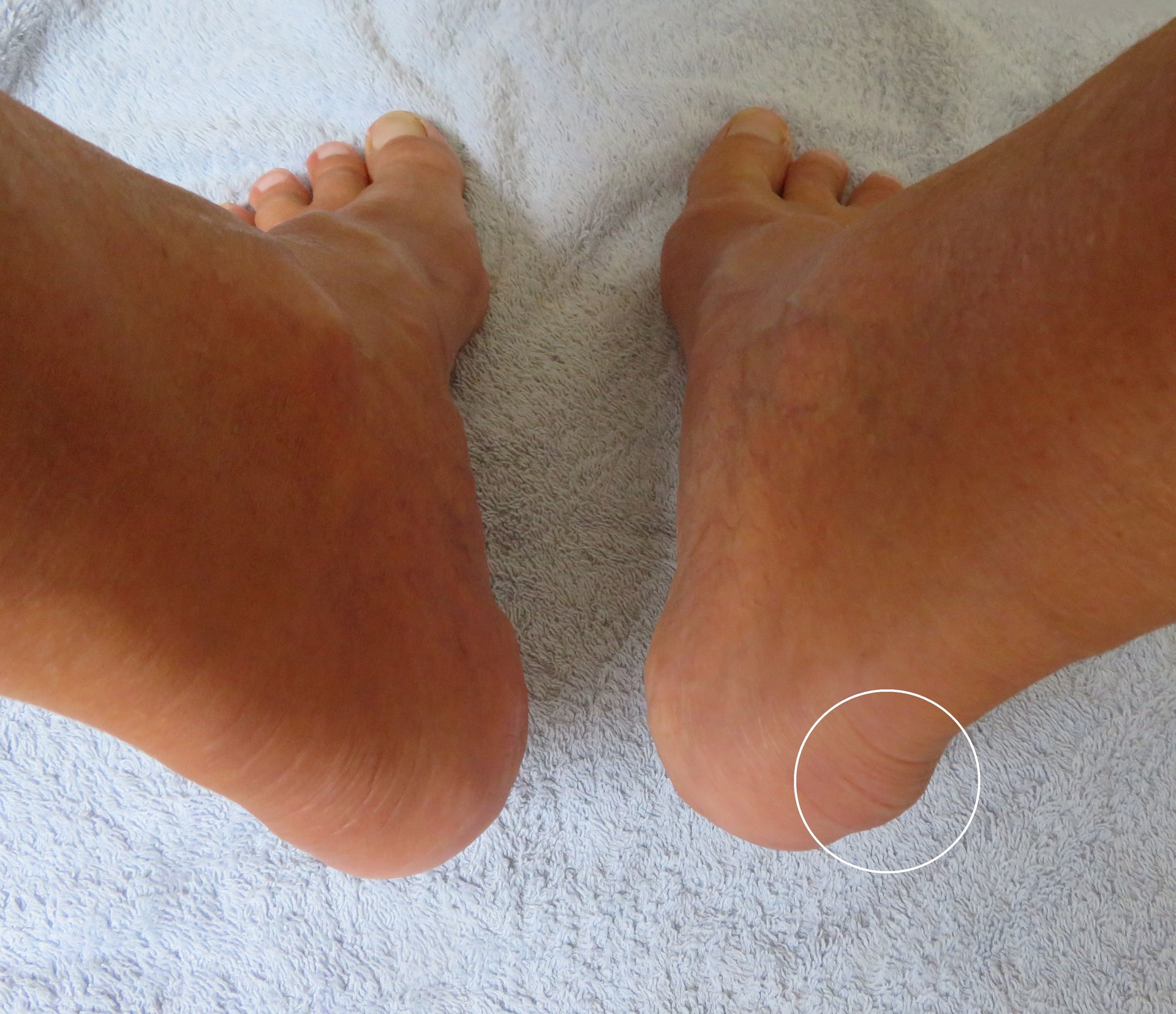
- Visible prominence on posterior heel ("pump bump")
- Sharp or aching pain at posterosuperior heel
- Worse with initial steps after rest (similar to plantar fasciitis)
- Aggravated by shoe counter pressure
- Relief with open-backed shoes or going barefoot
- May have associated morning stiffness
- Difficulty wearing dress shoes or athletic shoes
- Altered gait to avoid heel strike
- Reduced exercise tolerance
- May limit sporting activities
Key History Questions
- How long have you had heel pain? (Determines conservative trial adequacy)
- What treatments have you tried? (NSAIDs, heel lifts, physiotherapy, injections)
- What type of shoes do you wear? (Rigid heel counter is key provocative factor)
- What sports or activities? (Running, skating, occupational demands)
Physical Examination
Inspection (Standing and Walking)
- Ask patient to demonstrate problematic footwear
- Observe heel counter rigidity and height
- Note wear pattern on shoe posterior aspect
- Assess from behind: valgus vs neutral vs varus
- Pes cavus (high arch) predisposes to Haglund's
- "Too many toes" sign suggests pes planus with valgus
- Posterosuperior calcaneal prominence visible and palpable
- Bilateral in 60% of symptomatic cases
- Overlying skin changes (erythema, callus, bursitis)
Palpation
- Achilles tendon - palpate full length for thickening, nodules, crepitus
- Retrocalcaneal bursa - tender anterior to Achilles, just superior to insertion
- Posterosuperior prominence - bony prominence palpable through Achilles
- Lateral compression test - squeeze heel medially and laterally to compress retrocalcaneal space (positive if painful)
- Direct posterior pressure - pain with pressure over prominence
- Achilles insertion tenderness - suggests concurrent insertional tendinopathy
Range of Motion
- Measure with knee extended (gastrocnemius tight) and flexed (isolated soleus)
- Normal: 10-15 degrees with knee extended
- Haglund's patients often have reduced dorsiflexion (less than 5 degrees)
- Silfverskiöld test differentiates gastrocnemius vs soleus contracture
- Assess inversion and eversion
- Rule out hindfoot arthritis as pain source
Special Tests
- Passively dorsiflex ankle while palpating retrocalcaneal space
- Positive if reproduces posterior heel pain
- Indicates bursal impingement
- Compress Achilles tendon between thumb and finger at insertion
- Positive if elicits sharp pain
- Suggests insertional Achilles tendinopathy
- Patient performs single-leg heel raise on affected side
- Pain or inability indicates Achilles dysfunction
- Assess number of repetitions compared to contralateral side
Investigations and Radiographic Assessment
Imaging Studies
Plain Radiographs (First-Line)
Standard Views:
- Lateral heel radiograph - primary diagnostic view
- AP foot - assess for concurrent pathology
- Axial calcaneal view - evaluate calcaneal width and shape
Lateral Radiograph Assessment - Parallel Pitch Lines:
The Fowler-Philip angle and parallel pitch lines quantify posterosuperior prominence:
- Posterior calcaneal line - tangent to posterior calcaneal border
- Inferior calcaneal line - tangent to plantar calcaneal surface
- Total calcaneal angle - angle between posterior and inferior lines (normal 44-69 degrees)
- Parallel pitch line - parallel to inferior calcaneal line, drawn from anterior process
- Posterosuperior angle - angle between posterior surface and parallel pitch line
- Posterosuperior angle greater than 75 degrees suggests Haglund's deformity
- Prominence extending above parallel pitch line indicates exostosis
- Parallel pitch line above superior calcaneal border diagnostic
- Retrocalcaneal bursitis (soft tissue opacity anterior to Achilles)
- Achilles tendon thickening (greater than 9mm at insertion)
- Insertional calcification (enthesopathy)
- Kager's fat pad obliteration (suggests inflammation)
Magnetic Resonance Imaging (MRI)
- Atypical presentation requiring diagnostic clarification
- Preoperative planning to assess Achilles tendon quality
- Failed surgery evaluation
- Suspicion of concurrent pathology
- Retrocalcaneal bursa - fluid signal on T2-weighted images (normal less than 2mm, abnormal greater than 3mm)
- Achilles tendinopathy - intratendinous signal changes, thickening, partial tears
- Bone marrow edema - posterosuperior calcaneus signal changes on STIR sequences
- Kager's fat pad edema - indicates active inflammation
- Enthesophyte formation - bone proliferation at insertion
Ultrasound
- Dynamic assessment during ankle motion
- Real-time visualization of bursa compression
- Guide for injection therapy
- Lower cost than MRI
- Retrocalcaneal bursa distension (greater than 3mm anteroposterior dimension)
- Achilles tendon thickening and hypoechoic areas (tendinopathy)
- Posterosuperior calcaneal prominence contour
- Power Doppler may show hypervascularity (active inflammation)
Differential Diagnosis
Posterior heel pain is not synonymous with Haglund's deformity. The key examination distinction is the location of maximal tenderness and its relationship to the Achilles insertion.
- Site of Pain
- Posterosuperior heel, anterior to Achilles
- Key Distinguishing Feature
- Prominent bony bump, pain on mediolateral squeeze of bursa
- First-Line Investigation
- Lateral weight-bearing radiograph (parallel pitch lines)
- Site of Pain
- Central tendon at the calcaneal insertion
- Key Distinguishing Feature
- Tenderness ON the insertion +/- enthesophyte/calcification
- First-Line Investigation
- Lateral radiograph; MRI/US for tendon quality
- Site of Pain
- 2-6 cm proximal to insertion (watershed)
- Key Distinguishing Feature
- Fusiform thickening that moves with the tendon (arc sign)
- First-Line Investigation
- Ultrasound or MRI
- Site of Pain
- Skin overlying posterior heel
- Key Distinguishing Feature
- Pump bump callus/erythema posterior to (not anterior to) tendon
- First-Line Investigation
- Clinical diagnosis
- Site of Pain
- Bilateral insertions, may have plantar fasciitis
- Key Distinguishing Feature
- Inflammatory features, young male, raised CRP, HLA-B27
- First-Line Investigation
- Inflammatory screen +/- MRI bone marrow oedema
- Site of Pain
- Posterior calcaneus in skeletally immature
- Key Distinguishing Feature
- Child/adolescent athlete, positive squeeze test of apophysis
- First-Line Investigation
- Clinical; radiograph to exclude other pathology
- Site of Pain
- Plantar heel / gap in tendon / posterior ankle
- Key Distinguishing Feature
- Different anatomical zone; Simmonds-Thompson for rupture
- First-Line Investigation
- Clinical +/- imaging as indicated
Management Algorithm
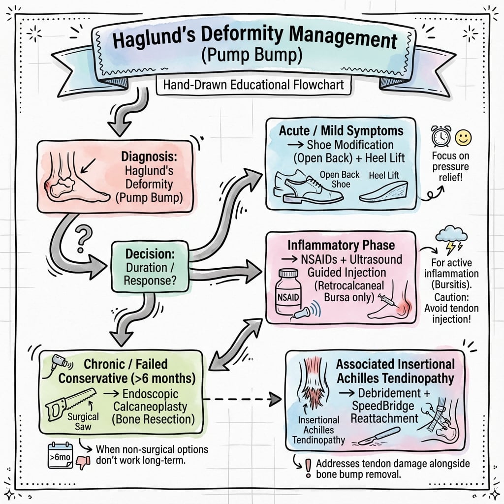
Conservative Management (First-Line)
Initial Treatment (0-6 Weeks)
- Reduce or eliminate provocative activities (running, jumping)
- Avoid rigid-backed footwear
- Open-backed shoes or sandals during acute phase
- Heel lifts (5-10mm) - reduce Achilles tension and posterior impingement
- Soft heel counters - remove or modify rigid back of shoe
- Wider toe box - accommodate orthotic if pes planus present
- Padded heel cups - cushion and offload posterior prominence
- NSAIDs for 2-4 weeks (ibuprofen 400mg three times daily, naproxen 500mg twice daily)
- Ice application 15-20 minutes three times daily
- Topical anti-inflammatory gel (diclofenac)
Physiotherapy Protocol (6 Weeks to 3 Months)
- Gastrocnemius stretch - knee extended, foot dorsiflexed, hold 30 seconds, 5 repetitions, 3 times daily
- Soleus stretch - knee flexed 20-30 degrees, foot dorsiflexed
- Combined stretch - slant board stretching
- Goal: achieve 10 degrees dorsiflexion with knee extended
- Bilateral heel raise to tiptoes on step
- Lower affected heel slowly below step level (eccentric phase)
- 3 sets of 15 repetitions, twice daily
- Perform with knee straight and knee bent (gastrocnemius and soleus isolation)
- Continue for 12 weeks minimum
- Ultrasound therapy (controversial benefit)
- Iontophoresis with dexamethasone
- Low-level laser therapy (limited evidence)
Orthotics and Bracing (3-6 Months)
- Indicated for pes planus or cavus foot deformity
- Medial posting for valgus hindfoot
- Arch support to normalize foot biomechanics
- Open heel design to avoid posterior compression
- Maintain ankle in neutral to 5 degrees dorsiflexion
- Prevent morning contracture
- Wear nightly for 3-6 months
- 80% compliance required for benefit
Advanced Conservative Therapies (3-6 Months)
- Indication: failed 3-6 months conservative treatment
- Protocol: 2000-4000 shocks per session, 3-5 sessions, 1-2 week intervals
- Energy flux density 0.08-0.28 mJ/mm²
- Success rate: 60-70% symptom improvement
- Mechanism: neovascularization stimulation, pain receptor desensitization
- Corticosteroid injection (retrocalcaneal bursa)
- Ultrasound-guided approach preferred
- Methylprednisolone 40mg or triamcinolone 40mg with 1ml lidocaine
- Maximum 2-3 injections lifetime (Achilles rupture risk)
- 60-70% short-term pain relief (3-6 months)
- Platelet-Rich Plasma (PRP)
- Emerging therapy for insertional tendinopathy
- May have lower rupture risk than corticosteroid
- Limited high-quality evidence for efficacy
- Typically 2-3 injections at 4-week intervals
Surgical Management
Indications for Surgery
- Failed comprehensive conservative management for 6 months minimum
- Documented compliance with physiotherapy protocol
- Significant functional impairment affecting quality of life
- Patient preference after informed consent regarding risks
- Severe deformity with skin breakdown
- Achilles partial tear requiring surgical repair
- Active infection
- Severe peripheral vascular disease
- Inadequate conservative trial (less than 3-6 months)
- Medical comorbidities precluding safe surgery
- Unrealistic patient expectations
Surgical Techniques - Evidence-Based Approaches
The surgical management of Haglund's deformity requires careful decision-making regarding approach and technique based on the specific components of the pathology.
Surgical Technique
Central Tendon-Splitting Approach
This is the preferred technique for most cases as it preserves the Achilles insertion while providing excellent access to the retrocalcaneal bursa and posterosuperior calcaneal prominence.
Advantages
- Preserves Achilles insertion and strength
- Direct visualization of bursa and bony prominence
- Single midline incision
- Lower risk of wound complications compared to lateral approaches
- Allows concurrent tendon debridement if needed
Disadvantages
- Risk of creating iatrogenic Achilles defect if excessive debridement
- Technically demanding to split tendon precisely
- Limited lateral exposure for extensive prominence
Patient Positioning
- Prone position on operating table
- Affected leg supported on bolster or bump
- Contralateral leg slightly flexed and abducted
- Pneumatic tourniquet on thigh (250-300 mmHg)
- All bony prominences padded
Surgical Technique Step-by-Step
- 6-8 cm longitudinal incision over midline posterior heel
- Centred over palpable Achilles insertion
- Extends from 6cm proximal to insertion to distal calcaneal tuberosity
- Careful dissection through subcutaneous tissue
- Identify sural nerve laterally and protect
- Incise Achilles tendon paratenon longitudinally in midline
- Reflect paratenon medially and laterally with flaps
- Identify central raphe of Achilles tendon
- Longitudinal split through Achilles tendon in midline
- Start 5-6cm proximal to insertion
- Extend distally to insertion on calcaneus
- Split approximately 50% of tendon thickness
- Use stay sutures in medial and lateral flaps for retraction
- Reflect medial and lateral Achilles flaps
- Excise thickened retrocalcaneal bursa completely
- Expose posterosuperior calcaneal prominence
- Identify insertional Achilles fibres on prominence
- Excise degenerative tendon tissue (yellow-brown discolored areas)
- Remove any intratendinous calcification
- Preserve greater than 50% of normal tendon insertion
- If greater than 50% excision needed, prepare for suture anchor repair
- Define prominence with Freer elevator
- Osteotomy with oscillating saw
- Remove wedge of bone parallel to anterior cortex
- Goal: create smooth posterior contour without prominence above parallel pitch line
- File sharp edges with rasp
- Irrigate thoroughly to remove bone debris
- Reapproximate Achilles tendon split with absorbable sutures (2-0 Vicryl)
- Running locked suture technique
- Ensure no gaps or defects in tendon
- Close paratenon over repair
- Layered subcutaneous closure
- Skin closure with nylon sutures or staples
This central tendon-splitting approach provides excellent outcomes while minimizing the major complications.
Complications
Surgical Complications
Early Complications (Less Than 6 Weeks)
- Delayed wound healing - most common complication
- Wound dehiscence - associated with excessive tension, poor tissue quality
- Superficial infection - requires antibiotics, local wound care
- Deep infection - rare but catastrophic (may lead to Achilles rupture)
- Hematoma formation - inadequate hemostasis
- Meticulous hemostasis before closure
- Avoid tension on skin closure
- Consider delayed weight-bearing in high-risk patients (diabetes, smoking, steroids)
- Protective splinting in equinus for 2 weeks
- Patient education regarding wound care and warning signs
- Small dehiscence: local wound care, dressing changes
- Large dehiscence: may require surgical debridement and secondary closure
- Infection: culture-directed antibiotics, surgical debridement if deep
- Numbness lateral heel and foot
- Painful neuroma formation
- Dysesthesias and allodynia
- Higher risk with lateral approach
- Direct visualization and protection of nerve
- Gentle soft tissue handling
- Maintain adequate distance from lateral Achilles border
- Most sensory deficits permanent but well-tolerated
- Painful neuroma: desensitization, nerve blocks, rarely excision
Late Complications (Greater Than 6 Weeks)
- Most feared complication with devastating functional impact
- Higher risk with complete detachment and reattachment
- May occur during rehabilitation phase (6-12 weeks)
- Associated with over-aggressive tendon debridement
- Greater than 50% tendon debridement
- Poor tissue quality (chronic degeneration)
- Corticosteroid injection within 3 months of surgery
- Premature aggressive rehabilitation
- Non-compliance with protected weight-bearing
- Non-operative: prolonged cast immobilization (limited outcomes in active patients)
- Operative: primary repair with augmentation (FHL transfer, turndown flap, allograft)
- Prolonged rehabilitation: 6-9 months return to activity
- Inadequate ostectomy (residual prominence)
- Continued mechanical irritation (footwear)
- Unaddressed concurrent pathology (hindfoot valgus, cavus foot)
- Scar tissue formation
- Adjacent segment pathology (plantar fasciitis)
- Investigate with MRI to identify specific cause
- Revision surgery if clear mechanical problem identified
- Manage soft tissue pain with physiotherapy, injections
- Reduced ankle dorsiflexion range
- Calf atrophy (especially with prolonged immobilisation)
- Persistent weakness with heel raise
- May improve with rehabilitation but often persistent deficit
- Progressive rehabilitation protocol
- Early range of motion when safe
- Eccentric strengthening emphasis
- Realistic patient expectations
Post-Operative Rehabilitation
Rehabilitation Protocol
Phase 1: Protection (Weeks 0-6)
- Bulky dressing with posterior splint in neutral for 2 weeks
- Below-knee walking boot at 2 weeks
- Weight-bearing as tolerated in boot
- Remove boot for gentle ankle pumps 3-4 times daily
- Below-knee cast in 20 degrees equinus for 4 weeks
- Non-weight-bearing or touch-down weight-bearing only
- Transition to neutral boot at 4 weeks
- Progressive weight-bearing weeks 4-6
Phase 2: Mobilisation (Weeks 6-12)
- Wean from boot to supportive shoe with heel lift
- Active ankle range of motion exercises
- Gentle Achilles stretching (avoid aggressive dorsiflexion)
- Stationary bicycle for cardiovascular fitness
- Pool exercises (non-impact)
- Progressive resistance exercises
- Eccentric Achilles strengthening (single-leg heel lowers)
- Proprioceptive training (wobble board, balance exercises)
- Gradual return to normal gait pattern
- Remove heel lift by 12 weeks
Phase 3: Strengthening (Weeks 12-26)
- Progressive calf strengthening program
- Isokinetic exercises
- Start low-impact activities (swimming, cycling)
- Begin straight-line jogging if strength adequate
- Sport-specific training
- Plyometric exercises (jumping, hopping)
- Agility drills
- Progressive running program
Phase 4: Return to Activity (Months 6-12)
- No pain with heel raises or hopping
- Calf strength greater than 85% of contralateral side (isokinetic testing)
- Full ankle range of motion
- Normal gait without limp
- Confidence in affected heel
- Low-impact sports first (cycling, swimming)
- Progress to higher-impact activities (running, court sports)
- Full unrestricted return typically 9-12 months for competitive athletes
Long-Term Outcomes
- 80-90% good to excellent outcomes at 2-year follow-up
- Satisfaction rates 85-95% when appropriate patient selection
- Return to pre-injury activity level: 70-80% of athletes
- Recurrence rate: 5-10% with proper surgical technique
- Adequate conservative trial before surgery (greater than 6 months)
- Isolated Haglund's without concurrent pathology
- Compliance with rehabilitation protocol
- Non-smoking status
- Absence of workers' compensation claim
The Silfverskiöld Test
The examination section measures ankle dorsiflexion "with knee extended and flexed" and names the Silfverskiöld test to "differentiate gastrocnemius vs soleus contracture" - but never explains how the test is performed or what a positive result means.
- The test. With the subtalar joint held in neutral, measure maximal ankle dorsiflexion first with the knee extended and then with the knee flexed to about 90 degrees. Because the gastrocnemius crosses the knee (the soleus does not), flexing the knee slackens the gastrocnemius.
- Interpretation. If dorsiflexion is limited with the knee extended but improves markedly (to neutral or beyond) with the knee flexed, the contracture lies in the gastrocnemius alone - an isolated gastrocnemius contracture, a positive Silfverskiöld test. If dorsiflexion stays restricted in both positions, the whole gastrocnemius-soleus complex (or the Achilles/capsule) is tight.
- Why it matters in Haglund's. A tight gastrocnemius is one of the topic's own listed risk factors, raising the tensile and compressive load of the insertion against the posterosuperior calcaneus. Identifying an isolated gastrocnemius contracture is what selects the patient for a gastrocnemius recession (next section) rather than a broader lengthening, and directs conservative care toward knee-extended gastrocnemius stretching.
Q: How is the Silfverskiöld test performed and interpreted? A: With the subtalar joint neutral, measure ankle dorsiflexion with the knee extended then flexed to about 90 degrees. If dorsiflexion is limited with the knee straight but improves with the knee bent, the gastrocnemius alone is tight (positive test) - bending the knee slackens the gastrocnemius, which crosses the knee, whereas the soleus does not. If it is restricted in both positions, the whole gastrocnemius-soleus/Achilles complex is contracted. An isolated gastrocnemius contracture is the indication for a gastrocnemius recession.
Gastrocnemius Recession for the Equinus Driver
The topic repeatedly names gastrocnemius-soleus contracture / tight Achilles as a driver of insertional Achilles and Haglund's disease and measures it with the Silfverskiöld test, yet the surgical options never include the operation that addresses it: gastrocnemius recession.
- The rationale. When an isolated gastrocnemius contracture (positive Silfverskiöld) is contributing, lengthening the gastrocnemius reduces the equinus pull and the compressive/tensile load on the diseased insertion without violating the Achilles insertion or the calcaneus - treating the underlying driver rather than only the local prominence.
- The Strayer procedure is the classic technique: through a posteromedial calf incision at the musculotendinous junction of the gastrocnemius, the gastrocnemius aponeurosis is transected and allowed to recede while the soleus is left intact; the sural nerve runs close by and must be protected. More proximal (Baumann, intramuscular) recessions are alternatives at different levels.
- How it fits. It is used as an adjunct - either as an isolated procedure in early insertional disease with a clear gastrocnemius contracture, or added to a debridement/ostectomy to offload the repair and reduce recurrence. It preserves plantarflexion strength better than a formal tendo-Achilles lengthening, but can cause temporary push-off weakness.
Q: When and how would a gastrocnemius recession help in Haglund's / insertional Achilles disease? A: When the Silfverskiöld test shows an isolated gastrocnemius contracture, a gastrocnemius recession lengthens the gastrocnemius to reduce the equinus pull and offload the diseased insertion without violating the Achilles or calcaneus. The Strayer technique transects the gastrocnemius aponeurosis at the musculotendinous junction (protecting the sural nerve), leaving the soleus intact. It is used as an isolated early procedure or as an adjunct to debridement/ostectomy to reduce recurrence, with less push-off weakness than a formal tendo-Achilles lengthening.
Guidelines, Registries & Global Practice
Global Epidemiology
Haglund's deformity occurs worldwide and is consistently linked to the same drivers regardless of region: running and endurance sport, rigid-heeled footwear, and occupations requiring stiff-backed boots (military, police, hospitality, healthcare). A female predominance (roughly 1.5-2:1) is reported in most series and is generally attributed to dress and fashion footwear. Peak presentation is in the active 20-40 year age group, with frequent bilateral involvement. A prominent posterosuperior calcaneus is an anatomical variant seen in a substantial minority of asymptomatic feet, so radiographic prominence must always be correlated with the clinical picture.
Side-by-Side Guidance
Haglund's deformity sits within the broader insertional Achilles tendinopathy literature; no single society publishes a dedicated standalone guideline, so recommendations are drawn from foot and ankle and sports-medicine consensus.
- Emphasis
- Structured non-operative trial first; central tendon-splitting workhorse
- Practical Recommendation
- Minimum 3-6 months conservative care; debride and exostectomy if refractory
- Emphasis
- Load management and modified eccentrics; caution with peritendinous steroid
- Practical Recommendation
- Physiotherapy-led; shockwave for recalcitrant cases before surgery
- Emphasis
- Activity modification, education and progressive loading
- Practical Recommendation
- Modify eccentric protocol to floor-level for insertional disease
- Emphasis
- Tendon-sparing dorsal closing-wedge (Zadek) osteotomy
- Practical Recommendation
- Consider where bony geometry dominates and insertion is healthy
Registry and Evidence Notes
Haglund's deformity involves no implant, so it does not feature in arthroplasty registries (NJR, AJRR, AOANJRR, SHAR). The evidence base is therefore built from single-centre series and a small number of randomised trials of conservative care (Rompe 2008; Mansur 2021) plus systematic reviews of osteotomy (Poutoglidou 2023). There is a recognised lack of randomised comparison between surgical techniques globally.
High- vs Limited-Resource Practice Variation
In well-resourced settings, ultrasound-guided injection, MRI for tendon mapping, shockwave devices, and endoscopic calcaneoplasty are routinely available, allowing a graded, imaging-guided pathway. In limited-resource settings, management relies on clinical diagnosis, plain radiographs, footwear modification, heel lifts and a supervised home eccentric programme; open debridement with exostectomy remains the default surgical option where endoscopic equipment is unavailable. The core principles - an adequate conservative trial, correlation of imaging with clinical findings, and tendon-preserving surgery - apply universally.
Controversies and Areas of Uncertainty
The evidence base for Haglund's deformity is dominated by Level III-IV retrospective series. There are no randomised trials comparing surgical techniques, and even the non-operative literature is conflicting - making this fertile ground for "what is the evidence?" viva questions.
Does Shockwave Add Anything?
The two best-quality trials reach opposite-sounding conclusions. Rompe (2008, Level I) found low-energy shockwave superior to floor-based eccentrics for insertional disease, whereas Mansur (2021, Level I, n=119) found that adding shockwave to a structured eccentric programme gave no extra benefit at 24 weeks. The reconciliation: shockwave is most useful as a substitute or adjunct when exercise alone is failing, not as a routine add-on. Position shockwave as an option, not a mandate.
Eccentric Protocol Must Be Modified at the Insertion
The classic Alfredson protocol drops the heel below the step into dorsiflexion, which compresses the diseased insertion against the calcaneus. For insertional disease the protocol is modified to floor-level eccentrics (no below-horizontal dorsiflexion). Quoting Alfredson without this caveat is a common trap - the original work was on mid-substance tendinopathy.
How Much Bone to Resect?
There is no validated target for the amount of calcaneal resection. Inadequate resection risks recurrence; over-resection risks weakening or avulsing the insertion and even calcaneal stress fracture. Most authors aim to bring the posterosuperior corner below the parallel pitch line while protecting the insertion footprint, but the "correct" amount remains opinion-based.
Tendon-Splitting vs Lateral vs Osteotomy
All decompress effectively. Central tendon-splitting allows quicker return to function (Anderson 2008) and is the workhorse for central/medial disease; the lateral approach avoids the tendon but risks the sural nerve. The Zadek dorsal closing-wedge osteotomy is a resurgent tendon-sparing alternative that corrects the underlying bony geometry with low (minor) complication rates (Poutoglidou 2023), but no trial has compared it head-to-head with debridement.
Endoscopic vs Open
Endoscopic calcaneoplasty (Jerosch 2007) shows good results with lower morbidity for isolated bony prominence, but cannot address significant intratendinous degeneration or large calcification. Patient selection - not surgeon enthusiasm - drives outcome.
Corticosteroid Injection
Retrocalcaneal corticosteroid gives short-term relief but is widely cautioned against because of Achilles rupture risk; ultrasound guidance to keep steroid out of the tendon and a strict injection limit are prudent. PRP is promoted as a lower-rupture-risk alternative but currently lacks high-quality supporting evidence in this specific condition.
Essential Mnemonics for Exams
HAGLUNDHAGLUND's Components
Hook:Think 'HAGLUND' - the eponymous name gives you the complete clinical picture and management pathway from diagnosis through treatment escalation.
PITCHParallel Pitch Lines Assessment
Hook:Remember 'PITCH' for the radiographic PITCH of the calcaneus - the angle lines that define the abnormal bony prominence requiring surgical resection.
WRAPSSurgical Complications to Discuss
Hook:The surgical wound needs WRAPS - protective dressings to prevent the major complications that examiners expect you to counsel patients about preoperatively.
Exam Viva Scenarios
Practise clinical reasoning and management decisions out loud
“A 32-year-old female runner presents with 8 months of posterior heel pain. She reports pain is worst when wearing dress shoes and after running. She has tried rest, ice, and NSAIDs without significant improvement. On examination, there is a prominent posterosuperior heel with tenderness over the Achilles insertion. She can perform single heel raise with pain. How would you manage this patient?”
“A 45-year-old male presents with 18 months of posterior heel pain. He has undergone 6 months of physiotherapy with eccentric exercises, wears custom orthotics, and has had two corticosteroid injections with temporary relief only. MRI shows prominent posterosuperior calcaneal exostosis, retrocalcaneal bursitis, and extensive insertional Achilles tendinopathy with large intratendinous calcification involving approximately 60% of the insertion. He requests surgical intervention. What is your surgical plan?”
“A 28-year-old figure skater presents with bilateral posterior heel pain for 5 months. She has been doing stretching exercises for 6 weeks and wearing heel lifts. She is frustrated and wants to know if she should have surgery now or continue conservative treatment. Lateral radiographs show bilateral Haglund's deformity with posterosuperior angle of 78 degrees on both sides. What do you advise?”
Definition
- Posterosuperior calcaneal prominence causing posterior heel pain
- Haglund's Triad: bony prominence + retrocalcaneal bursitis + insertional Achilles tendinopathy
- Also known as 'pump bump' (associated with rigid-backed footwear)
- Prevalence 20-30% general population, symptomatic in subset
Clinical Diagnosis
- Posterior heel pain worse with shoe counter pressure
- Visible and palpable posterosuperior calcaneal prominence
- Tenderness anterior to Achilles (retrocalcaneal bursa)
- Lateral compression test positive (pain with mediolateral squeeze)
- May have reduced ankle dorsiflexion (tight Achilles)
Radiographic Assessment
- Lateral heel X-ray: parallel pitch lines method
- Posterosuperior angle greater than 75° diagnostic
- Total calcaneal angle normal 44-69°
- Prominence above parallel pitch line indicates Haglund's
- MRI shows retrocalcaneal bursa (greater than 3mm abnormal), Achilles signal changes
Conservative Management (First-Line)
- Success rate 70-80% with comprehensive protocol
- Heel lifts, soft heel counter shoes, activity modification
- Eccentric Achilles strengthening (Alfredson protocol)
- Custom orthotics for hindfoot malalignment
- ESWT for refractory cases (60-70% improvement)
- Minimum 3-6 month trial before surgical consideration
Surgical Indications
- Failed 6 months comprehensive conservative management
- Significant functional impairment affecting quality of life
- Documented compliance with physiotherapy
- Radiographic confirmation of deformity
Surgical Technique - Central Tendon-Splitting (Preferred)
- Preserves Achilles insertion, lower complication rate
- Midline posterior incision 6-8cm
- Split Achilles longitudinally 50% thickness
- Excise retrocalcaneal bursa completely
- Calcaneal ostectomy to remove prominence
- Debride degenerative tendon if less than 50% involved
- Reapproximate tendon split with absorbable sutures
Alternative - Achilles Detachment with Repair
- Indication: greater than 50% insertional pathology
- Complete detachment, extensive debridement, calcification removal
- Decorticate insertion footprint, place 2-4 suture anchors
- Krakow suture reattachment with ankle in plantarflexion
- Consider FHL augmentation for large defects
- Equinus cast 4 weeks non-weight-bearing
Complications
- Wound healing problems 10-15% (most common)
- Achilles rupture 2-5% (central split) vs 8-10% (detachment)
- Sural nerve injury 5-10% (lateral approach) vs 1-2% (central)
- Persistent pain/recurrence 5-10% (inadequate ostectomy)
- Infection, stiffness, prolonged rehabilitation
Post-Operative Rehabilitation
- Central splitting: boot 2 weeks, weight-bearing as tolerated, wean by 6 weeks
- Detachment: equinus cast 4 weeks NWB, neutral boot 4-6 weeks, wean by 8-12 weeks
- Progressive strengthening 12-26 weeks (eccentric emphasis)
- Return to sport 6-12 months (9 months average)
- Success rate 80-90% good/excellent outcomes
Exam Approach Stem
- This is Haglund's deformity presenting with [describe triad components]
- I would confirm diagnosis with lateral heel radiograph (parallel pitch lines)
- First-line management is conservative (70-80% success): heel lifts, eccentric exercises, orthotics, minimum 3-6 months
- Consider ESWT for refractory cases before surgery
- Surgical indications: failed comprehensive conservative care 6+ months
- Preferred technique: central tendon-splitting approach (preserves insertion)
- Counsel about wound healing, Achilles rupture risk, 6-12 month recovery
Evidence Base
Parallel Pitch Lines - Original Radiographic Description
- Original description of the parallel pitch line method to quantify the posterosuperior calcaneal bursal projection
- Haglund syndrome characterised by prominent bursal projection, retrocalcaneal bursitis, Achilles thickening and a posterior soft-tissue convexity (pump bump)
- Parallel pitch lines measured in 10 symptomatic feet and 78 control feet
- A bursal projection breaching the upper parallel pitch line identifies a prominent (positive) calcaneus
- Method helps distinguish local mechanical from systemic (inflammatory) causes of posterior heel pain
Eccentric Loading vs Shockwave for Insertional Tendinopathy (RCT)
- 50 patients with chronic recalcitrant insertional Achilles tendinopathy randomised to eccentric loading or low-energy shockwave therapy
- At 4 months VISA-A improved from 53 to 63 with eccentric loading versus 53 to 80 with shockwave therapy
- 28% (7/25) much improved/recovered with eccentric loading versus 64% (16/25) with shockwave
- Standard floor-based eccentric protocol was inferior to shockwave for insertional disease
- Insertional tendinopathy responds less well to classic Alfredson eccentrics than mid-substance disease
Shockwave Adds No Benefit to Eccentrics (Level I RCT)
- Double-blind placebo-controlled RCT of 119 patients with insertional Achilles tendinopathy
- Eccentric exercises plus radial shockwave versus eccentric exercises plus sham shockwave
- No between-group difference in VISA-A at 24 weeks (63.2 vs 62.3; p=0.876)
- Both groups improved significantly over the study period
- Shockwave group had higher initial failure but lower recurrence than sham
Central Tendon-Splitting Approach - Outcomes
- 22 heels (21 patients) treated by midline central tendon-splitting debridement and calcaneal exostectomy
- Disease involved the middle third of the insertion in 21 of 22 cases
- 82% (18/22) overall satisfaction; 77% would have the operation again
- 20 of 22 returned to work or routine activity by 3 months, but only 13 of 22 were completely pain-free
- Three required tendon reinsertion via drill holes; one needed plantaris augmentation
Tendon-Splitting vs Lateral Approach (Comparative)
- 31 feet (tendon-splitting) compared with 35 feet (lateral approach) for refractory retrocalcaneal bursitis
- AOFAS improved 43 to 81 (tendon-splitting) and 54 to 86 (lateral); both relieved pain
- Median return to normal function 4.1 months (tendon-splitting) versus 6.4 months (lateral)
- Both osteotomy approaches gave durable symptomatic relief
- Tendon-splitting allowed quicker functional recovery
Retrocalcaneal Decompression - Bursitis vs Calcific Tendinosis
- 16 feet with retrocalcaneal bursitis compared with 22 feet with calcific insertional tendinosis after decompression
- Calcific insertional tendinosis patients were older and took nearly twice as long to reach maximal improvement
- Calcific insertional tendinosis had lower satisfaction and more residual shoewear restriction
- Radiographic recurrence did not correlate with symptomatic outcome
- Minimum 2-year follow-up using AOFAS hindfoot scores
Endoscopic Calcaneoplasty
- 81 patients with isolated posterosuperior calcaneal exostosis (no cavus/varus) treated endoscopically
- Ogilvie-Harris score good in 34 and excellent in 41 of 81 patients; only 6 fair/poor
- All patients had failed at least 6 months of conservative treatment
- Only minor complications; reduced morbidity and operating time versus open surgery
- Mean follow-up 35 months with radiographically sufficient resection in all
Zadek (Dorsal Closing-Wedge) Osteotomy - Meta-analysis
- 10 studies with 232 patients undergoing Zadek (Keck and Kelly) dorsal closing-wedge calcaneal osteotomy for insertional disease
- Functional scores and pain improved significantly after osteotomy (p less than 0.00001)
- 22 reported complications, all minor - most commonly superficial infection and sural nerve paraesthesia
- The osteotomy reduces the calcaneal pitch and internally rotates the prominence away from the tendon
- No randomised trials exist comparing osteotomy with conventional debridement
References
References
-
Kang S, Thordarson DB, Charlton TP. Insertional Achilles tendinitis and Haglund's deformity. Foot Ankle Int. 2012;33(6):487-491.
-
Pavlov H, Heneghan MA, Hersh A, Goldman AB, Vigorita V. The Haglund syndrome: initial and differential diagnosis. Radiology. 1982;144(1):83-88.
-
Watson AD, Anderson RB, Davis WH. Comparison of results of retrocalcaneal decompression for retrocalcaneal bursitis and insertional Achilles tendinosis with calcific spur. Foot Ankle Int. 2000;21(8):638-642.
-
Brunner J, Anderson J, O'Malley M, Bohne W, Deland J, Kennedy J. Physician and patient based outcomes following surgical resection of Haglund's deformity. Acta Orthop Belg. 2005;71(6):718-723.
-
Alfredson H, Lorentzon R. Chronic Achilles tendinosis: recommendations for treatment and prevention. Sports Med. 2000;29(2):135-146.
-
Chuckpaiwong B, Berkson EM, Theodore GH. Extracorporeal shock wave for chronic proximal plantar fasciitis: 225 patients with results and outcome predictors. J Foot Ankle Surg. 2009;48(2):148-155.
-
Jerosch J, Schunck J, Sokkar SH. Endoscopic calcaneoplasty (ECP) as a surgical treatment of Haglund's syndrome. Knee Surg Sports Traumatol Arthrosc. 2007;15(7):927-934.
-
Scholten PE, van Dijk CN. Endoscopic calcaneoplasty. Foot Ankle Clin. 2006;11(2):439-446.
-
Leitze Z, Sella EJ, Aversa JM. Endoscopic decompression of the retrocalcaneal space. J Bone Joint Surg Am. 2003;85(8):1488-1496.
-
Anderson JA, Suero E, O'Loughlin PF, Kennedy JG. Surgery for retrocalcaneal bursitis: a tendon-splitting versus a lateral approach. Clin Orthop Relat Res. 2008;466(7):1678-1682.
-
Nicholson CW, Berlet GC, Lee TH. Prediction of the success of nonoperative treatment of insertional Achilles tendinosis based on MRI. Foot Ankle Int. 2007;28(4):472-477.
-
McGarvey WC, Palumbo RC, Baxter DE, Leibman BD. Insertional Achilles tendinosis: surgical treatment through a central tendon splitting approach. Foot Ankle Int. 2002;23(1):19-25.
-
Den Hartog BD. Flexor hallucis longus transfer for chronic Achilles tendonosis. Foot Ankle Int. 2003;24(3):233-237.
-
Elias I, Raikin SM, Besser MP, Nazarian LN. Outcomes of chronic insertional Achilles tendinosis using FHL autograft through single incision. Foot Ankle Int. 2009;30(3):197-204.
-
Mansur NSB, Matsunaga FT, Carrazzone OL, et al. Shockwave therapy plus eccentric exercises versus isolated eccentric exercises for Achilles insertional tendinopathy: a double-blinded randomized clinical trial. J Bone Joint Surg Am. 2021;103(14):1295-1302.
-
Rompe JD, Furia J, Maffulli N. Eccentric loading compared with shock wave treatment for chronic insertional Achilles tendinopathy: a randomized, controlled trial. J Bone Joint Surg Am. 2008;90(1):52-61.
-
Poutoglidou F, Drummond I, Patel A, Malagelada F, Jeyaseelan L, Parker L. Clinical outcomes and complications of the Zadek calcaneal osteotomy in insertional Achilles tendinopathy: a systematic review and meta-analysis. Foot Ankle Surg. 2023;29(4):298-305.
Suggested Reading
- Wapner KL, Pavlock GS, Hecht PJ, Naselli F, Walther R. Repair of chronic Achilles tendon rupture with flexor hallucis longus tendon transfer. Foot Ankle. 1993;14(8):443-449.
- Myerson MS, McGarvey W. Disorders of the Achilles tendon insertion and Achilles tendinitis. Instr Course Lect. 1999;48:211-218.