First MTP Joint Arthritis | Coughlin-Shurnas Classification | Grading-Based Treatment Algorithm
- Grading (Coughlin-Shurnas) determines treatment: Grade 1-2 = cheilectomy, Grade 3-4 = arthrodesis
- Cheilectomy requires at least 30 degrees dorsiflexion to be effective
- Arthrodesis fusion position: 10-15 degrees valgus, 15-20 degrees dorsiflexion, neutral rotation
- Cheilectomy contraindicated if cartilage loss extends beyond dorsal third of joint
- First MTP arthrodesis has 90-95% fusion rate with high satisfaction
- “Distinguish from hallux valgus (lateral deviation vs stiffness/pain on dorsiflexion)
- “Grind test (compression + rotation) reproduces pain from joint arthritis
- “Dorsal osteophyte causes impingement in toe-off phase of gait
- “Failed cheilectomy can proceed to arthrodesis without major compromise
Degenerative arthritis of first MTP joint. Primary (70%) or secondary to trauma, gout, inflammatory arthropathy. Dorsal osteophyte blocks dorsiflexion needed for toe-off.
Coughlin-Shurnas grading (0-4) based on radiographic changes and ROM. Grade 1-2 = cheilectomy, Grade 3-4 = arthrodesis. Hattrup-Johnson simpler (3 grades) but less granular.
Conservative first for all grades. Surgical: cheilectomy if dorsal disease only, arthrodesis for advanced disease, arthroplasty controversial due to high failure.
Cheilectomy requires adequate cartilage on plantar surface. If cartilage loss extends beyond dorsal third, proceed directly to arthrodesis. Check intraoperatively.
- Coughlin-Shurnas Grade
- Grade 0-1: Dorsal spurring, mild JSN
- First-Line Surgical
- Cheilectomy (30% dorsal head + osteophyte)
- Key Pearl
- 80-90% good results, preserves joint
- Coughlin-Shurnas Grade
- Grade 2: 50-75% JSN, moderate ROM loss
- First-Line Surgical
- Cheilectomy vs Interposition arthroplasty
- Key Pearl
- Intraop assessment crucial - check cartilage
- Coughlin-Shurnas Grade
- Grade 3: Over 75% JSN, severe stiffness
- First-Line Surgical
- First MTP arthrodesis (gold standard)
- Key Pearl
- 90-95% fusion, 85-90% satisfaction
- Coughlin-Shurnas Grade
- Grade 4: Advanced disease plus deformity
- First-Line Surgical
- First MTP arthrodesis
- Key Pearl
- Correct alignment: 10-15° valgus, 15-20° dorsiflexion
DORSALCheilectomy Indications
Hook:DORSAL disease = cheilectomy removes the DORSAL bump!
VDNFirst MTP Arthrodesis Fusion Position
Hook:VDN - Very Deliberate Numbers for fusion position!
Overview and Epidemiology
Hallux rigidus is the most common arthritic condition of the foot, second only to hallux valgus as a disorder of the first MTP joint. Unlike hallux valgus (deformity-driven), hallux rigidus is pain and stiffness-driven, significantly affecting gait and quality of life. Treatment is grading-based with predictable outcomes.
- Age: Bimodal - adolescent (osteochondritis) and 40-60 years (degenerative)
- Gender: Males twice as common as females
- Bilateral: 50-80% of cases
- Occupation: Higher in athletes, dancers, manual laborers
- Primary (70%): Idiopathic, likely multifactorial (genetics, mechanics, anatomy)
- Secondary (30%): Trauma, inflammatory arthritis (gout, RA), osteochondritis dissecans
- Gait impact: Painful toe-off, compensatory external foot progression angle
- Function loss: Unable to squat, difficulty with stairs, impaired running
Pathophysiology and Mechanisms
The first MTP joint undergoes 2-3 times body weight during normal gait, increasing to 8 times body weight with running. Normal dorsiflexion of 65-75 degrees is required for toe-off. Hallux rigidus reduces this to typically under 30 degrees, forcing compensatory mechanisms that alter gait mechanics.
- Normal Anatomy
- Smooth, covers entire joint surface
- Hallux Rigidus Changes
- Erosion starts dorsal, progresses plantar
- Clinical Significance
- Dorsal-only disease amenable to cheilectomy
- Normal Anatomy
- Allows 65-75° dorsiflexion
- Hallux Rigidus Changes
- Contracted, fibrotic, thickened
- Clinical Significance
- Capsular release improves ROM post-cheilectomy
- Normal Anatomy
- Glides smoothly under metatarsal head
- Hallux Rigidus Changes
- Arthritic changes in advanced disease
- Clinical Significance
- Consider sesamoid debridement if involved
- Normal Anatomy
- Absent
- Hallux Rigidus Changes
- Progressive spurring blocking extension
- Clinical Significance
- Primary cause of impingement pain
- Heel strike: Foot plantigrade
- Mid-stance: First MTP joint neutral
- Toe-off: Requires 65-75° dorsiflexion
- Push-off: 60% body weight through hallux
- Toe-off altered: Cannot achieve normal dorsiflexion
- External rotation: Foot turns out to avoid MTP dorsiflexion
- Lateral weight shift: Loads lesser toes abnormally
- Pain cycle: Dorsal impingement reinforces stiffness
Classification Systems
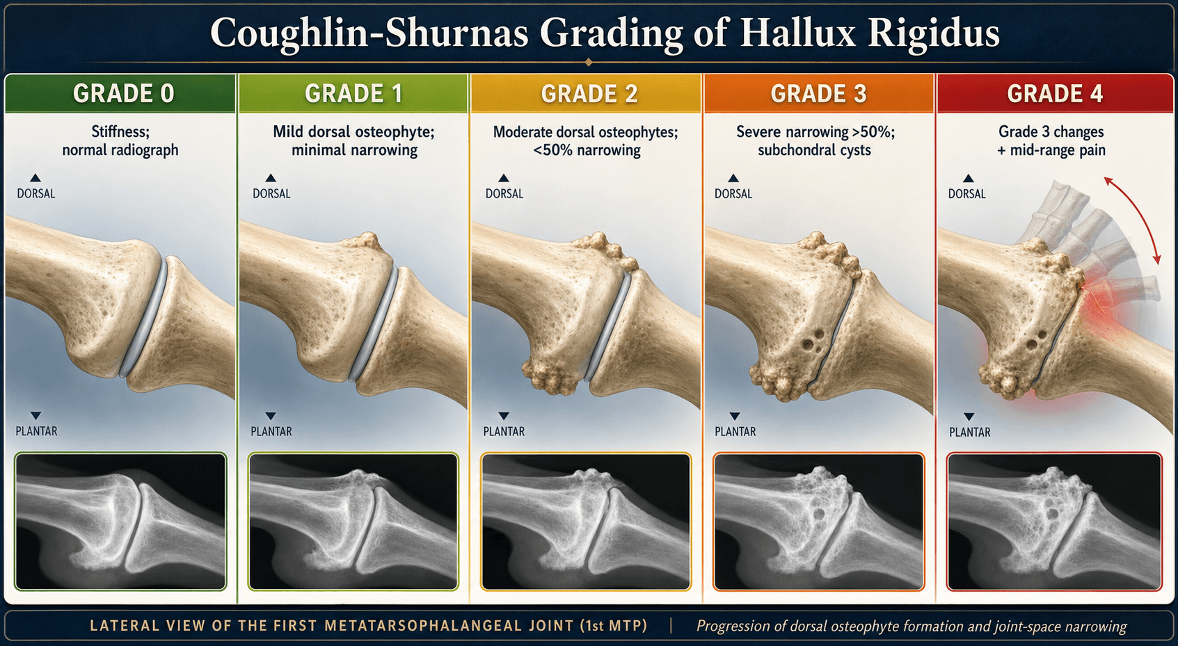
Coughlin-Shurnas Classification (Most Commonly Used)
- Radiographic Findings
- Dorsal osteophyte, no JSN
- Clinical ROM
- 10-20% loss, over 60° dorsiflexion
- Treatment
- Conservative, consider cheilectomy if symptomatic
- Radiographic Findings
- Mild spurring, 20-50% JSN, minimal sclerosis
- Clinical ROM
- 20-50% loss, 40-60° dorsiflexion
- Treatment
- Cheilectomy first-line, excellent results
- Radiographic Findings
- Moderate spurring, 50-75% JSN, subchondral sclerosis
- Clinical ROM
- 50-75% loss, 20-40° dorsiflexion
- Treatment
- Cheilectomy if plantar cartilage OK, or interposition
- Radiographic Findings
- Severe spurring, over 75% JSN, cysts, loose bodies
- Clinical ROM
- Over 75% loss, under 20° dorsiflexion
- Treatment
- Arthrodesis gold standard
- Radiographic Findings
- Grade 3 changes plus hallux valgus or varus
- Clinical ROM
- Severe stiffness plus deformity
- Treatment
- Arthrodesis with deformity correction
The critical decision point is Grade 2: if intraoperative assessment shows cartilage preservation on the plantar surface, cheilectomy can succeed. If cartilage loss is circumferential, proceed directly to arthrodesis. Do not compromise with inadequate debridement.
This classification system correlates well with treatment outcomes and provides clear decision-making framework.
CARPHallux Rigidus Classification Systems
Hook:CARP - like a fish mouth that can't open (rigid joint)!
Clinical Assessment
- Pain: Dorsal MTP, worse with toe-off, stairs, squatting
- Stiffness: Progressive loss of dorsiflexion
- Gait: External foot progression angle to avoid dorsiflexion
- Footwear: Difficulty with heels, dress shoes, athletic shoes
- Activities: Reduced running, dancing, sports participation
- Previous treatments: Orthotics, injections, activity modification
- Look: Dorsal prominence, skin irritation over osteophyte
- Feel: Tenderness over dorsal MTP, osteophyte palpable
- Move: Measure dorsiflexion (normal 65-75°), grind test positive
- Deformity: Assess for hallux valgus/varus component (Coughlin-Shurnas Grade 4)
- Gait: Observe toe-off phase, external rotation compensation
- Neurovascular: Ensure intact (dorsalis pedis, sensation)
Compress the first MTP joint while rotating the hallux. Pain reproduction indicates intra-articular pathology (arthritis). Compare with dorsal impingement pain (pain only at end-range dorsiflexion). Grind test specificity distinguishes arthritis from isolated dorsal impingement.
- Hallux Rigidus
- Pain and stiffness
- Hallux Valgus
- Deformity and bunion pain
- Turf Toe
- Acute traumatic pain
- Hallux Rigidus
- Dorsal osteophyte, usually straight alignment
- Hallux Valgus
- Lateral deviation, medial eminence
- Turf Toe
- Swelling, ecchymosis
- Hallux Rigidus
- Restricted dorsiflexion, painful
- Hallux Valgus
- Variable, often normal early
- Turf Toe
- All motion painful acutely
- Hallux Rigidus
- Dorsal osteophyte, JSN, sclerosis
- Hallux Valgus
- Hallux valgus angle, 1-2 IM angle
- Turf Toe
- Often normal, may show avulsion
Investigations
Imaging Protocol
Essential views for grading and planning. AP shows joint space narrowing, medial/lateral osteophytes. Lateral shows dorsal osteophyte (key for cheilectomy planning), assess dorsal 30% of metatarsal head.
Sesamoid assessment. Evaluates sesamoid arthritis which may require debridement at surgery. More sensitive than AP for lateral osteophytes.
CT: Preoperative planning for complex deformity or failed surgery. MRI: If concern for osteochondritis dissecans (young patient) or to assess cartilage (not routine).
- Early (Grade 1): Dorsal osteophyte, maintained joint space
- Moderate (Grade 2): Flattening of metatarsal head, 50% JSN
- Advanced (Grade 3): Severe JSN, subchondral sclerosis, cysts
- Late (Grade 4): Near ankylosis, loose bodies, deformity
- Measure dorsal osteophyte: Extent of resection for cheilectomy
- Assess joint space: Plantar cartilage preservation?
- Check sesamoids: Arthritic changes requiring debridement?
- Measure alignment: Hallux valgus/varus for fusion correction

Non-Operative Management
All patients should trial non-operative management before surgery, unless severe pain or functional limitation. Success rates vary: 20-30% achieve satisfactory symptom control with conservative measures. Duration of trial: 3-6 months.
Conservative Treatment Algorithm
Wide toe box, stiff sole. Rigid sole reduces MTP dorsiflexion demand. Rocker-bottom sole shifts toe-off proximal. Avoid high heels (increase dorsiflexion demand).
Morton's extension orthotic (carbon fiber plate extending to hallux tip) prevents MTP dorsiflexion. Turf toe plate similar effect. Padding over dorsal osteophyte for shoe pressure.
Avoid high-impact activities, running, jumping. Low-impact alternatives: cycling, swimming. Occupational modifications for prolonged standing/walking.
NSAIDs for pain and inflammation. Oral or topical. Caution in elderly, renal disease. Not disease-modifying, symptom control only.
Corticosteroid (+ local anesthetic). Diagnostic: confirms intra-articular source. Therapeutic: 3-6 months relief common. Maximum 2-3 injections. Consider hyaluronic acid (less evidence).
Indications for surgical referral: Failure of 3-6 months conservative treatment, severe pain limiting daily activities, progressive deformity (Grade 4), significant gait disturbance affecting work/recreation. Emphasize to patients that surgery is elective but highly effective.
Management Algorithm
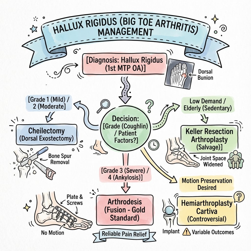
Cheilectomy - Joint-Preserving Procedure
Indications:
- Coughlin-Shurnas Grade 1-2 (mild to moderate arthritis)
- Dorsal osteophyte causing impingement
- Preserved plantar cartilage (critical!)
- At least 30 degrees dorsiflexion remaining
- Failed conservative management
Contraindications:
- Circumferential cartilage loss (intraop finding)
- Severe stiffness (under 20 degrees dorsiflexion)
- Grade 3-4 disease
- Sesamoid arthritis
Cheilectomy Technique
Dorsal longitudinal incision over first MTP joint, 3-4 cm. Protect dorsal sensory nerves (medial and lateral cutaneous branches). Incise capsule longitudinally, preserve for repair.
Intraoperative cartilage evaluation. Plantarflex hallux to expose dorsal metatarsal head. Assess cartilage: if intact on plantar two-thirds, proceed with cheilectomy. If circumferential loss, convert to arthrodesis.
Remove dorsal 25-30% of metatarsal head. Use oscillating saw or osteotome. Resect from medial to lateral, ensuring complete removal of dorsal ridge. Smooth with rongeur. Remove phalangeal osteophytes.
Release dorsal capsular adhesions to improve dorsiflexion. Gentle manipulation to achieve at least 60-70 degrees dorsiflexion. Avoid forced manipulation (fracture risk).
Repair capsule loosely (over-tightening limits dorsiflexion). Subcuticular skin closure. Soft dressing, wooden shoe or post-op shoe for 2 weeks.
- 30% rule: Remove dorsal 30% to decompress joint
- Check motion: Aim for 60-70° intraoperative dorsiflexion
- Preserve plantar cortex: Critical for stability
- Early mobilization: Start ROM at 2 weeks
- Under-resection: Inadequate decompression, recurrent impingement
- Over-resection: Metatarsal fracture, instability, transfer metatarsalgia
- Missed plantar disease: Poor outcome, consider conversion
- Forced manipulation: Fracture, damage plantar cartilage
Grade 1-2 disease: 80-90% good-excellent results at 5 years. Pain relief predictable, ROM improvement variable (average 20-30 degree gain). Satisfaction high. Durability: 70-80% avoid further surgery at 10 years. Failed cheilectomy can proceed to arthrodesis without compromise.
These outcomes make cheilectomy an excellent first-line option for appropriate candidates.

Surgical Technique
Patient Positioning
Setup Checklist
Supine on standard operating table. Ankle bump to internally rotate leg, expose medial aspect of first MTP joint. Contralateral limb: flat on table or frog-leg position.
Thigh or ankle tourniquet. Ankle preferred for better access. Exsanguinate with Esmarch or elevation. Inflate to 250-300 mmHg (ankle) or 100 mmHg above systolic (thigh).
Foot and ankle free-draped. Expose from toes to mid-calf. Ensure C-arm access for lateral and AP views of first MTP joint.
Standard positioning allows both cheilectomy and arthrodesis through same approach.
Moberg Osteotomy (Dorsal Closing-Wedge Phalangeal Osteotomy)
The controversies name a "Moberg osteotomy" added to cheilectomy, and the Lau-Daniels evidence used a "cheilectomy and phalangeal osteotomy", but the procedure and its rationale are never developed.
- What it is. A dorsal closing-wedge osteotomy of the base of the hallux proximal phalanx - a small dorsally-based wedge is removed and closed to tilt the toe into extension.
- Why it works. Hallux rigidus loses dorsiflexion but usually retains plantarflexion. The Moberg does not create new joint motion; it rotates the retained arc dorsally, shifting the toe's functional position into more extension so the patient achieves usable dorsiflexion for toe-off without forcing the arthritic dorsal joint - effectively "borrowing" surplus plantarflexion for dorsiflexion.
- How it is used. It is an adjunct to cheilectomy (cheilectomy removes the dorsal impingement; the Moberg adds functional dorsiflexion) for Grade 1-2 disease, especially the younger patient left with limited dorsiflexion but a good plantarflexion arc after cheilectomy alone. It is fixed with a small staple, screw or suture.
- The caveat. It slightly shortens the phalanx and its independent benefit over cheilectomy alone is not firmly established (a genuine controversy), so it is offered selectively rather than routinely.
Q: What is a Moberg osteotomy and why add it to a cheilectomy for hallux rigidus? A: A dorsal closing-wedge osteotomy of the proximal phalanx base that tilts the hallux into extension. Because hallux rigidus loses dorsiflexion but keeps plantarflexion, the Moberg rotates the retained arc dorsally, giving functional dorsiflexion for toe-off without creating new joint motion - a useful adjunct to cheilectomy in Grade 1-2 disease (especially younger patients). It shortens the phalanx slightly and its independent benefit over cheilectomy alone is unproven.
Synthetic Cartilage (Hydrogel) Implant
The Baumhauer RCT card, the guidelines table ("Cartiva synthetic implant") and the controversies all name a synthetic cartilage implant as a motion-sparing option, but what it is and how it is used is never described.
- What it is. A small (about 8-10 mm) polyvinyl-alcohol (PVA) hydrogel implant (Cartiva) that mimics articular cartilage - a resilient, water-containing cylinder that resurfaces the metatarsal head.
- How it is implanted. Through a dorsal approach and cheilectomy, a cylindrical recess is drilled in the first metatarsal head and the hydrogel plug is press-fit so its surface sits slightly proud to articulate against the phalangeal base - a partial resurfacing that keeps the joint mobile, with no cement or fixation.
- The evidence and role. In the multicentre non-inferiority RCT (Baumhauer) it was statistically equivalent to arthrodesis at 2 years for pain and function, gained about 6 degrees (27%) of active dorsiflexion, and under 10% required conversion to fusion by 2 years, with no fragmentation, wear or bone loss. It is a motion-sparing alternative for advanced disease in a patient who prioritises retaining MTP motion and accepts a higher revision risk.
- The caveat. The trial was industry-sponsored with only 2-year follow-up, and post-market series report higher early revision, subsidence and cyst formation than the trial - so arthrodesis remains the most durable, lowest-cost end-stage option and the implant is offered selectively.
Q: What is the synthetic cartilage (Cartiva) implant and what is its role in hallux rigidus? A: A small polyvinyl-alcohol hydrogel plug that resurfaces the first metatarsal head - drilled and press-fit after a cheilectomy to keep the joint mobile. The multicentre non-inferiority RCT (Baumhauer) found it equivalent to arthrodesis at 2 years (about 6 degrees dorsiflexion gain, under 10% converted to fusion). It is a motion-sparing option for advanced disease in a motion-priority patient, but with only 2-year trial data and higher real-world revision/subsidence, arthrodesis remains the durable gold standard.
Complications
- Incidence
- 15-20% at 5-10 years
- Risk Factors
- Under-resection, progression of arthritis, Grade 3-4 disease
- Management
- Revision cheilectomy if residual osteophyte, or convert to arthrodesis
- Incidence
- 10-15% (most common complication)
- Risk Factors
- Technical error, inadequate fluoroscopy, poor positioning technique
- Management
- If symptomatic: revision arthrodesis with osteotomy
- Incidence
- 5-10%
- Risk Factors
- Smoking, diabetes, inadequate fixation, poor bone quality
- Management
- Revision arthrodesis with bone graft, biologics, rigid fixation
- Incidence
- 10-15%
- Risk Factors
- Prominent plate, low-profile skin, patient factors
- Management
- Hardware removal after union (typically 6-12 months)
- Incidence
- 5-10%
- Risk Factors
- Over-resection (cheilectomy), malunion (arthrodesis)
- Management
- Orthotics, metatarsal pads, rarely osteotomy
- Incidence
- 1-3%
- Risk Factors
- Diabetes, smoking, immunosuppression
- Management
- Antibiotics, wound care, rarely debridement or hardware removal
- Incidence
- 5-10% temporary, 1-2% permanent
- Risk Factors
- Dorsal medial/lateral cutaneous nerves
- Management
- Usually resolves, neuropathic pain management if persistent
Malunion is the most common significant complication of first MTP arthrodesis. Prevention requires meticulous intraoperative technique: use sterile block to simulate weight-bearing, check hallux alignment (should point between 2nd-3rd toes), ensure 15-20 degrees dorsiflexion (1-2 cm ground clearance), confirm 10-15 degrees valgus. Fluoroscopy in multiple planes before final fixation. Do not accept suboptimal position - reposition and re-fix if needed.
Postoperative Care and Rehabilitation
Cheilectomy Rehabilitation
Dressing and footwear: Soft dressing, wooden shoe or post-op shoe. Weight-bearing: Immediate weight-bearing as tolerated in protective shoe. Activity: Elevate foot, ice, minimal walking. Pain control: Oral analgesics, NSAIDs after 48 hours.
ROM exercises: Start gentle dorsiflexion exercises at 2 weeks (critical!). Manual stretching, active ROM. Goal: regain 60-70 degrees. Footwear: Transition to stiff-soled athletic shoe. Weight-bearing: Full weight-bearing. Activity: Walking, avoid running/jumping.
Strengthening: Toe curls, marble pick-up, resistance band dorsiflexion. Proprioception: Balance exercises. Activity: Gradual return to sports, impact activities. Footwear: Normal shoes, avoid high heels initially.
Return to sport: Running, jumping, cutting at 3 months if ROM adequate. Long-term: Expect 90% recovery by 6 months. Maintain ROM with daily stretching. Avoid excessive high heels long-term.
Early ROM exercises are critical to cheilectomy success. Start at 2 weeks - capsular adhesions form quickly. Goal: 60-70 degrees dorsiflexion. Aggressive physiotherapy improves outcomes. Stiffness post-cheilectomy often reflects inadequate rehabilitation, not surgical failure.
Early mobilization maximizes the motion-preservation benefit of cheilectomy.
Outcomes and Prognosis
- Best For
- Grade 1-2, dorsal disease
- Success Rate
- 80-90% satisfaction at 5 years
- Advantages
- Motion preserved, simple procedure, low morbidity
- Disadvantages
- May fail (15-20% at 10 years), arthritis progression
- Best For
- Grade 2, young patient
- Success Rate
- 60-80% satisfaction at 5 years
- Advantages
- Motion preserved, no implant
- Disadvantages
- Higher failure than cheilectomy or arthrodesis, limited evidence
- Best For
- Grade 3-4, failed cheilectomy
- Success Rate
- 85-90% satisfaction long-term
- Advantages
- Predictable pain relief, durable, low revision rate
- Disadvantages
- Loss of motion, malunion risk, longer recovery
- Best For
- Limited role (elderly, low demand)
- Success Rate
- 60-80% at 5 years, 60% at 10 years
- Advantages
- Motion preserved (theoretically)
- Disadvantages
- High failure, loosening, revision difficult, not recommended
Cheilectomy: Grade 3-4 disease (wrong procedure), circumferential cartilage loss, inadequate debridement. Arthrodesis: Malunion (affects function, satisfaction), nonunion (5-10%), smoking. General: Inflammatory arthropathy, worker's compensation, unrealistic patient expectations. Patient selection and meticulous technique are critical to optimal outcomes.
Guidelines, Registries & Global Practice
Hallux rigidus is the most common arthritic condition of the foot and the second most common disorder of the first MTP joint after hallux valgus. Population studies estimate symptomatic disease in roughly 2.5% of adults over 50 years, with bilateral involvement in 50-80%. Peak incidence is in the fifth and sixth decades, with a bimodal pattern (adolescent osteochondral and adult degenerative). These figures are broadly consistent across high-income populations; data from low- and middle-income settings are sparse.
- Stance on Grading
- Coughlin-Shurnas 5-grade system most widely cited
- Joint-Preserving Surgery
- Cheilectomy for Grade 1-2 and selected Grade 3; synthetic cartilage implant an accepted alternative (FDA-approved)
- End-Stage Disease
- Arthrodesis is gold standard for advanced disease
- Stance on Grading
- Grading guides management; emphasis on shared decision-making
- Joint-Preserving Surgery
- Cheilectomy first-line for early disease; cautious adoption of synthetic implants pending long-term data
- End-Stage Disease
- Arthrodesis preferred for end-stage; replacement reserved for selected low-demand patients
- Stance on Grading
- Focus on fixation principles rather than grading
- Joint-Preserving Surgery
- Joint preparation to bleeding bone; congruent surfaces
- End-Stage Disease
- Rigid compression (lag screw plus dorsal plate or crossed screws) targeting 10-15 deg valgus, 10-15 deg dorsiflexion
- Stance on Grading
- Supports validated grading and patient-reported outcomes
- Joint-Preserving Surgery
- Motion-sparing options offered to younger, higher-demand patients after counselling
- End-Stage Disease
- Arthrodesis remains the most durable, cost-effective end-stage option
- No dedicated joint registry captures first MTP implants the way hip/knee registries do, so durability data rely on RCTs and case series
- Cartiva synthetic implant: equivalent to fusion at 2 years (Baumhauer RCT) but post-market series report higher early revision and subsidence
- Constrained total joint replacements: historically poor (Gibson RCT - loosening), largely abandoned
- Fusion union rates consistently 90-100% across series with rigid compression fixation
- High-resource: ready access to weight-bearing radiographs, locking plates, synthetic implants, formal physiotherapy and rocker-sole footwear
- Limited-resource: greater reliance on conservative care (stiff-soled footwear, activity modification); fusion favoured over implants given cost and lack of revision infrastructure
- Universal principles: grade-directed treatment, conservative trial first, fusion as the reliable end-stage solution regardless of setting
- Implant selection should reflect availability of revision capability, not novelty
Key documentation requirements:
- Conservative management trial documented (footwear, orthotics, injections, duration)
- Informed consent: procedure options (cheilectomy vs arthrodesis vs motion-sparing implant), fusion position (permanent loss of motion), outcomes (satisfaction and revision rates), complications (malunion, nonunion, infection, nerve injury)
- Realistic expectations: cheilectomy may fail (15-20% at 10 years); arthrodesis sacrifices motion but gives reliable pain relief
- Intra-operative decision-making: if converting from cheilectomy to arthrodesis based on cartilage status, document the finding and rationale
- Malunion prevention: document intra-operative checks (sterile block, fluoroscopy, alignment confirmation)
Common litigation themes: malunion (position not verified intra-operatively), deep infection, sensory nerve injury (medial dorsal cutaneous nerve), and unrealistic expectations of retained motion after arthrodesis.
Controversies and Areas of Uncertainty
The central debate is whether synthetic cartilage implants or interposition arthroplasty justify their higher revision risk to preserve motion in active patients. RCT data (Baumhauer) show 2-year non-inferiority for the hydrogel implant, but durability beyond 5 years and real-world revision rates remain contested. Fusion remains the most predictable, lowest-cost option.
Coughlin-Shurnas showed cheilectomy can succeed in SELECTED Grade 3 joints with greater than 50% cartilage remaining, blurring the simple "Grade 3 equals fusion" rule. The decision is ultimately intra-operative, based on cartilage assessment, not radiographs alone.
Earlier teaching favoured dorsal plate-plus-lag-screw over crossed screws, but the 2025 meta-analysis (Lim) found no difference in union or complications, only marginally faster fusion with plating. Construct choice is now driven by bone quality, cost and surgeon preference.
Texts quote 15-20 degrees of dorsiflexion relative to the ground, but AO and several series favour 10-15 degrees; excessive dorsiflexion causes interphalangeal joint overload and shoe-wear difficulty, while insufficient dorsiflexion causes pulp pressure. The functional reference (foot plantigrade on a block) matters more than an absolute number.
A dorsal closing-wedge phalangeal (Moberg) osteotomy is increasingly added to cheilectomy to augment effective dorsiflexion, but its independent contribution to outcome versus cheilectomy alone is not firmly established.
Radiographic grade correlates poorly with symptoms and with intra-operative cartilage status (Coughlin-Shurnas). Whether advanced imaging (MRI/CT cartilage mapping) should refine pre-operative decision-making, rather than intra-operative inspection, remains unresolved.
MCQ Practice Points
Q: What is the normal dorsiflexion range of the first MTP joint required for normal gait? A: 65-75 degrees. This range is required for toe-off phase of gait. Hallux rigidus typically reduces this to under 30 degrees, causing compensatory gait alterations (external foot progression angle, lateral weight shift).
Q: What are the key features distinguishing Coughlin-Shurnas Grade 2 from Grade 3 hallux rigidus? A: Grade 2: 50-75% joint space narrowing, moderate dorsal/lateral osteophytes, 20-50% ROM loss. Treatment: cheilectomy if plantar cartilage OK. Grade 3: Over 75% joint space narrowing, severe osteophytes, subchondral cysts, over 75% ROM loss (under 20 degrees dorsiflexion). Treatment: arthrodesis. The distinction guides surgical decision-making.
Q: What is the critical intraoperative decision point during cheilectomy for Grade 2 hallux rigidus? A: Assessment of plantar cartilage status. If cartilage is preserved on the plantar two-thirds of the joint, proceed with cheilectomy (30% dorsal head resection). If cartilage loss is circumferential, convert to arthrodesis. Do not compromise with inadequate debridement - this leads to poor outcomes.
Q: What is the optimal fusion position for first MTP arthrodesis? A: VDN mnemonic: Valgus 10-15 degrees (relative to first metatarsal axis), Dorsiflexion 15-20 degrees (relative to ground with foot plantigrade), Neutral rotation. Check alignment with foot on sterile block - hallux should clear ground by 1-2 cm and point between 2nd-3rd toes. Malunion is the most common complication and is position-dependent.
Q: What are the evidence-based success rates for cheilectomy vs arthrodesis in hallux rigidus? A: Cheilectomy (Grade 1-2): 80-90% satisfaction at 5 years, 70-80% avoid further surgery at 10 years. Arthrodesis: 90-95% fusion rate, 85-90% patient satisfaction long-term. Arthrodesis has slightly higher satisfaction but sacrifices motion. Both are evidence-based, appropriate procedures when used for correct indications.
Q: What is the most common significant complication of first MTP arthrodesis and how is it prevented? A: Malunion (10-15%) is the most common complication. Prevention requires meticulous intraoperative technique: use sterile block to simulate weight-bearing, check hallux alignment (between 2nd-3rd toes), ensure 15-20 degrees dorsiflexion (1-2 cm ground clearance), confirm 10-15 degrees valgus, fluoroscopy in multiple planes before final fixation. Do not accept suboptimal position.
Exam Viva Scenarios
Practise clinical reasoning and management decisions out loud
“A 55-year-old male accountant presents with 2 years of progressive pain and stiffness in his right great toe. Pain is worse with walking, particularly when pushing off. He has tried wider shoes and ibuprofen with minimal relief. On examination, there is a dorsal prominence at the first MTP joint, tenderness on palpation, and dorsiflexion limited to 25 degrees (plantarflexion full). Grind test is positive. Weight-bearing radiographs show a dorsal osteophyte, 60% joint space narrowing, and mild subchondral sclerosis. What is your assessment and management?”
“A 62-year-old female with severe hallux rigidus (Coughlin-Shurnas Grade 3, over 75% joint space loss, dorsiflexion 10 degrees) has failed conservative management and wants definitive treatment. You plan a first MTP arthrodesis. Walk me through your surgical technique, focusing on achieving optimal fusion position.”
“A 48-year-old male underwent cheilectomy for Grade 2 hallux rigidus 18 months ago. He returns with recurrent pain and stiffness. Dorsiflexion is now only 20 degrees. Radiographs show progression to Grade 3 disease with near-complete joint space loss and a small residual dorsal osteophyte. How do you manage this patient?”
Key Anatomy
- First MTP joint: 2-3x body weight in gait, 8x with running
- Normal dorsiflexion: 65-75 degrees (required for toe-off)
- Hallux rigidus: Typically under 30 degrees dorsiflexion
- Dorsal osteophyte: Blocks dorsiflexion, causes impingement pain
Coughlin-Shurnas Classification
- Grade 0: Dorsal osteophyte, no JSN = Conservative
- Grade 1: 20-50% JSN, mild spurring = Cheilectomy
- Grade 2: 50-75% JSN, moderate spurring = Cheilectomy or Interposition
- Grade 3: Over 75% JSN, severe changes = Arthrodesis
- Grade 4: Grade 3 plus hallux valgus/varus = Arthrodesis
Treatment Algorithm
- Conservative first (all grades): Stiff shoes, orthotics, NSAIDs, injections
- Cheilectomy: Grade 1-2, plantar cartilage preserved, 80-90% satisfaction
- Arthrodesis: Grade 3-4, failed cheilectomy, 90-95% fusion rate
- Arthroplasty: Limited role, high failure rates (20-40% at 10 years)
Surgical Pearls
- Cheilectomy: Remove dorsal 30% metatarsal head, achieve 60-70° intraop dorsiflexion
- Arthrodesis position (VDN): 10-15° Valgus, 15-20° Dorsiflexion, Neutral rotation
- Use sterile block to simulate weight-bearing when checking fusion position
- Plate fixation: 95% union vs screws 88% (meta-analysis)
- Intraop cartilage assessment determines cheilectomy vs arthrodesis
Complications
- Malunion (arthrodesis): 10-15%, most common, position-dependent
- Nonunion (arthrodesis): 5-10%, smoking major risk factor
- Recurrent pain (cheilectomy): 15-20% at 10 years, disease progression
- Hardware irritation: 10-15%, may require removal after union
- Transfer metatarsalgia: Over-resection or malunion
Evidence Base and Key Trials
Coughlin & Shurnas - The Landmark Grading and Long-Term Outcome Study
- Single-surgeon series of 110 patients (114 joints) followed over a 19-year period; mean follow-up 9.6 years (cheilectomy) and 6.7 years (arthrodesis)
- Introduced the now widely used 5-grade (0-4) clinical-radiographic classification combining ROM, pain and radiographic change
- 80 patients (93 feet) had cheilectomy, 30 patients (34 feet) had arthrodesis
- 92% (86/93) of cheilectomies were successful for pain and function; cheilectomy reliable for Grade 1-2 and SELECTED Grade 3
- Grade 4, or Grade 3 with less than 50% of metatarsal head cartilage remaining at surgery, should be treated with arthrodesis
- No association found between hallux rigidus and first-ray hypermobility or metatarsus primus elevatus
Cheilectomy vs Interpositional Arthroplasty - Procedure-Specific Outcomes
- Retrospective comparison: 19 patients (24 feet) Grade 2 disease treated with cheilectomy vs 11 patients (11 feet) Grade 3 disease treated with interpositional arthroplasty
- Cheilectomy satisfaction 87.5% vs interpositional arthroplasty 72.7%; mean AOFAS 77.3 vs 71.6
- Great-toe weakness reported in 72.7% of interposition patients vs only 16.7% of cheilectomy patients
- Pedobarography showed reduced great-toe load and weight transfer to lesser metatarsals in all patients, greatest after interposition
- Authors conclude interposition is a salvage procedure with less reliable results than cheilectomy
Arthrodesis vs Total Joint Replacement - Randomised Controlled Trial
- RCT of 63 patients (77 toes): 38 toes arthrodesis vs 39 toes total replacement arthroplasty, single surgeon, 24-month follow-up
- Pain improved in both groups but significantly more after arthrodesis (p = 0.01)
- All 38 arthrodeses united (mean dorsiflexion 26 degrees) with few complications
- 6 of 39 arthroplasty implants required removal for phalangeal component loosening; remaining implants gained poor motion
- Cost ratio 2:1 in favour of arthrodesis; arthrodesis preferred even when implant failures were excluded
Synthetic Cartilage Implant vs Arthrodesis - Multicentre Non-inferiority RCT
- Prospective randomised (2:1) non-inferiority trial across 12 centres in Canada and the UK; 152 hydrogel synthetic cartilage implants vs 50 arthrodeses, advanced-stage disease
- VAS pain and FAAM sport/ADL scores improved significantly in both groups at 12 and 24 months with statistical equivalence on the composite outcome
- Implant gained 6.2 degrees (27.3%) active dorsiflexion, maintained at 24 months
- Secondary surgery: 11.2% implant vs 12.0% arthrodesis; under 10% of implants required conversion to arthrodesis at 2 years
- No cases of implant fragmentation, wear or bone loss
Crossed Screws vs Plate-and-Screw Fixation - Meta-Analysis
- Systematic review and meta-analysis: 9 comparative studies, 976 patients (1,035 toes)
- NO significant difference in union rates between crossed screws and plate-plus-interfragmentary-screw (OR 0.75, p = 0.29)
- No significant difference in overall complications, revision, hardware removal or malunion
- Plate-and-screw construct gave a significantly shorter time to fusion (mean difference 0.51 weeks, p = 0.02)
- Choice should also weigh indication, bone quality and cost
Interposition Arthroplasty - Contemporary Systematic Review
- Systematic review of 20 studies, 498 patients (539 feet), mean follow-up 4.5 years
- Autogenous first MTP capsular tissue was the commonest interposition material (60% of studies)
- Mean improvements in standardised scores exceeded the minimal clinically important difference in most studies
- Progression to further surgery in only 3.8% of toes
- Transfer metatarsalgia was the commonest complication (up to 57.9% in one series)
- 85% of included studies were Level IV evidence