Hartofilakidis Classification of Congenital Hip Disease in Adults
The Hartofilakidis classification has three types, not four (unlike Crowe which has four grades). Dysplasia (Type 1) means the femoral head is contained in a shallow true acetabulum. Low dislocation (Type 2) means the femoral head articulates with a false acetabulum overlapping the superior true socket. High dislocation (Type 3) means the femoral head is fully dislocated onto the ilium. Examiners expect you to describe the acetabular deficiency pattern and the femoral implications for each type, and to contrast this system with the Crowe classification. The key surgical rule is the same as Crowe: always reconstruct the acetabulum in the true anatomic position.
The Hartofilakidis Classification System
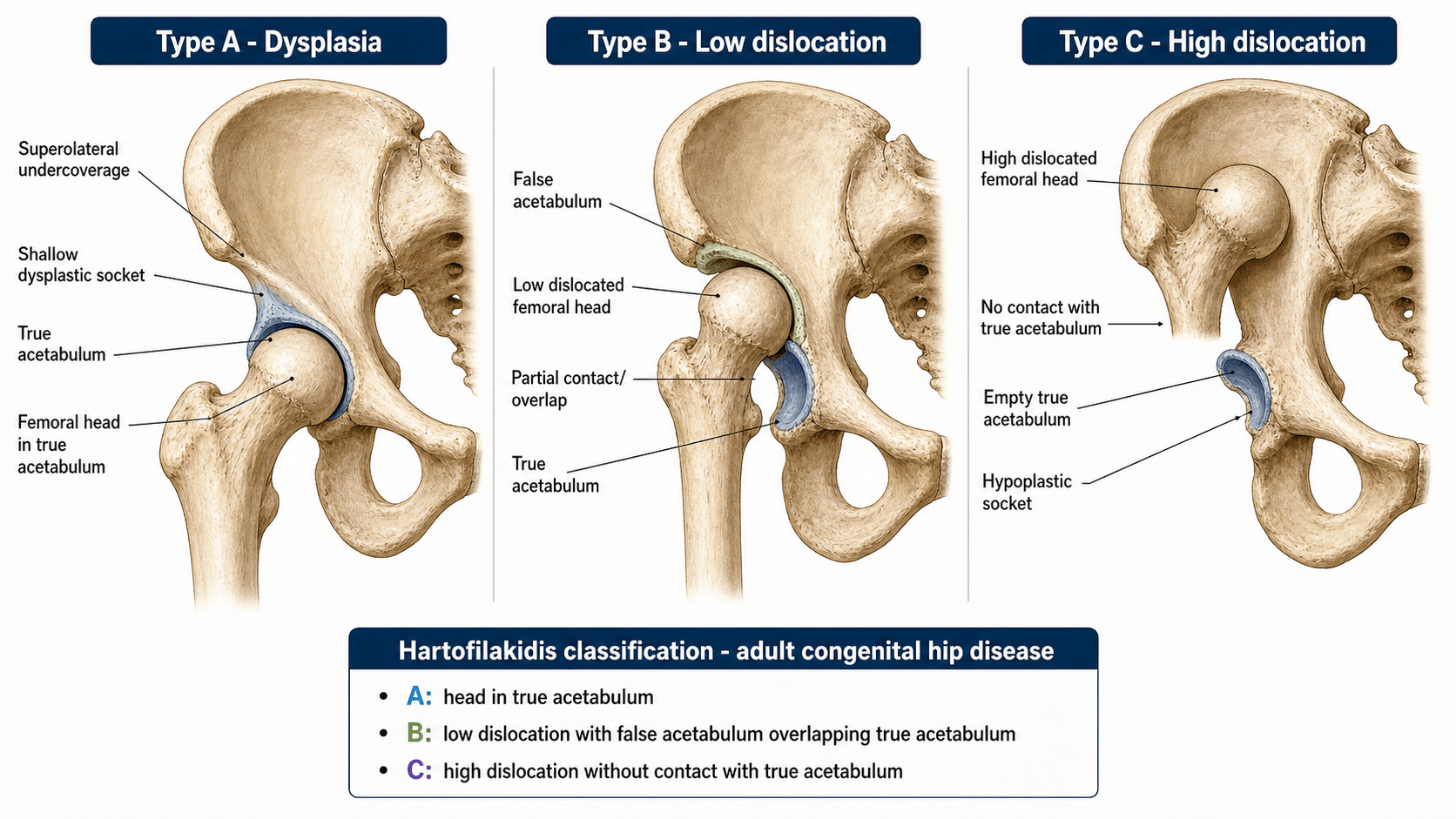
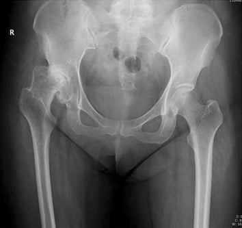
The Hartofilakidis classification was described in 1996 based on a series of over 200 total hip arthroplasties performed for congenital hip disease in adults. Unlike the Crowe system, which quantifies femoral head migration as a percentage, Hartofilakidis groups patients by the anatomical relationship between the femoral head and the true acetabulum. This makes the system clinically intuitive: the surgeon classifies what they see on the radiograph without needing to measure or calculate percentages.
- Femoral head position
- Contained within the true acetabulum but the socket is shallow with deficient superior and anterolateral coverage
- Acetabular characteristics
- Shallow true acetabulum; segmental deficiency of the superior rim; anteverted and lateralised; bone stock in the medial wall and posterior column is preserved
- Femoral characteristics
- Femoral head is smaller than normal; neck is short; anteversion is increased; the canal may be narrow but the diaphysis is usually normally shaped
- Femoral head position
- Articulates with a false acetabulum that partially overlaps the superior rim of the true acetabulum; the head is partially displaced
- Acetabular characteristics
- The false acetabulum erodes the superior rim of the true socket; segmental bone loss is greater than in dysplasia; the true acetabulum remains identifiable but is hypoplastic
- Femoral characteristics
- Femoral head is small and deformed; anteversion is markedly increased (often 40 to 60 degrees); the canal is narrow; the greater trochanter may be posteriorly positioned
- Femoral head position
- Completely dislocated from the true acetabulum; the femoral head lies against the lateral iliac wing above the true socket
- Acetabular characteristics
- The true acetabulum is severely hypoplastic — narrow, shallow, and anteverted; it may be a mere slit; bone stock is severely deficient anteriorly, superiorly, and posteriorly
- Femoral characteristics
- Femoral head is very small or absent; the femoral neck is extremely anteverted; the canal is extremely narrow and straight; the greater trochanter lies posterior to the femoral shaft
Contained — Contacting — Completely goneThe three types
Hook:Contained, Contacting, Completely gone — three Cs for three types in order of severity. Once the head leaves the socket entirely (Completely gone), you need a shortening osteotomy.
The distinguishing feature between Type 2 (low dislocation) and Type 3 (high dislocation) is whether the false acetabulum overlaps the true socket. In low dislocation, the femoral head sits in a false socket that partially shares the superior rim of the true acetabulum. In high dislocation, the femoral head has migrated proximally onto the ilium with no contact with the true socket at all. This distinction matters because the acetabular bone stock available for reconstruction is dramatically different.
The topic classifies the radiograph, but examiners also expect the clinical context:
- Presentation: typically a younger adult (often a woman), with activity-related groin/hip pain from labral and rim overload, a limp and a leg-length discrepancy (in unilateral disease); bilateral high dislocation gives a waddling (bilateral Trendelenburg) gait with marked lumbar hyperlordosis. A high dislocation can be surprisingly mobile and less painful early because there is no true bony articulation.
- Natural history: the shallow dysplastic socket concentrates contact stress on the deficient superolateral rim and labrum, causing labral tears and premature secondary osteoarthritis (often by the fourth or fifth decade); with progressive subluxation a false acetabulum forms (low dislocation) and ultimately the head escapes onto the ilium (high dislocation).
- Biomechanics — why true-socket reconstruction matters: the superolaterally migrated centre of rotation shortens the abductor lever arm, causing abductor insufficiency and the Trendelenburg gait, and raises the joint reactive force. Restoring the centre of rotation to the true (medial, inferior) position lengthens the abductor lever arm and lowers joint force — the biomechanical reason every type is reconstructed in the true acetabulum.
Acetabular Deficiency by Type
Each Hartofilakidis type produces a characteristic pattern of acetabular bone deficiency. Understanding the deficiency pattern is essential for selecting the correct reconstruction technique at THA.
- Acetabular deficiency pattern
- Segmental deficiency of the superior and anterolateral rim; the medial wall and posterior column are intact; the socket is anteverted and lateralised
- Reconstruction technique
- Acetabuloplasty with a small socket placed medially in the true position; rim augmentation with autograft or allograft if coverage is less than 70 percent
- Key consideration
- Lateralising the cup or using a high hip centre increases contact forces and loosening risk — reconstruct in the true position
- Acetabular deficiency pattern
- Segmental deficiency is more extensive; the false acetabulum has eroded the superior rim; the true socket is small but identifiable with preserved medial wall
- Reconstruction technique
- Reconstruct in the true acetabulum with a small socket; structural bone graft (femoral head autograft) to restore the superior rim; consider reinforcement ring if bone stock is poor
- Key consideration
- The temptation to place the cup in the larger false acetabulum must be resisted — it lateralises the centre of rotation and fails
- Acetabular deficiency pattern
- Severe global deficiency; the true acetabulum is a narrow slit with minimal bone stock anteriorly, superiorly, and posteriorly; medial wall may be the only usable bone
- Reconstruction technique
- Major reconstruction required: medial wall protrusio technique, bulk structural allograft, oblong cup, or custom triflange component; the true acetabulum is used but may need extreme medialisation
- Key consideration
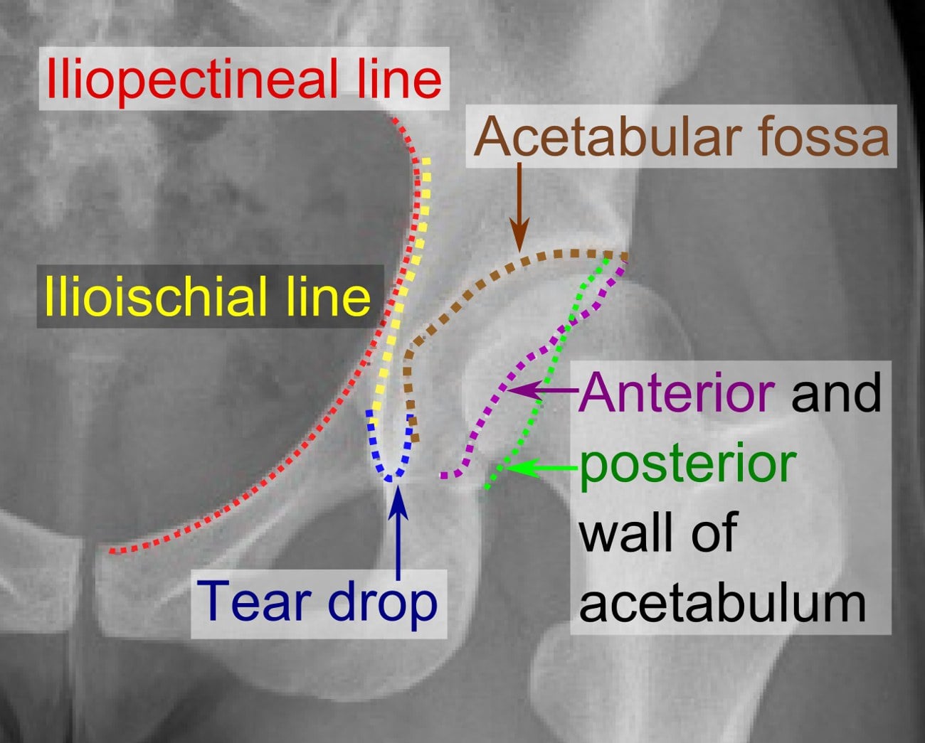
- The true acetabulum may be difficult to identify intraoperatively — use the transverse acetabular ligament and the teardrop as landmarks; pre-operative CT is essential
Always reconstruct the acetabulum in the true anatomic position, regardless of type. Placing the cup in the false acetabulum (especially in Type 2 and 3) is a well-documented cause of early failure because it lateralises the centre of rotation, increases joint reactive forces, and provides inadequate bone for fixation. In the exam, stating that you would reconstruct in the true acetabulum is a non-negotiable answer.
Worth naming, because it comes from the original 1996 paper and is the technique most associated with this classification. When the acetabulum is so deficient that an appropriately sized cup cannot be covered (typically low and high dislocation), cotyloplasty deliberately creates a controlled comminuted fracture of the medial acetabular wall to medialise the socket (a controlled protrusio), then autografts the medial defect with morsellised bone (commonly from the resected femoral head) and seats a small cup in the true position. The medialisation gains bony coverage without lateralising the centre of rotation, and the graft reconstitutes the medial wall over time. In the original series of 66 patients (86 hips) the acetabular components had a cumulative success of about 100% at 5 years and roughly 93% at 10 years. Modern practice often substitutes a cementless cup with or without bulk/structural graft, but the principle — medialise into the true socket rather than lateralise into the false one — is unchanged.
Femoral Management by Type
The femoral side of the reconstruction is equally important. The degree of femoral deformity, canal narrowing, and anteversion increases with each type and directly affects stem selection and the need for shortening osteotomy.
- Dysplasia (Type 1)
- Small but present
- Low dislocation (Type 2)
- Small and deformed
- High dislocation (Type 3)
- Very small or absent; may be a rudimentary nubbin
- Dysplasia (Type 1)
- Increased (20 to 30 degrees)
- Low dislocation (Type 2)
- Markedly increased (40 to 60 degrees)
- High dislocation (Type 3)
- Extremely increased (may exceed 60 degrees)
- Dysplasia (Type 1)
- May be narrow
- Low dislocation (Type 2)
- Narrow and straight
- High dislocation (Type 3)
- Very narrow, straight; may require a 7 to 9 mm stem
- Dysplasia (Type 1)
- Not required
- Low dislocation (Type 2)
- May be needed if the leg length discrepancy is significant or reduction is tight
- High dislocation (Type 3)
- Mandatory — subtrochanteric shortening of 2 to 6 cm to bring the femur down to the true socket without nerve tension
- Dysplasia (Type 1)
- Standard or slightly small cementless stem; modular if version adjustment needed
- Low dislocation (Type 2)
- Small tapered cementless stem; modular stem to correct version; shortening osteotomy if performed
- High dislocation (Type 3)
- Very small tapered cementless stem; modular or custom stem for version correction; shortening osteotomy is mandatory
Size — Straighten — ShortenFemoral management — three priorities
Hook:Three S's in order: Size the stem, Straighten the version, Shorten the femur. Each step is more critical as the type increases from 1 to 3.
The femoral canal in high dislocation (Type 3) can be as narrow as 7 to 9 mm. Always measure the canal diameter on pre-operative CT and have extra-small tapered stems available. Intraoperative fracture of the femur during broaching is a recognised complication in these extremely narrow canals — consider prophylactic cerclage wiring or a fracture stem.
Hartofilakidis versus Crowe — When to Use Each
Both classifications are valid and widely used. They are complementary rather than competing systems. Understanding when each is preferred helps in exam answers and clinical practice.
- Hartofilakidis
- 3 types (dysplasia, low dislocation, high dislocation)
- Crowe
- 4 grades (I, II, III, IV)
- Hartofilakidis
- Anatomical relationship of the femoral head to the true acetabulum — descriptive, no calculation needed
- Crowe
- Percentage of femoral head migration measured on an AP pelvis radiograph — requires calculation
- Hartofilakidis
- Acetabular deficiency pattern and reconstruction strategy
- Crowe
- Femoral head migration distance and THA complexity grading
- Hartofilakidis
- Substantial to almost-perfect agreement in the head-to-head Yiannakopoulos study — comparable to Crowe, NOT clearly superior
- Crowe
- Substantial to almost-perfect agreement in the same study; both systems are reproducible when a standardised technique is used
- Hartofilakidis
- More commonly used in Europe (Greek and UK centres); codified in the EFORT literature
- Crowe
- More commonly used in North America; codified in the AAOS literature
- Hartofilakidis
- Intuitive at the operating theatre — the surgeon sees the anatomy and classifies immediately
- Crowe
- Quantitative and reproducible for research — the measurement provides a number that can be compared across studies
Hartofilakidis = Head-to-Socket; Crowe = Calculate percentageHow to remember the difference
Hook:Hartofilakidis is qualitative (describe what you see); Crowe is quantitative (measure a percentage). Use both in the exam for full marks.
In the exam, mentioning both classifications demonstrates depth of knowledge. When shown a dysplastic hip radiograph, classify it by both systems: state the Hartofilakidis type (anatomical description) and the Crowe grade (percentage calculation). Examiners value this dual classification because it shows you understand the acetabular side (Hartofilakidis) and the femoral migration severity (Crowe) simultaneously.
Limitations and Modern Context
- It does not quantify bone stock. The Hartofilakidis types describe the pattern of deficiency qualitatively but do not measure the volume or quality of the remaining acetabular bone. For operative planning, supplement with CT-based 3D templating to quantify bone stock and plan graft or implant size.
- The boundary between low and high dislocation can be ambiguous. Some hips sit at the transition between Type 2 and Type 3, where the femoral head is in contact with a false acetabulum that barely overlaps the true socket. In these cases, the surgical strategy should be guided by the severity of acetabular bone deficiency rather than the type label alone.
- It does not account for femoral deformity severity. Two Type 3 hips may have vastly different femoral canal diameters and anteversion angles. The classification groups the acetabular side but the femoral side requires independent assessment (CT measurement of canal diameter, anteversion, and the need for shortening osteotomy).
- Modern 3D planning and patient-specific instrumentation have reduced the reliance on any single classification system. Pre-operative CT with 3D reconstruction allows direct visualisation and measurement of both the acetabular and femoral sides, making the Hartofilakidis and Crowe classifications complementary descriptors rather than stand-alone decision tools.
- Previous childhood surgery (open reduction, Salter osteotomy, Pemberton or Chiari osteotomy) alters the anatomy independently of the Hartofilakidis type. Scar tissue, altered pelvic morphology, and retained hardware increase surgical complexity and should be documented separately.
- The classification is validated for THA planning and does not directly apply to hip preservation surgery (periacetabular osteotomy, Bernese PAO) where the goal is to improve coverage without arthroplasty.
Exam Viva
Practise clinical reasoning and management decisions out loud
“A 48-year-old woman presents with progressive left hip pain. She has a known history of untreated developmental dysplasia. The AP pelvis radiograph shows a shallow left acetabulum with a small femoral head that is contained within the true socket but has deficient superior coverage. There is secondary osteoarthritis. The right hip is normal. How would you classify this and plan her total hip arthroplasty?”
“A 42-year-old woman with bilateral congenital hip disease presents with severe bilateral hip pain and a waddling gait. The AP pelvis radiograph shows both femoral heads are completely dislocated and lie against the lateral iliac wing. The true acetabula are barely identifiable — narrow slits. She has a leg length discrepancy of approximately 4 cm bilaterally. How would you classify and manage her?”
The three types
- Type 1 (Dysplasia): femoral head contained within a shallow true acetabulum; deficient superior coverage
- Type 2 (Low dislocation): femoral head articulates with a false acetabulum overlapping the superior true socket rim
- Type 3 (High dislocation): femoral head completely dislocated onto the ilium; true acetabulum is a narrow slit
Acetabular reconstruction by type
- Type 1: small socket in the true acetabulum with rim graft if coverage is less than 70 percent
- Type 2: true acetabular reconstruction with structural graft (femoral head autograft) to restore the superior rim
- Type 3: major reconstruction — medial protrusio, bulk allograft, oblong cup, or custom triflange; always in the true position
- Universal rule: never place the cup in the false acetabulum — it lateralises the joint and fails
Femoral management by type
- Type 1: small cementless stem, modular for version; no shortening osteotomy
- Type 2: narrow tapered stem, modular for anteversion correction; shortening osteotomy if leg length discrepancy is significant
- Type 3: very small tapered stem, mandatory subtrochanteric shortening osteotomy (2 to 6 cm), sciatic nerve protection
- Pre-operative CT for all types: canal diameter, anteversion, bone stock assessment
Hartofilakidis versus Crowe
- Hartofilakidis: 3 types, anatomical (head-to-socket relationship), focuses on acetabular deficiency; reliability comparable to Crowe (not superior)
- Crowe: 4 grades, quantitative (percentage migration), focuses on femoral displacement, widely used in North America
- Both are complementary — classify by both systems in the exam for maximum credit
- Both share the same surgical principle: reconstruct the acetabulum in the true anatomic position
Evidence Base
The Hartofilakidis system rests on a single-surgeon body of work (Hartofilakidis 1996, Karachalios & Hartofilakidis 2010, Lampropoulou-Adamidou & Hartofilakidis 2019) — internally consistent and definitive for the classification itself, but Level 4 in design and not externally validated for outcomes. The one independent comparative test of reproducibility (Yiannakopoulos 2008) found the Hartofilakidis and Crowe systems comparably reliable — neither is clearly superior, so the choice between them is about what you want to describe (acetabular anatomy vs femoral migration percentage), not about reliability. The outcome message that survives across all of it is consistent and exam-critical: reconstruct the acetabulum in the TRUE position, and the fixation method (cemented, hybrid, cementless) matters far less than getting the centre of rotation right.
Congenital hip disease in adults. Classification of acetabular deficiencies and operative treatment with acetabuloplasty combined with total hip arthroplasty
- Proposed the three-type classification of adult congenital hip disease: dysplasia, low dislocation, and high dislocation
- Demonstrated that acetabular reconstruction in the true anatomic position with acetabuloplasty was feasible and durable across all three types
- Reported that femoral shortening osteotomy was essential for high dislocations to achieve safe reduction without nerve injury
Inter- and intra-observer variability of the Crowe and Hartofilakidis classification systems for congenital hip disease in adults
- Independent head-to-head test of reproducibility: both the Crowe and Hartofilakidis classifications showed substantial-to-almost-perfect inter- and intra-observer agreement
- Agreement was comparable between the two systems — the study did NOT show that either is meaningfully more reliable than the other
- A standardised radiographic measurement technique improved reproducibility for both systems
Congenital hip disease in adults: terminology, classification, pre-operative planning and management
- Comprehensive review of the Hartofilakidis classification terminology, type definitions, and pre-operative planning strategy
- Detailed guidance on acetabular reconstruction techniques for each type with emphasis on true acetabular positioning
- Established a systematic approach to femoral management including the role of shortening osteotomy in high dislocations