Sports Medicine | FAI Treatment | Labral Repair | Traction Essential | Nerve Protection Critical
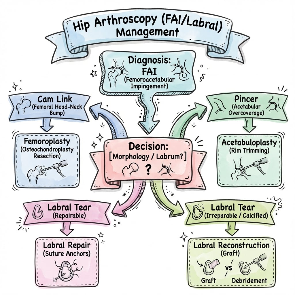
- Cam = femoral-based (alpha angle greater than 55°); Pincer = acetabular-based (overcoverage)
- Mixed FAI is most common (80%) - address both cam and pincer components
- Traction essential for central compartment - 25-50lbs force, break seal with internal rotation
- Maximum 2 hours traction - risk of nerve injury (pudendal, sciatic) increases significantly
- Lateral femoral cutaneous nerve at risk with anterolateral portal
- “Alpha angle greater than 55° on axial MRI indicates cam morphology
- “Positive anterior impingement test (FADIR) is most sensitive clinical sign
- “Always break the vacuum seal with longitudinal traction plus internal rotation before distraction
- “Lateral femoral cutaneous nerve crosses 1-2cm distal and medial to ASIS
Keep traction time and force to a minimum - traction time greater than 60 minutes significantly raises the complication rate (Larson 2016). Pudendal (perineal) numbness occurs in about 1.4% and the lateral femoral cutaneous nerve is the most commonly affected nerve overall (16.5%, persisting beyond 6 months in only 1.6%). Use a well-padded perineal post and release traction intermittently in prolonged cases.
Lateral femoral cutaneous nerve crosses 1-2cm distal and medial to ASIS - at risk with anterolateral portal. Superior gluteal nerve can be injured with proximal portal placement. Establish portals under fluoroscopic guidance with air arthrogram.
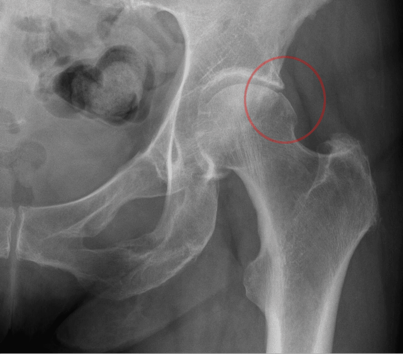
Cam morphology defined as alpha angle greater than 55° on axial MRI or cross-table lateral radiograph. Measured as angle between femoral neck axis and point where head-neck junction exceeds head radius. Normal is less than 50°.
Central compartment (under traction): labrum, acetabular cartilage, ligamentum teres, femoral head. Peripheral compartment (without traction): head-neck junction, medial synovial fold, zona orbicularis. Both compartments need assessment.
- Pathoanatomy
- Femoral head-neck asphericity (bump)
- Imaging Finding
- Alpha angle greater than 55°
- Surgical Treatment
- Osteochondroplasty (bump resection)
- Pathoanatomy
- Acetabular overcoverage
- Imaging Finding
- CE angle greater than 40°, crossover sign
- Surgical Treatment
- Rim trimming plus or minus labral repair
- Pathoanatomy
- Both cam and pincer (80% of cases)
- Imaging Finding
- Combined findings
- Surgical Treatment
- Address both components
CAM vs PINCERFAI Types - 'CAM vs PINCER'
Hook:CAM = bump on femoral head (young males). PINCER = rim pinches labrum (middle-aged females)!
AAPHip Arthroscopy Portals - 'AAP'
Hook:AAP = Anterolateral, Anterior, Posterolateral - the three standard hip arthroscopy portals!
SFLPNerves at Risk - 'SFLP'
Hook:SFLP = Sciatic, Femoral (rare), Lateral cutaneous (most common), Pudendal (traction)!
2-25-6Traction Safety - '2-25-6'
Hook:Remember 2-25-6: 2 hours max, 25lbs minimum, 6mm distraction needed!
Overview
Hip arthroscopy is a minimally invasive surgical technique for diagnosing and treating intra-articular and periarticular hip pathology. The primary indication is femoroacetabular impingement (FAI) with associated labral tears, though indications continue to expand.
- Imaging morphology is common and often asymptomatic: a systematic review of 2114 asymptomatic hips found cam morphology in 37% overall - 54.8% in athletes versus 23.1% in the general population (Frank 2015)
- Symptomatic FAI (FAI syndrome): Presents in athletes and active individuals, typically 20-45 years; diagnosis requires symptoms plus signs plus imaging (Warwick Agreement 2016)
- Sex distribution: Cam more common in males; Pincer more common in females; Mixed most common overall
- Athletic association: High-risk flexion-loading sports include ice hockey, football/soccer, martial arts, ballet and Australian Rules football
- Cam mechanism: Aspherical femoral head-neck junction causes outside-in abrasion of acetabular cartilage during flexion and internal rotation. The abnormal convexity shears against the labrum and cartilage.
- Pincer mechanism: Acetabular overcoverage causes labral crush injury and countrecoup lesions on posterior acetabulum. Overcoverage may be global (coxa profunda) or focal (acetabular retroversion).
- Natural history: Untreated FAI leads to progressive cartilage damage and eventual osteoarthritis
- Femoroacetabular impingement (cam, pincer, or mixed)
- Labral tears
- Chondral lesions (debridement, microfracture)
- Ligamentum teres tears
- Loose bodies
- Synovial disorders (PVNS, synovial chondromatosis)
- Hip instability (capsular plication)
- Iliopsoas tendon release
Pathophysiology and Mechanisms
The hip is a ball-and-socket joint with inherent bony stability enhanced by the acetabular labrum, capsule, and ligaments. The femoral head is covered by hyaline cartilage except at the fovea (ligamentum teres attachment).
- Fibrocartilaginous ring attached to the acetabular rim
- Deepens the acetabulum by 21% and increases surface area by 28%
- Creates a suction seal that provides joint stability
- Blood supply from superior gluteal, inferior gluteal, and obturator vessels
- Vascularity is better peripherally than centrally (important for repair)
- Iliofemoral ligament (Y-ligament of Bigelow): strongest ligament, anterior
- Pubofemoral ligament: inferior, limits abduction
- Ischiofemoral ligament: posterior, limits internal rotation
- Zona orbicularis: circular fibers around femoral neck
- Femoral neurovascular bundle: anterior, protected by iliopsoas
- Lateral femoral cutaneous nerve: crosses 1-2cm distal and medial to ASIS
- Superior gluteal nerve: exits above piriformis, at risk with proximal portals
- Sciatic nerve: posterior, at risk with traction and posterior portals
- Abnormal bump at femoral head-neck junction (anterosuperior most common)
- Results from developmental abnormality during skeletal maturation
- Alpha angle greater than 55° is diagnostic
- Causes outside-in cartilage abrasion during flexion/internal rotation
- Acetabular overcoverage (global or focal)
- Center-edge angle greater than 40° suggests overcoverage
- Crossover sign on AP pelvis indicates focal retroversion
- Causes labral crush injury and countrecoup posterior cartilage damage
Classification
Cam-type FAI results from femoral-based pathology where an aspherical femoral head-neck junction (bump) impinges against the acetabular labrum and cartilage during hip flexion and internal rotation.
Key features: Alpha angle greater than 55° on axial imaging. Bump typically anterosuperior. More common in young athletic males. Causes outside-in cartilage abrasion pattern. Treatment involves osteochondroplasty (bump resection) to restore sphericity.
- Type 1: Detachment of labrum from acetabular cartilage (most common)
- Type 2: Cleavage plane within labral substance
- Grade 0: Normal cartilage
- Grade 1: Softening (malacia)
- Grade 2: Partial thickness defect
- Grade 3: Full thickness defect
- Grade 4: Exposed subchondral bone
- Focal pincer: Retroversion of superior acetabulum
- Global pincer: Coxa profunda/protrusio acetabuli
Clinical Assessment
Classic FAI presentation is a young athletic patient with activity-related groin pain, often with clicking, catching, or giving way. Pain typically insidious onset, worse with prolonged sitting, pivoting activities, and hip flexion.
Common symptoms: Deep anterior groin pain (C-sign where patient cups hand over greater trochanter). Pain with prolonged sitting (theater sign). Pain with hip flexion activities (squatting, stairs). Mechanical symptoms (clicking, catching, locking).
Observe gait for Trendelenburg or antalgic pattern. Note pelvic obliquity and limb length. Assess hip flexion contracture.
Compare to contralateral side. FAI typically shows decreased internal rotation in flexion. Measure flexion, extension, abduction, adduction, internal and external rotation.
The FADIR (anterior impingement) test is the most sensitive clinical test for FAI. Position: supine with hip and knee at 90° flexion, then passively adduct and internally rotate. Positive test reproduces the patient's groin pain. While sensitive (95%), it has low specificity - must correlate with imaging.
Differential Diagnosis of Young-Adult Groin / Hip Pain:
Because cam and pincer morphology is common in asymptomatic people (37% asymptomatic cam in athletes/general population; Frank 2015), FAI syndrome is a clinical diagnosis of exclusion as much as inclusion. Actively exclude these mimics before attributing pain to impingement.
- Typical patient / pain
- Young active adult, deep anterior groin (C-sign), worse sitting/pivoting
- Discriminating feature
- Positive FADIR; cam/pincer plus symptoms; greater than 50% relief from intra-articular injection
- Typical patient / pain
- Often female, antero-lateral pain, instability/fatigue, micro-instability
- Discriminating feature
- Lateral centre-edge angle less than 20-25°; under-coverage (opposite of pincer)
- Typical patient / pain
- Athlete, lower abdominal/adductor-origin pain with sprinting and Valsalva
- Discriminating feature
- Tender pubic tubercle/adductor origin; pain NOT reproduced by FADIR; negative injection
- Typical patient / pain
- Older patient, stiffness, reduced overall ROM
- Discriminating feature
- Joint space narrowing, osteophytes (Tönnis 2 or more); poor arthroscopy candidate
- Typical patient / pain
- Back pain, dermatomal radiation, neurological signs
- Discriminating feature
- Pain not localised to groin; negative hip provocation; spinal signs positive
- Typical patient / pain
- Lateral hip pain, tender over trochanter, pain lying on side
- Discriminating feature
- Lateral (not groin) tenderness; pain on single-leg stance/resisted abduction
Investigations
Essential initial view. Assess center-edge angle, crossover sign, posterior wall sign. Must have proper positioning (coccyx 1-3cm above symphysis).
Cross-table lateral (Dunn view) or frog-leg lateral. Assess alpha angle, head-neck offset.
Evaluates labrum, cartilage, and periarticular soft tissues. Axial oblique views best for alpha angle measurement.
Gold standard for labral pathology. Intra-articular gadolinium improves labral tear detection. Sensitivity greater than 90% for labral tears.
Series of views radiating from femoral neck axis. Best for comprehensive assessment of cam lesion location.
Provides excellent bony detail of cam and pincer morphology. Useful for surgical planning. Less radiation with low-dose protocols.
Intra-articular injection of local anesthetic under fluoroscopic guidance can confirm intra-articular source of pain. Greater than 50% pain relief suggests intra-articular pathology.
Non-Operative Management
Indications for Non-Operative Treatment:
- Mild symptoms with minimal functional limitation
- Patient preference
- Significant osteoarthritis (relative contraindication to arthroscopy)
- Medical contraindications to surgery
- Initial management while diagnosis is being confirmed
Non-Operative Protocol:
Activity modification: Avoid aggravating positions (deep flexion, prolonged sitting). Physical therapy: Core and hip strengthening, focus on gluteal activation. NSAIDs: For symptomatic relief. Education: Explain pathophysiology and natural history.
Advance rehabilitation: Progressive hip strengthening program. Sport-specific modification: Identify and modify aggravating activities. Injection therapy: Consider intra-articular corticosteroid if indicated. Reassess: Evaluate response to conservative treatment.
Evaluate response: If improved, continue activity modification and maintenance program. If symptoms persist: Consider surgical intervention if appropriate candidate. Shared decision-making: Discuss risks and benefits of surgery vs continued non-operative care.
While intra-articular corticosteroid injections may provide temporary relief, they do not address the underlying mechanical problem. Some evidence suggests repeated injections may negatively affect cartilage and outcomes of subsequent surgery. Use judiciously and primarily as a diagnostic tool.
Management Algorithm
Positive FADIR test with anterior groin pain in a young active patient. Correlate with imaging findings of cam (alpha greater than 55°) or pincer (CE greater than 40°) morphology. MR arthrogram to assess labrum.
Greater than 50% pain relief with intra-articular local anesthetic confirms intra-articular source and predicts good surgical outcome.
Key Decision Points:
- Joint space greater than 2mm → Good candidate for arthroscopy
- Joint space less than 2mm with arthritis → Consider arthroplasty
- Positive impingement test + positive injection → Best outcomes
- Mixed FAI → Must address both components
Surgical Management
Supine positioning on fracture table is most common. Patient supine with perineal post well-padded. Operative limb in traction boot. Contralateral limb in padded post or abducted in leg holder.
Advantages: Familiar position, easier fluoroscopy, better anterior access. Disadvantages: Perineal post pressure risk.
- Force: 25-50 pounds typically needed
- Technique: Apply longitudinal traction, then internal rotation to break vacuum seal
- Confirmation: Fluoroscopic confirmation of 6mm distraction minimum
- Time limit: Maximum 2 hours - release if prolonged case
- Anterolateral (AL): Primary viewing portal, 1cm anterior and superior to greater trochanter tip
- Anterior: Primary working portal, intersection of ASIS horizontal line and GT vertical line
- Posterolateral (PL): 1cm posterior and superior to GT, supplementary viewing/working
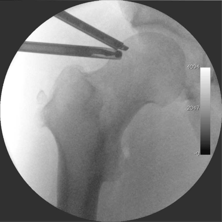
Establish portals under fluoroscopic guidance with air arthrogram. Use spinal needle to confirm trajectory. Make skin incision, then blunt trocar insertion to avoid nerve injury.
- Labral repair: Suture anchors for peripheral labral tears
- Labral debridement: For non-repairable tears
- Acetabular rim trimming: For pincer lesions
- Microfracture: For focal cartilage defects
- Ligamentum teres debridement: For symptomatic tears
- Loose body removal
- Osteochondroplasty: Cam lesion resection to restore head-neck offset
- Capsulotomy management: Repair or plication as needed
- Iliopsoas release: If symptomatic internal snapping hip
The text mentions "capsulotomy management" and instability from an unrepaired capsulotomy, but the examinable detail is which capsulotomy and whether to close it:
- Capsulotomy types: access to the central compartment requires dividing the iliofemoral ligament. An interportal capsulotomy (a transverse cut connecting the anterolateral and anterior portals) is the basic exposure; a T-capsulotomy adds a longitudinal limb down the femoral neck for wider peripheral-compartment/cam access but divides more of the iliofemoral ligament.
- Why it matters: the iliofemoral ligament is the primary anterior stabiliser of the hip, so an unrepaired (especially T-) capsulotomy can produce anterior microinstability - a recognised cause of persistent pain/failure and of the rare frank dislocation after hip arthroscopy.
- The closure controversy: routine capsular closure/plication is increasingly advocated, and the evidence and consensus most strongly support closure in higher-risk hips - borderline dysplasia, ligamentous laxity/hypermobility, and revision surgery - where capsular competence is critical. In the normal, well-covered hip the benefit of routine closure over selective closure is still debated, but most hip-preservation surgeons now repair the capsule.
Exam point: know interportal vs T-capsulotomy, that the divided iliofemoral ligament is the key anterior stabiliser, and that capsular closure/plication is particularly important in borderline dysplasia, laxity and revision to avoid iatrogenic microinstability.
Complications
Nerve Injuries:
- Mechanism
- Anterior/anterolateral portal injury
- Clinical Finding
- Meralgia paraesthetica (lateral thigh numbness) - most common (16.5%)
- Prevention
- Portal placement 1-2cm from ASIS, blunt dissection
- Mechanism
- Perineal post pressure / traction (about 1.4%)
- Clinical Finding
- Perineal numbness, sexual dysfunction
- Prevention
- Well-padded post, intermittent release, minimal traction time
- Mechanism
- Excessive traction, posterior portals
- Clinical Finding
- Posterior thigh numbness, foot drop
- Prevention
- Limit traction force and time, careful posterior portals
- Mechanism
- Proximal portal placement
- Clinical Finding
- Gluteus medius weakness, Trendelenburg
- Prevention
- Stay distal to piriformis, avoid proximal portals
Other Complications:
In the largest prospective multicentre series (1615 hips), the overall complication rate was 8.3% with mostly low-grade events; rates below are from that series unless stated otherwise (Larson 2016).
- Fluid extravasation: Risk of abdominal compartment syndrome with prolonged cases (no extra-abdominal extravasation observed in the 1615-hip series)
- Femoral neck fracture / stress fracture: 0.1%; risk from excessive osteochondroplasty (avoid resecting more than ~30% of neck circumference)
- Instability: From excessive rim trimming or unrepaired capsulotomy (no iatrogenic instability in the 1615-hip series)
- Iatrogenic chondral injury: 1.2%; iatrogenic labral puncture: 0.9%
- Heterotopic ossification: 0.8%; NSAID prophylaxis reduces incidence
- Avascular necrosis: Rare (none in the 1615-hip series), associated with lateral epiphyseal vessel damage
- Superficial portal infection: 1.1%
- DVT 0.1% / PE 0.1%: Standard VTE risk-stratified prophylaxis
The lateral femoral cutaneous nerve is the most commonly affected nerve (16.5% transient disturbance, persisting beyond 6 months in only 1.6%); pudendal (perineal) numbness is less common at about 1.4%. The single most important modifiable factor is traction: time greater than 60 minutes significantly raises the complication rate, and pudendal neurapraxia is linked to longer traction. Prevention: well-padded wide perineal post; minimal traction force and time; intermittent release in prolonged cases; consider post-less or lateral positioning.
Rehabilitation
Post-Operative Protocol:
Weight-bearing: Typically 20lbs flat-foot weight-bearing for 2-4 weeks (longer if microfracture). ROM restrictions: Limit hip flexion to 90°, avoid FADIR position for 4-6 weeks. CPM: Consider continuous passive motion machine. Goals: Protect repair, control inflammation, maintain ROM.
Progress weight-bearing: Advance to full as tolerated. ROM: Progress to full ROM. Strengthening: Aquatic therapy, stationary bike, hip strengthening (avoid impingement positions). Goals: Restore ROM, begin strengthening, normalize gait.
Activities: Progress to elliptical, swimming, functional exercises. Strengthening: Progressive resistance training. Sport-specific: Begin sport-specific training at 12+ weeks. Return to sport: Typically 4-6 months depending on procedure and sport.
- Protect repair for 4-6 weeks with ROM restrictions
- Avoid FADIR position during healing
- Full return to sport: 4-6 months
- Protected weight-bearing 6-8 weeks
- CPM encouraged for cartilage healing
- Return to sport: 6-12 months
- Earlier weight-bearing (2 weeks)
- Less ROM restriction needed
- Return to sport: 3-4 months
Outcomes and Prognosis
Short-Term Outcomes:
- Patient satisfaction: 85-90% satisfaction at 2 years
- Return to sport: 80-90% return to sport, 70-80% at same level
- Pain improvement: Significant improvement in pain scores (70-80% reduction)
- Functional improvement: Improved modified Harris Hip Score in 85%+
Long-Term Outcomes:
- Short-term Outcome
- Excellent (over 90% good/excellent)
- Long-term Outcome
- Over 90% hip preservation at 10 years
- Short-term Outcome
- Good (75-85% good/excellent)
- Long-term Outcome
- 70-80% hip preservation at 10 years
- Short-term Outcome
- Fair (50-60% good/excellent)
- Long-term Outcome
- 50-60% hip preservation at 10 years
- Young age (less than 40 years)
- Minimal osteoarthritis (Tonnis 0-1)
- Preserved joint space (greater than 2mm)
- Labral repair (vs debridement)
- Positive injection response pre-operatively
- Advanced osteoarthritis
- Joint space less than 2mm
- Full-thickness cartilage loss
- Age greater than 50 years
- Significant acetabular dysplasia
Guidelines, Registries & Global Practice
Global epidemiology. FAI morphology is common and frequently asymptomatic, so imaging must always be correlated with symptoms and signs. A PRISMA systematic review of 2114 asymptomatic hips (mean age 25 years) found asymptomatic cam morphology in 37% overall - 54.8% in athletes versus 23.1% in the general population - and the mean alpha angle in asymptomatic hips was 54° (Frank 2015). This is the single most important framing fact: morphology alone is not disease.
The diagnosis is a triad, not a radiograph. The 2016 Warwick Agreement (international consensus, 25 endorsing societies) introduced the term FAI syndrome, requiring symptoms + clinical signs + imaging findings together. Imaging morphology without symptoms is not FAI syndrome and should not be treated (Warwick 2016).
Guidance side by side
- Core position
- FAI syndrome = symptoms + signs + imaging; first-line either conservative care or surgery after shared decision-making
- Basis / evidence level
- Consensus statement (Level V)
- Core position
- Open and arthroscopic FAI surgery may be used with standard arrangements for governance, consent and audit; recognises evidence is still maturing
- Basis / evidence level
- Interventional procedures guidance
- Core position
- Arthroscopy gives a clinically meaningful but modest benefit over structured physiotherapy at 12 months (iHOT-33 +6.8)
- Basis / evidence level
- Level I RCT
- Core position
- Endorse arthroscopic management for appropriately selected FAI syndrome with preserved joint space and minimal arthritis
- Basis / evidence level
- Society endorsement / expert consensus
Patient selection drives outcome (global, evidence-led). Across health systems the same predictors apply: preserved joint space (2mm or more) and labral repair predict better results, while advanced arthritis (Tönnis 2 or more) and joint space less than 2mm predict failure and conversion to total hip replacement (Philippon 2009). The decisive modifiable intra-operative safety factor is traction - time greater than 60 minutes raises the complication rate (Larson 2016).
Registries and practice variation. Hip arthroscopy is a hip-preservation rather than an arthroplasty procedure, so it is not captured by the major joint replacement registries (NJR England & Wales, AOANJRR Australia, AJRR USA, SHAR Sweden). These registries are nonetheless relevant downstream: a meaningful minority of arthroscopy patients with pre-existing chondral wear later convert to total hip arthroplasty, and registry analyses are used to compare outcomes of THA after prior hip arthroscopy. Volume and access vary widely - arthroscopy is concentrated in high-resource centres with fluoroscopy and traction tables and a learning curve effect on complications, whereas in many regions open surgical hip dislocation or non-operative care predominate. FAI syndrome is recognised worldwide in flexion-loading sports (football/soccer, ice hockey, Australian Rules football, dance, martial arts), reflecting the higher cam prevalence seen in athletes.
Special Considerations
- Center-edge angle 20-25° is borderline
- May benefit from arthroscopy if FAI component present
- Higher failure rate than non-dysplastic hips
- Consider periacetabular osteotomy (PAO) if significant dysplasia
- Success rates lower than primary (60-70%)
- Most common reasons for failure: inadequate cam resection, missed pincer, untreated labral tear
- Better outcomes if clear identifiable pathology for revision
- FAI morphology develops during skeletal maturation
- Arthroscopy may be considered in skeletally mature adolescents
- Must ensure physes are closed before aggressive osteochondroplasty
- Indicated when labrum is deficient and cannot be repaired
- Options: iliotibial band autograft, tensor fascia lata, allograft
- Emerging technique with promising early results
- Can result from excessive capsulotomy or rim trimming
- Capsular plication may be needed
- Important to balance impingement correction with stability preservation
This whole topic addresses intra-articular FAI (cam/pincer), but an examiner may probe extra-articular impingement, which can coexist with or mimic FAI and is missed if you only look at the joint:
- Subspine (AIIS) impingement: a prominent or low-lying anterior inferior iliac spine (often after an AIIS apophyseal avulsion that heals with overgrowth, or a congenitally low AIIS) impinges on the distal femoral neck in terminal flexion. It causes anterior hip pain and a limited, painful flexion arc; it is assessed on the AP/false-profile radiograph and 3D CT (Hetsroni AIIS morphology types) and treated by arthroscopic AIIS/subspine decompression, often at the same sitting as cam/pincer correction.
- Other extra-articular types to be aware of: ischiofemoral impingement (quadratus femoris between ischium and lesser trochanter), greater trochanteric-pelvic (ischiofemoral/trochanteric) impingement, and iliopsoas impingement on the labrum (anterior 3-o'clock labral tear from a tight psoas).
Exam point: if anterior impingement pain persists despite a "normal" cam/pincer correction, think extra-articular impingement - especially subspine/AIIS impingement (often post-AIIS-avulsion), diagnosed on 3D CT and treated by AIIS decompression.
MCQ Practice Points
Q: What are the radiographic definitions of Cam and Pincer FAI? A: Cam FAI is defined by an alpha angle greater than 55 degrees (femoral side). Pincer FAI is defined by a Center-Edge (CE) angle greater than 40 degrees or a crossover sign (acetabular side).
Q: Which nerve is most commonly injured during hip arthroscopy? A: The Lateral Femoral Cutaneous Nerve (LFCN) is the most commonly affected nerve overall (16.5% transient disturbance in large series, persisting beyond 6 months in only 1.6%), typically from the anterior/anterolateral portal. The Pudendal nerve (perineal numbness, about 1.4%) is the classic traction-related injury and is the high-yield answer for "traction complication".
Q: What are the traction safety limits? A: Traction should be limited to less than 2 hours duration and minimum necessary force (typically 25-50lbs) to reduce neurapraxia risk.
Q: What joint space width predicts poor outcomes? A: Joint space less than 2mm is a strong predictor of failure and conversion to Total Hip Arthroplasty (50% within 2 years).
Q: What is the most sensitive physical exam test for FAI? A: The FADIR test (Flexion, Adduction, Internal Rotation). It is highly sensitive (95%) but has low specificity.
Clinical Decision Scenarios
Practise clinical reasoning and management decisions out loud
“A 25-year-old male professional soccer player presents with 6 months of right groin pain that is worse with kicking and sprinting. He describes a deep anterior hip pain that is aggravated by prolonged sitting.”
“You are planning hip arthroscopy on a patient with confirmed FAI and labral tear. The examiner asks you to describe your portal placement and the nerves at risk.”
“You are shown radiographs of a hip. The examiner asks you to differentiate between cam and pincer morphology and describe how you would measure the relevant angles.”
“During hip arthroscopy, the anesthetist informs you that you have been operating for 2.5 hours with traction continuously applied. They ask about traction-related complications.”
“A 55-year-old woman presents with hip pain and imaging shows FAI with a joint space of 1.5mm and Tonnis grade 2 osteoarthritis. She is keen for hip arthroscopy to avoid hip replacement.”
FAI Classification
- Cam = femoral bump, alpha greater than 55°, young males
- Pincer = acetabular overcoverage, CE greater than 40°, middle-aged females
- Mixed = most common (80%), address both components
- Crossover sign = focal acetabular retroversion
Traction Safety
- Maximum 2 hours traction time
- 25-50lbs force typically needed
- Minimum 6mm joint distraction
- Break seal with IR before distraction
Nerves at Risk
- LFCN: most common nerve injury (16.5% transient); pudendal ~1.4%
- Lateral femoral cutaneous: anterolateral portal (meralgia)
- Sciatic: excessive traction, posterior portals
- Superior gluteal: proximal portal placement
Portal Placement
- Anterolateral: 1cm anterior/superior to GT tip (viewing)
- Anterior: ASIS horizontal meets GT vertical (working)
- Posterolateral: 1cm posterior/superior to GT
- Always establish under fluoroscopic guidance
Key Numbers
- Alpha angle: greater than 55° = cam
- CE angle: greater than 40° = pincer
- Joint space: less than 2mm = poor outcome
- 2 hours max traction, 25lbs min force, 6mm distraction
Exam Pearls
- FADIR test most sensitive for FAI
- Joint space greater than 2mm critical for good outcomes
- Labral repair preferred over debridement
- LFCN most commonly affected nerve overall (16.5%); pudendal (~1.4%) is the classic traction injury
Evidence Base
Hip Arthroscopy vs Best Conservative Care - UK FASHIoN RCT
- Pragmatic multicentre RCT, 348 patients with FAI syndrome at 23 UK NHS hospitals
- Hip arthroscopy versus personalised hip therapy (physiotherapist-led conservative care)
- Both groups improved; arthroscopy superior at 12 months
- Adjusted mean iHOT-33 difference 6.8 points (95% CI 1.7-12.0) favouring arthroscopy
- Difference exceeded the minimum clinically important difference (6.1 points)
Warwick Agreement on FAI Syndrome (International Consensus)
- International multidisciplinary consensus, 22 panellists from 9 countries, endorsed by 25 societies
- Introduced the term 'FAI syndrome' - requires symptoms PLUS signs PLUS imaging findings
- Imaging morphology alone (cam/pincer) is NOT a diagnosis without symptoms
- Accepted treatments: conservative care, rehabilitation, and arthroscopic or open surgery
Labral Refixation vs Debridement
- Comparative cohort, 94 hips with pincer/combined FAI, mean 3.5-year follow-up
- Good-to-excellent results in 92% of refixation versus 68% of debridement hips (p=0.004)
- Modified Harris Hip Score, SF-12 and VAS pain all significantly better after refixation
- Supports labral preservation to retain the suction-seal function
Alpha Angle and Cam Morphology
- Defined the alpha angle on oblique axial MRI to quantify head-neck concavity
- Mean alpha angle 74° in symptomatic impingement hips versus 42° in asymptomatic controls (p less than 0.001)
- Good inter-observer reproducibility across four observers
- Established the radiographic basis for diagnosing cam morphology
Outcomes Based on Pre-operative Joint Space
- Prospective series, 112 hips, minimum 2-year follow-up
- Modified Harris Hip Score improved from 58 to 84 (mean gain 24 points)
- Joint space narrowing of 2mm or more independently predicted a better outcome (p=0.005)
- Labral repair (versus debridement) also predicted a better outcome (p=0.032)
Complications After Hip Arthroscopy - Prospective Multicentre Series
- 1615 consecutive hips, 4 centres, validated complication grading scheme
- Overall complication rate 8.3% (excluding transient periportal numbness)
- Lateral femoral cutaneous nerve disturbance 16.5% (persisting beyond 6 months in only 1.6%)
- Pudendal (perineal) numbness 1.4%; traction time greater than 60 minutes raised complication risk
- Pudendal neurapraxia hips had longer traction (61.5 vs 43.8 minutes, p less than 0.001)