Duverney Fracture | Stable Pelvic Injury
- Definition: Fracture of the iliac wing not involving the pelvic ring or acetabulum.
- Stability: STABLE injury. Pelvic ring intact. Weight-bearing through sacrum/acetabulum.
- Mechanism: Direct lateral trauma (Side impact MVA, Fall onto side, Crush).
- Treatment: Conservative - Protected weight-bearing, Analgesia, Physio.
- Risk: Hip abductor weakness (Gluteus medius/minimus attachment).
- “Duverney fracture is STABLE because the pelvic ring is intact.
- “Direct lateral force is the mechanism (vs AP or LC for ring injuries).
- “Must rule out extension to SI joint or acetabulum on CT.
- “Hip abductor weakness may occur (Gluteus Med/Min origin from iliac wing).
Overview and Epidemiology
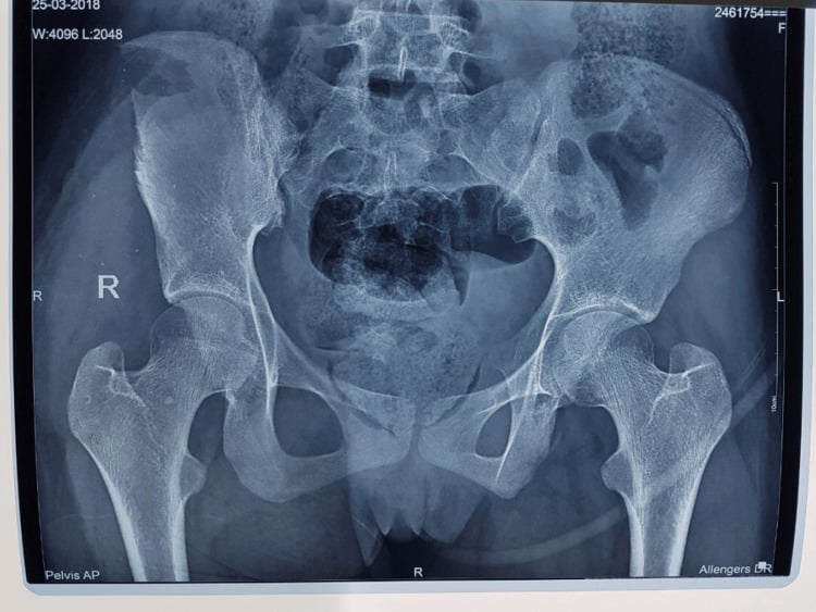
An iliac wing fracture, the Duverney fracture, is an isolated fracture of the iliac ala that does not disrupt the pelvic ring. The fracture stays within the iliac bone and does not extend to the SI joint, the acetabulum or the contralateral side. It is named after Joseph Guichard Duverney (1648-1730), a French anatomist.
Mechanism. Direct lateral trauma: a side-impact motor vehicle accident, a fall onto the side, or a crush injury.
Who. It is seen at all ages and is often part of polytrauma. It is less common than pelvic ring injuries.
The Two Populations: High-Energy Duverney vs Fragility Fracture of the Pelvis
The classic Duverney fracture is a young, high-energy injury from a direct lateral blow, and the concern is excluding ring or acetabular extension and associated visceral injury. Alongside it sits a second and increasingly common population, the elderly fragility fracture of the pelvis (FFP), classified by the Rommens system. The distinction changes the whole management emphasis.
The fragility patient. The far more common FFP is a low-energy or insufficiency injury in an osteoporotic older adult, often after a trivial fall or with no clear trauma at all, presenting with hip, groin or low-back pain. Incidence in those over 60 is roughly 224 per 100,000 per year and rising. Persistent pain after low-energy trauma requires MRI or CT even when the iliac wing is not the primary fracture.
The Rommens FFP classification. Severity is instability:
- FFP I - isolated anterior ring fracture
- FFP II - non-displaced posterior ring fracture
- FFP III/IV - displaced or bilateral posterior instability
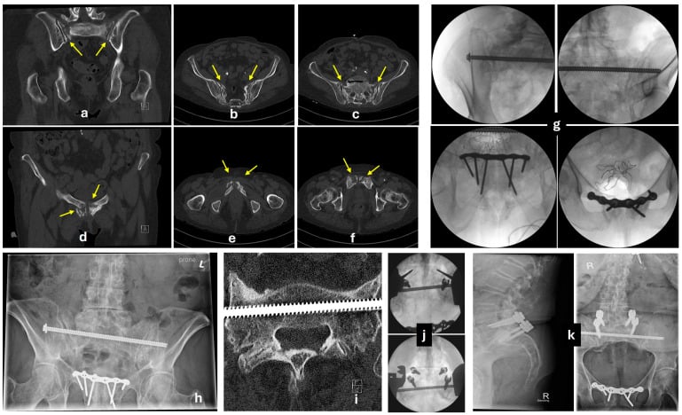
FFP I and II are both usually stable and treated conservatively. FFP III and IV usually need surgical stabilisation, for example sacroiliac or transsacral screws, or sacroplasty.
Why the emphasis differs. In the older adult the danger is not haemorrhage: these are usually stable fractures in frail patients, and one-year mortality is a striking 9.5 to 27 percent, a marker of frailty rather than of the fracture. Reduced mobility and independence are reported alongside that mortality rather than shown to cause it, because frailty drives both, but the management follows either way: immobility is harmful, and pain is what prevents mobilising.
The goal is therefore early mobilisation with adequate analgesia, not bed rest, together with bone-health and osteoporosis work-up and falls prevention. That is the opposite priority to the polytrauma resuscitation of the young high-energy injury. The sacral insufficiency component is covered in the sacral insufficiency fracture topic.
Anatomy and Pathophysiology
The iliac wing. The ala is a broad, fan-shaped bone that gives attachment to:
- Gluteus medius and minimus, the hip abductors, which arise from its outer surface
- Iliacus, a hip flexor
- The abdominal wall muscles: external and internal oblique and transversus abdominis
A fracture through the gluteal origin can cause abductor dysfunction.
Why it is stable. The pelvic ring is the sacrum, the two innominate bones (ilium, ischium and pubis) and the joints that join them, the SI joints and the pubic symphysis. A Duverney fracture is confined to the wing and leaves the ring, SI joints and symphysis intact. Weight-bearing passes through the sacrum and the unfractured columns.
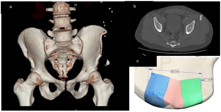
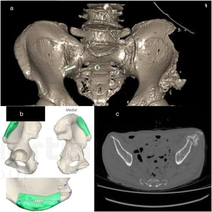
The donor site. The anterior iliac crest is also a bone-graft donor site, and it can fracture after harvest.
Surgical Danger Anatomy of the Iliac Wing
The abductor nerve. Gluteus medius and minimus are supplied by the superior gluteal nerve (L4-S1), which exits the pelvis through the greater sciatic foramen above piriformis with the superior gluteal vessels and runs forward between the two muscles. Persistent abductor weakness after the fracture reflects injury to the muscle origin and, rarely, to this nerve, and produces a Trendelenburg gait.
Structures at risk during wing and crest fixation.
- Superior gluteal neurovascular bundle at the greater sciatic notch - tethered as it exits the notch, so aggressive posterior dissection or screws near the notch risk troublesome bleeding (retraction into the pelvis) and abductor denervation
- L5 nerve root and lumbosacral trunk, which cross the ala just medial to the SI joint, and the iliac vessels on the inner table - at risk with medial dissection or screw penetration
- Lateral femoral cutaneous nerve at or near the ASIS during anterior approaches - its injury causes meralgia paraesthetica (covered in that topic) and explains the lateral-thigh numbness among the complications
Classification
Isolated or not. The simple classification is binary, and CT is essential to confirm that the fracture is isolated:
- Isolated Duverney - confined to the iliac wing, ring intact
- With ring extension - reaches the SI joint, pubic rami or acetabulum, and is no longer purely a Duverney fracture
Once the fracture reaches the ring it is managed as a ring injury, and its stability is that of the ring pattern: the crescent (LC-II) is unstable, whereas an LC-I is usually stable.
Young-Burgess context. Iliac wing fractures are sometimes seen as part of lateral compression (LC) injuries:
- LC-I - rami and sacral compression; the iliac wing may be indirectly involved
- LC-II - rami and an iliac wing fracture (the crescent pattern); the ring is disrupted
- LC-III - an LC-I or LC-II injury with a contralateral AP injury
A true Duverney fracture is not an LC injury, because the ring is intact.
- Iliac Wing (Duverney)
- STABLE
- Pelvic Ring (LC/AP/VS)
- UNSTABLE (varies)
- Iliac Wing (Duverney)
- Yes
- Pelvic Ring (LC/AP/VS)
- No
- Iliac Wing (Duverney)
- Direct Lateral
- Pelvic Ring (LC/AP/VS)
- AP, LC, VS, CM
- Iliac Wing (Duverney)
- Conservative
- Pelvic Ring (LC/AP/VS)
- Often ORIF / Ex-Fix
- Iliac Wing (Duverney)
- Low
- Pelvic Ring (LC/AP/VS)
- High
Differential diagnosis. Several injuries mimic an isolated iliac wing fracture.
- Distinguishing Feature
- Posterior wing fracture extends INTO the SI joint
- Key Test
- CT - SI joint disruption
- Stability
- UNSTABLE
- Distinguishing Feature
- Anterior rami + sacral ala impaction
- Key Test
- CT - sacral fracture line
- Stability
- Usually stable
- Distinguishing Feature
- Fracture line reaches the joint surface
- Key Test
- CT - articular involvement
- Stability
- Joint-threatening
- Distinguishing Feature
- Adolescent, sprinting/kicking, growth plate
- Key Test
- Plain film + age
- Stability
- Stable
- Distinguishing Feature
- No fracture line on imaging
- Key Test
- CT/MRI negative for fracture
- Stability
- Stable
- Distinguishing Feature
- Confined to ala; ring, SI joint, acetabulum intact
- Key Test
- CT confirms isolation
- Stability
- STABLE
Clinical Assessment
History. After the mechanism above, pain is felt over the lateral pelvis and hip.
Examination. Bruising over the lateral pelvis and flank and tenderness over the iliac crest localise the injury. Compression and distraction may elicit pain even though the ring is stable, and hip abduction is painful. Check the sciatic, femoral and lateral cutaneous nerves.
The abdomen. Direct lateral trauma can also cause intra-abdominal injury, so examine the abdomen.
Investigations
Radiographs. The AP pelvis may show the wing fracture but can miss subtle injuries.
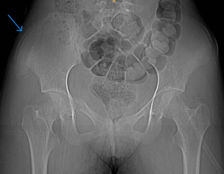
CT pelvis is essential. It confirms the fracture pattern and rules out extension to the ring or acetabulum. The fracture is a Duverney fracture only when CT shows:
- A fracture line through the iliac wing
- Intact SI joints
- Intact pubic rami and symphysis
- No acetabular involvement
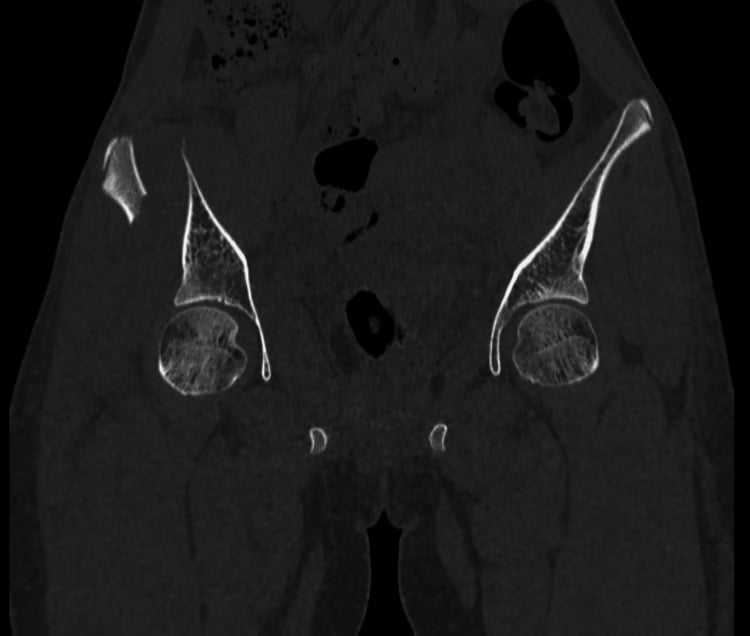
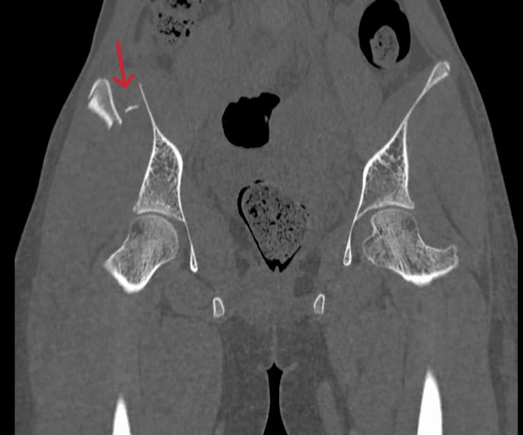
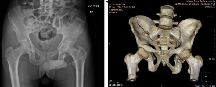
CT abdomen, or FAST, is added when there is concern for intra-abdominal injury.
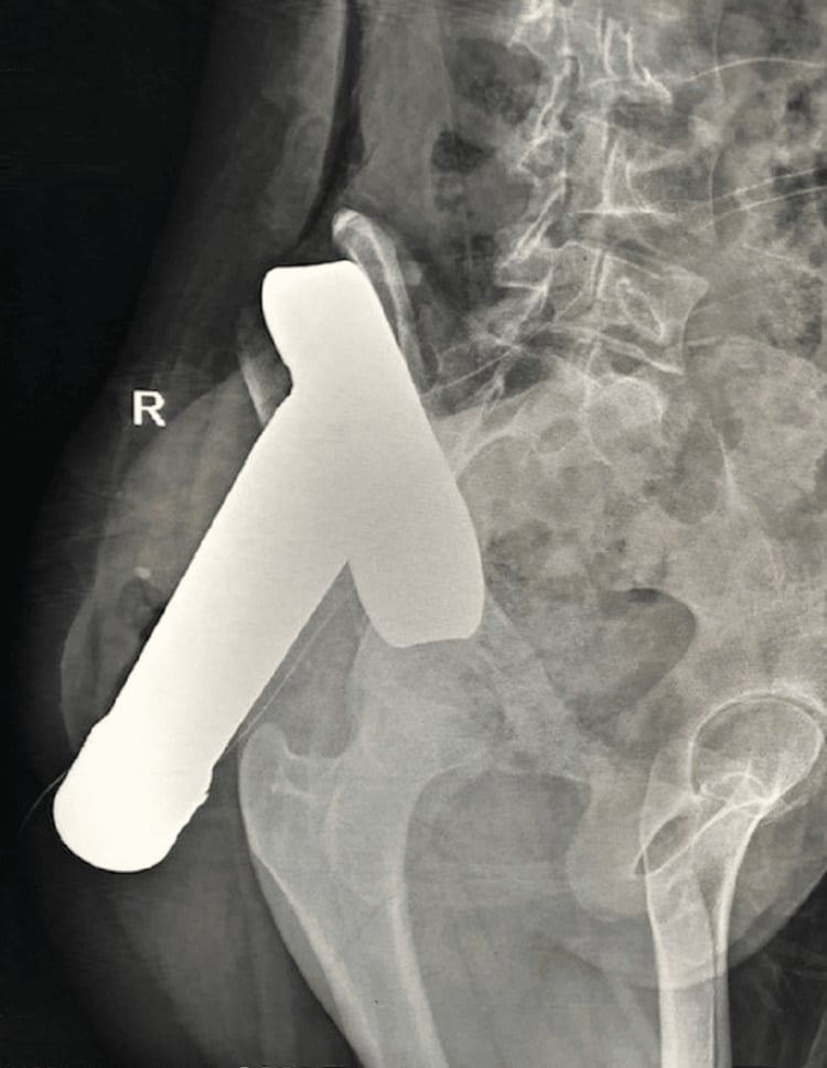
Management Algorithm
Conservative care is the standard for the isolated Duverney fracture, and most heal well with it:
- Rest - bed rest for 1-2 days if needed for pain
- Weight-bearing - protected (non- or touch-toe weight-bearing) at first, progressing from weeks 2-4 to full weight-bearing as tolerated by week 6
- Analgesia - multimodal: NSAIDs, paracetamol, opioids as required
- DVT prophylaxis - mechanical and chemical, until mobile
- Physiotherapy - hip range of motion, and abductor strengthening when pain allows
- Follow-up - clinical assessment and an X-ray at 4-6 weeks
How much weight, and when. No high-level evidence dictates a single protocol, and practice ranges from immediate weight-bearing as tolerated to a short non-weight-bearing period. Cohort data (Gaski, Soni) show good outcomes regardless of weight-bearing status for stable injuries, so pain and comfort can guide progression. Most iliac wing fractures are treated conservatively with weight-bearing as tolerated.
Surgery is rare. The indications:
- Extension to the pelvic ring (SI joint, rami) - treat as a ring injury
- Extension to the acetabulum - ORIF of the acetabulum
- A large displaced fragment affecting hip abductor function (rare)
- Significant comminution requiring stabilisation
- Associated injuries requiring a surgical approach
- Open fracture
Surgical Technique
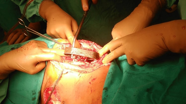
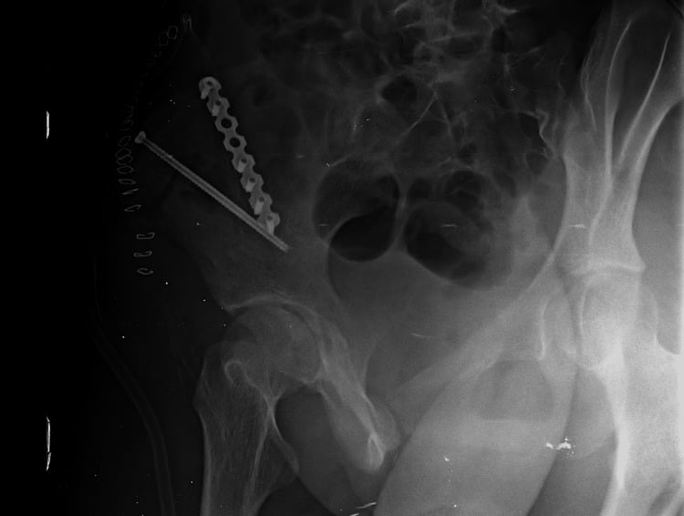
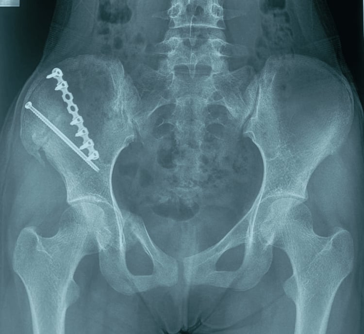
Approach. The wing is fixed through the lateral window of the ilioinguinal approach or, alternatively, a direct lateral approach, preserving the gluteal muscle origins where possible. Acetabular extension is approached anteriorly (ilioinguinal or anterior IC).
Fixation.
- Reduce the fracture fragments
- Apply a reconstruction plate along the iliac crest
- Use 3.5mm cortical or locking screws
- Close in layers over a drain
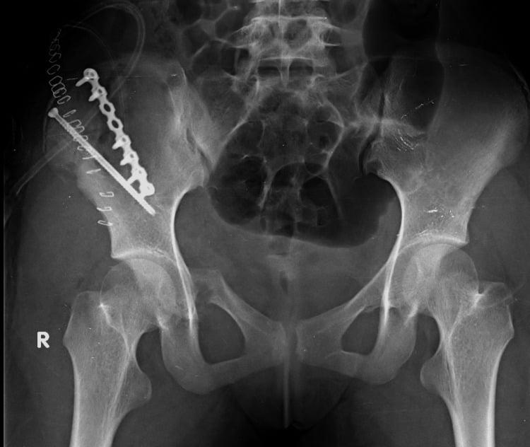
When other injuries drive surgery. Associated injuries dictate the surgical approach and timing.
- Acetabular extension - treat as an acetabular fracture (see the dedicated topic); the wing component is addressed during the approach
- SI joint disruption - makes this an unstable ring injury requiring SI fixation, with percutaneous screws or open
- Pelvic ring injury - follow the pelvic ring management algorithm; the wing fragment is stabilised as part of the reconstruction
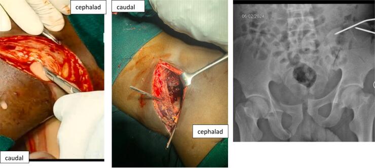
The Morel-Lavallée lesion. This closed internal degloving occurs when a shearing force strips subcutaneous fat off the deep fascia, leaving a fluctuant cavity of haemolysed blood, liquefied fat and serous fluid. Its classic site, the greater trochanter, flank and buttock, sits directly over the iliac wing and over the incision used to approach it, so it is the soft-tissue lesion most likely to complicate operating on this fracture.
Look for it before planning the approach. The skin is soft, boggy and abnormally mobile over a fluctuant swelling, with overlying bruising, reduced sensation from injury to the cutaneous nerves, and sometimes a palpable rim once a capsule forms. It is frequently missed in the trauma bay because the skin is intact and attention is on the fracture.
Imaging. MRI is the definitive study; ultrasound or the CT already obtained for the pelvis will usually show it. Recognising a capsule matters, because an established capsule will not resolve with compression alone.
It changes the operation, not just the wound care. Do not make an elective incision through an unaddressed lesion. Tseng and Tornetta drained and débrided percutaneously within three days, and although only 3 of 16 cultures grew organisms, they still performed percutaneous posterior fixation at the same sitting and deferred every open procedure until at least 24 hours after the drain came out.
Complications
- Risk Factor
- Gluteal origin injury
- Management
- Physiotherapy
- Risk Factor
- Large displacement
- Management
- Rarely symptomatic
- Risk Factor
- Immobility
- Management
- Prophylaxis
- Risk Factor
- Direct trauma; anterior approach at the ASIS
- Management
- Usually resolves
- Risk Factor
- Associated
- Management
- Trauma workup
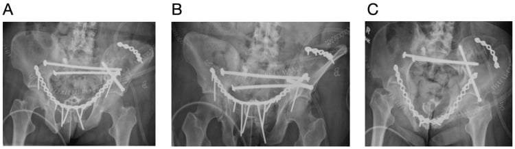
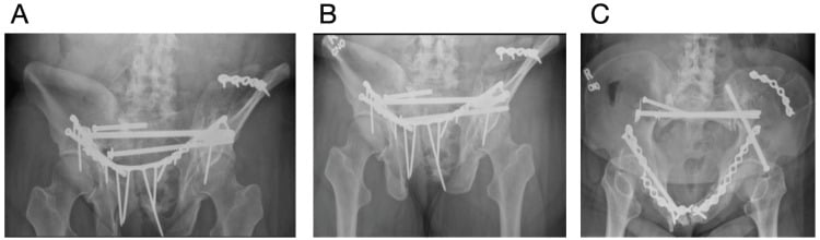
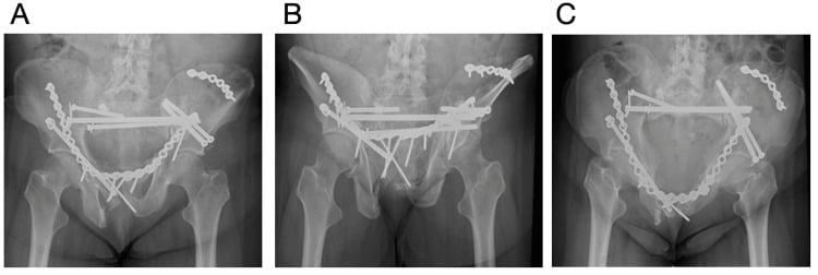
Fixation failure. Constructs for fragility fractures of the pelvis can fail and need revision.
Postoperative Care
After fixation, weight-bearing follows the operative protocol, usually touch-toe weight-bearing for 6-8 weeks, with wound care and DVT prophylaxis. Conservative care follows the protocol above.
Outcomes
The isolated Duverney fracture has excellent outcomes with conservative care. With ring involvement, the outcome depends on the stability of the ring as a whole. Hip abductor recovery is usually good with rehabilitation.
Guidelines, Registries & Global Practice
Global epidemiology:
- Isolated iliac wing (Duverney) fractures are uncommon relative to pelvic ring injuries and are typically high-energy (side-impact MVC, fall onto side, crush) in younger patients.
- In adults over 60, anterior/iliac fragility fractures are far more common: estimated incidence of osteoporotic pelvic fracture of around 224 per 100,000 per year and rising, with one-year mortality of 9.5-27% driven by loss of mobility (Oberkircher, Dtsch Arztebl Int 2018).
Side-by-side guidance:
- Imaging
- AP pelvis + CT
- Stable wing/anterior injury
- Type A (stable, ring intact) - conservative
- Surgical trigger
- Type B/C ring instability
- Imaging
- CT in major trauma pathway
- Stable wing/anterior injury
- Mobilise, analgesia, VTE prophylaxis
- Surgical trigger
- Haemodynamic instability or ring displacement
- Imaging
- CT defines ring involvement
- Stable wing/anterior injury
- Non-operative for isolated stable patterns
- Surgical trigger
- Open book / vertical shear / bleeding pelvis
- Imaging
- CT +/- MRI in elderly
- Stable wing/anterior injury
- FFP I-II usually conservative
- Surgical trigger
- FFP III-IV instability
Registries and practice variation:
- Pelvic and trauma registries (e.g. UK TARN, German Pelvic Trauma Registry, regional trauma databases) consistently show isolated wing fractures carry low transfusion and mortality compared with ring disruptions - underpinning conservative care.
- High-resource settings: routine whole-body or pelvic CT confirms isolation, enabling confident early mobilisation and same-admission discharge.
- Limited-resource settings: where CT is not readily available, inlet/outlet and Judet views plus careful clinical stability testing guide management; a low threshold for referral applies if SI-joint or acetabular extension cannot be excluded.
- Physiotherapy: early hip-abductor rehabilitation is the shared standard worldwide.
Controversies and Areas of Uncertainty
Is the wing fracture truly isolated? The central controversy is diagnostic rather than therapeutic. A posterior wing fracture that reaches the SI joint is a crescent (LC-II) injury in the Day classification, which is unstable and surgical, so the label "Duverney" should only be applied after CT excludes SI, ramus and acetabular extension.
Surgery for the displaced fragment. Whether a large, displaced wing fragment with hip-abductor compromise benefits from fixation is decided case by case on expert opinion alone; there are no comparative trials. Most are still managed conservatively.
Borderline lateral-compression injuries. The genuine operative-versus-non-operative debate (the TULIP feasibility RCT) concerns LC1 injuries with a complete sacral fracture, the unstable end of the spectrum, not the isolated wing fracture.
Viva Scenarios
Clinical Decision Scenarios
Practise clinical reasoning and management decisions out loud
“What is your diagnosis and management?”
“Explain the stability of Duverney fracture.”
“Explain the cause and management.”
MCQ Practice Points
Q: Why is a Duverney fracture stable? A: The pelvic ring is intact. The fracture is confined to the iliac wing and does not disrupt the SI joint, symphysis, or rami.
Q: What is the typical mechanism for Duverney fracture? A: Direct lateral trauma (e.g., side-impact MVA, fall onto side, crush injury).
Q: What is the treatment for isolated Duverney fracture? A: Conservative - Protected weight-bearing, Analgesia, DVT prophylaxis, Physiotherapy.
Q: What imaging is essential for iliac wing fractures? A: CT Pelvis - to confirm the fracture is isolated and does not extend to the SI joint, rami, or acetabulum.
Q: What muscle weakness can occur with iliac wing fractures? A: Hip abductor weakness (Gluteus Medius/Minimus) due to injury at their origin on the iliac wing.
Definition
- Duverney = Iliac Wing fracture
- Ring INTACT (key criterion)
- STABLE fracture pattern
- AO Type A (Stable Pelvic Ring)
Mechanism
- Direct lateral trauma
- Side-impact MVA
- Fall onto side
- Crush/compression injury
Treatment
- Conservative standard
- Protected WB
- DVT prophylaxis
- Physio (Abductors)
Check
- CT for ring integrity
- SI joint extension?
- Acetabular extension?
- Intra-abdominal injury?
Evidence Base
Crescent Fracture-Dislocation: Day Classification
- Defined the crescent fracture-dislocation as a lateral-compression injury combining SI joint disruption with a posterior iliac wing fracture - the key unstable mimic of a benign wing fracture.
- Three types by crescent fragment size: Type I (large fragment, under one-third of SI joint dislocated), Type II (intermediate, one- to two-thirds), Type III (small fragment, most of joint dislocated).
- 16 patients managed by this scheme achieved good functional results at approximately 2 years; classification guides surgical approach and reduction.
Nonoperative LC1 with Complete Sacral Fracture
- 104 intermediate-severity LC1 injuries (complete sacral fracture, under 1 cm displacement); 50 assessed at mean 33 months.
- Mean Majeed score 85.5 (33 excellent, 9 good, 5 fair, 3 poor); no fracture displaced over 1 cm on follow-up radiographs.
- Patients with concurrent lower-limb injury had significantly lower Majeed scores (p = 0.01); weight-bearing status did not affect outcome.
LC1 Non-operative Functional Outcome (Majeed)
- 46 LC1 injuries with incomplete sacral fracture managed non-operatively (non-weight-bearing 3 weeks, then weight-bearing as tolerated).
- Mean Majeed score 82.6 (27 excellent, 19 good); no mortality in any subgroup.
- Best functional outcome reached within 12 months and did not change thereafter.
Mechanism Predicts Pelvic Trauma Severity (Young-Burgess)
- In complete pelvic ring disruptions, APC-mechanism injuries required far more transfusion than vertical-shear (15/18 needed 10 or more units vs 11/14 of VS needing under 10).
- APC group: 39% mortality and higher multi-organ failure vs 0% mortality in VS group.
- Confirms the Young-Burgess principle that injury mechanism drives haemorrhage risk and outcome.
Haemorrhage in Major Pelvic Disruption
- Of 4712 pelvic fractures, only 7.3% required angiography; overall mortality among those was 18%.
- Haemorrhage was the recorded cause in 35.5% of deaths and sepsis/multi-organ failure in a further 43.5%; the authors argue the latter were also driven by delayed haemostasis.
- Median time to angiography was 286 minutes - nearly five hours - prompting the case for preperitoneal packing and aortic balloon occlusion.
Osteoporotic / Fragility Pelvic Fractures (FFP)
- Estimated incidence of osteoporotic pelvic fracture over age 60 of 224 per 100,000 per year and rising.
- Rommens FFP classification: isolated anterior (FFP I) and non-displaced posterior (FFP II) injuries are usually stable and treated conservatively; FFP III/IV are unstable and usually need surgery.
- Retrospective series report reduced mobility and independence, and separately a one-year mortality of 9.5-27%; the paper associates the two rather than showing that immobility causes the deaths.
Operative vs Non-operative LC1 (TULIP) - published protocol only, no results
- Protocol for a feasibility study that would randomise LC1 injuries with complete sacral fracture to operative or non-operative care, in order to design a definitive trial.
- Acknowledges equipoise: some complete sacral fractures may displace over time, and surgery may allow faster pain-free mobilisation.
- Highlights that the operative threshold sits at the unstable end of the lateral-compression spectrum, not the isolated wing fracture.
Morel-Lavallée Lesion Over the Operative Field (percutaneous drainage)
- 19 closed degloving lesions drained and débrided percutaneously within three days of injury through two 2 cm incisions, with brush débridement, pulsed lavage and a suction drain removed once output fell below 30 mL/24 h.
- 3 of 16 cultures taken at débridement grew organisms - in a closed injury with intact skin.
- Percutaneous posterior pelvic fixation was performed at the same sitting in 7 of 9 surgically treated pelvic fractures; all open procedures, including every acetabular fracture, were deferred until at least 24 hours after drain removal.
- No deep infection and no skin débridement at a minimum of six months.