Friction at 30° Flexion | Hip Abductor Weakness | Activity Modification Key | Rarely Surgical
- Lateral femoral epicondyle friction occurs at 30 degrees of knee flexion during foot strike
- Hip abductor weakness (gluteus medius) is the primary biomechanical cause
- Noble compression test is pathognomonic - pain at 30 degrees with compression
- Activity modification and eccentric strengthening are cornerstones of treatment
- Surgery (ITB release/lengthening) is rarely needed - less than 10% of cases
- “Most common cause of lateral knee pain in distance runners (12% of running injuries)
- “Friction occurs 2cm proximal to lateral femoral epicondyle at 30 degrees flexion
- “Ober test assesses ITB tightness - hip abduction contracture indicates positive
- “Downhill running and track running (always turning same direction) increase risk
ITB friction occurs at 30 degrees of knee flexion over the lateral femoral epicondyle. At angles greater than 30 degrees, the ITB moves posterior to the axis of rotation. This repetitive friction causes inflammation and pain.
Gluteus medius weakness is the root cause in 95% of cases. Weak hip abductors allow contralateral pelvic drop, increasing ITB tension. Always assess hip strength - this guides rehabilitation.
Pathognomonic clinical test: Apply pressure 2cm proximal to lateral epicondyle while passively extending knee from 90 degrees. Pain at 30 degrees flexion is positive - reproduces the friction zone pain.
More than 90% respond to non-operative management with activity modification, eccentric strengthening, and biomechanical correction. Surgery is a last resort after 6-12 months of failed conservative care.
- Clinical Picture
- Pain after running only, resolves in minutes
- Treatment
- Activity modification + ITB stretching
- Key Pearl
- Catch it early - excellent prognosis
- Clinical Picture
- Pain during run but doesn't stop activity
- Treatment
- Structured rehab + hip strengthening
- Key Pearl
- Address biomechanics - prevent progression
- Clinical Picture
- Pain limits running distance
- Treatment
- Cease running + intensive PT
- Key Pearl
- Running break essential for healing
- Clinical Picture
- Pain with stairs, prolonged sitting
- Treatment
- Complete rest + cortisone injection
- Key Pearl
- Consider MRI to exclude other pathology
- Clinical Picture
- Failed 6-12 months conservative care
- Treatment
- ITB release/lengthening surgery
- Key Pearl
- Less than 10% require surgery
FLEXITB - Friction Zone Anatomy
Hook:When the knee goes into FLEX-ion past 30 degrees, the ITB moves and friction occurs
REHABREHAB - Conservative Treatment Framework
Hook:REHAB is the pathway - surgery rarely needed if you follow this framework
30-2-1030-2-10 Rule
Hook:The 30-2-10 rule: where it hurts (30 degrees, 2cm proximal) and how rarely you operate (10%)
Overview and Epidemiology
Iliotibial band (ITB) syndrome is the second most common running injury after patellofemoral pain syndrome, accounting for approximately 12% of all running-related injuries. It presents as lateral knee pain caused by repetitive friction of the ITB over the lateral femoral epicondyle during the gait cycle.
Epidemiology:
- Distance runners are predominantly affected (especially marathon and ultramarathon)
- Cyclists are second most common (particularly with improper bike fit)
- Military recruits during basic training
- Athletes in cutting sports (basketball, soccer) less commonly
ITB syndrome is sometimes called "runner's knee" (though this term more commonly refers to patellofemoral pain). The distinction is important - ITB syndrome is lateral knee pain, while patellofemoral pain is anterior. In the exam, always clarify pain location.
Global burden (key figures):
- ITB syndrome is the most common cause of lateral knee pain in runners worldwide and the second most common overuse running injury overall after patellofemoral pain
- Reported incidence in running cohorts ranges from 5% to 14% of all running injuries (van der Worp systematic review)
- Pooled prevalence across running populations is approximately 8% (Kakouris systematic review)
- Also seen in cyclists (poor bike fit, excessive saddle height), rowers, military recruits, and endurance/multisport athletes globally
Why Downhill Running and the Key Risk Factors Worsen ITB Syndrome
The topic lists downhill running, genu varum, overstriding and cambered surfaces as risk factors, and a viva followUp asks specifically "why does downhill running worsen ITB syndrome?" - but the mechanisms are never explained.
- Downhill running. Two linked reasons. First, running downhill the knee is less flexed at foot strike (a straighter knee at contact), so it sits closer to the roughly 30 degree impingement zone at the moment of peak load - the ITB is compressed against the epicondyle exactly when the braking load is highest. Second, downhill running greatly increases the eccentric braking load and stance time, so each stride transmits more force through the ITB. (This is why Noble's original series noted the pain is "more severe running downhill".)
- Slow running / overstriding. Counter-intuitively, ITB syndrome is often worse at slower speeds and with overstriding: a slower cadence with a longer stride keeps the knee near the impingement angle for longer during stance, whereas faster running flexes the knee past the friction zone more quickly.
- Genu varum and tibial internal rotation. A varus (bow-legged) knee and excessive foot pronation with tibial internal rotation both increase the tension and compression of the ITB over the lateral epicondyle.
- Cambered surfaces and track direction. Running the same side of a cambered road (or always the same direction on a track) loads the down-slope limb into relative hip adduction, mimicking pelvic drop and raising ITB strain - hence varying the surface and alternating track direction.
Q: Why does downhill running worsen ITB syndrome? A: Running downhill the knee is less flexed at foot strike, so it sits closer to the roughly 30 degree impingement zone just as the eccentric braking load (increased downhill) peaks - maximal ITB compression against the lateral epicondyle at the moment of maximal load, over a longer stance time. The same logic explains why ITB syndrome is often worse at slower speeds / overstriding (the knee lingers near the friction angle) and why genu varum, tibial internal rotation and cambered surfaces aggravate it.
RUNNERSRUNNERS - Risk Factors for ITB Syndrome
Hook:RUNNERS get ITB syndrome - remember the risk factors every distance athlete faces
Pathophysiology and Mechanisms
ITB Anatomy
The iliotibial band is a thickened fascial structure extending from the iliac crest to the lateral tibia (Gerdy's tubercle).
- Tensor fasciae latae (anterior)
- Gluteus maximus (posterior, 75% of fibers)
- Iliac crest
- Gerdy's tubercle on anterolateral proximal tibia
- Lateral femoral epicondyle (NO anatomical attachment but friction zone)
- Lateral patellar retinaculum
The ITB does not attach to the lateral femoral epicondyle - it glides over it. At knee flexion angles less than 30 degrees, the ITB is anterior to the axis of rotation (extends the knee). At angles greater than 30 degrees, it moves posterior (flexes the knee). This repetitive anterior-posterior movement at 30 degrees during running creates friction.
Biomechanics of Friction
- ITB Position
- Anterior to epicondyle
- Function
- Knee extensor
- Clinical Significance
- No friction, ITB under tension
- ITB Position
- Moving posterior
- Function
- Transitioning
- Clinical Significance
- FRICTION ZONE - impingement occurs
- ITB Position
- Posterior to epicondyle
- Function
- Knee flexor
- Clinical Significance
- No friction, ITB relaxed
During running gait:
- Foot strike - knee at approximately 20-30 degrees flexion → ITB compressed against epicondyle
- Mid-stance - knee extends → ITB slides anteriorly
- Toe-off - knee flexes → ITB slides posteriorly
- Swing phase - repetitive anterior-posterior gliding
This occurs approximately 1,000 times per mile of running.
Hip Abductor Role
Primary hip abductor - prevents contralateral pelvic drop during single-leg stance (mid-stance of gait). Weakness leads to Trendelenburg gait pattern with increased hip adduction on stance leg.
Hip adduction increases ITB tension over lateral femoral epicondyle. Weak gluteus medius → pelvic drop → increased hip adduction → increased ITB strain → friction syndrome. This is the biomechanical cascade.
The Trendelenburg test (single-leg stance causes contralateral pelvic drop) is often positive in ITB syndrome patients. This indicates gluteus medius weakness. In the exam, always assess hip abductor strength - it's the key to understanding and treating ITB syndrome.
Classification Systems
Clinical Severity Stages
ITB syndrome is typically classified by symptom severity and functional limitation.
- Symptoms
- Pain after running, resolves within minutes
- Functional Impact
- No limitation of distance or speed
- Treatment Approach
- Activity modification, ITB stretching, continue running
- Symptoms
- Pain during running but tolerable
- Functional Impact
- Can complete runs but painful
- Treatment Approach
- Structured rehabilitation, reduce mileage/intensity
- Symptoms
- Pain limits running distance
- Functional Impact
- Cannot complete planned distance
- Treatment Approach
- Cease running, intensive physiotherapy
- Symptoms
- Pain with daily activities (stairs, sitting)
- Functional Impact
- Unable to run, affects quality of life
- Treatment Approach
- Complete rest, consider cortisone injection
Catching ITB syndrome at Stage 1 and implementing proper rehabilitation prevents progression to chronic, disabling pain. Most runners who progress to Stage 3-4 either ignored early symptoms or attempted to "run through" the pain.
Clinical Presentation and Assessment
History
- Location: Lateral knee, 2-3cm proximal to joint line
- Quality: Sharp, burning, or aching
- Onset: Gradual over days-weeks
- Timing: During running, especially on hills or turns
- Relieving: Rest, stopping running
- Recent training changes: Increased mileage, intensity, hills
- Surface: Track (always same direction), camber, downhill
- Footwear: New shoes, worn-out shoes
- Previous ITB issues: Recurrence common if rehab incomplete
- Cross-training: Cycling (also risk factor)
Red flag questions (exclude other pathology):
- Locking or catching (meniscal tear)
- Giving way (ligament injury)
- Night pain (tumor, infection)
- Systemic symptoms (inflammatory arthropathy)
Physical Examination
Systematic Examination
- Gait: Trendelenburg gait pattern (hip drops on opposite side during stance)
- Alignment: Genu varum (bow-legged) increases ITB tension
- Muscle wasting: Gluteus medius atrophy (chronic cases)
- Point tenderness: 2cm proximal to lateral femoral epicondyle
- ITB tightness: Palpate along entire ITB from iliac crest to Gerdy tubercle
- Lateral joint line: Ensure not meniscal tenderness
- Noble compression test (pathognomonic)
- Ober test (ITB tightness)
- Trendelenburg test (hip abductor weakness)
- Single-leg squat (biomechanical assessment)
- Knee ROM: Usually full (pain may limit terminal extension)
- Hip ROM: Assess for limited abduction (tight ITB)
- Hip abduction strength: Gluteus medius manual muscle testing (often 3-4/5)
- Knee stability: ACL/PCL/collateral ligaments (exclude instability)
Noble Compression Test (Pathognomonic)
Technique:
- Patient supine or standing
- Flex knee to 90 degrees
- Apply pressure with thumb 2cm proximal to lateral femoral epicondyle
- While maintaining pressure, passively extend the knee
- Positive test: Pain at approximately 30 degrees of flexion (friction zone)
The Noble compression test has high specificity (greater than 90%) for ITB syndrome. Pain at the 30-degree position reproduces the exact biomechanical impingement that occurs during running. This is the single most important clinical test.
Ober Test (ITB Tightness)
Technique:
- Patient side-lying (affected side up)
- Flex lower knee to 90 degrees for stability
- Flex upper hip and knee to 90 degrees
- Extend upper hip (bring leg in line with trunk)
- Abduct upper hip, then release and let gravity adduct
- Positive test: Leg remains abducted (does not fall to table) - indicates ITB contracture
Interpretation: Positive Ober test indicates ITB tightness but is not specific for ITB syndrome (many asymptomatic individuals have positive Ober).
Trendelenburg Test (Hip Abductor Strength)
Technique:
- Patient stands on affected leg (single-leg stance)
- Observe pelvis from behind
- Positive test: Contralateral pelvis drops (weak gluteus medius cannot stabilize pelvis)
Significance: Positive Trendelenburg indicates the biomechanical cause of ITB syndrome. This must be addressed in rehabilitation.
Differential Diagnosis of Lateral Knee Pain
- Onset / Mechanism
- Gradual, overuse (training error, downhill/track)
- Pain Location
- 2-3cm proximal to lateral joint line over epicondyle
- Discriminating Features
- Positive Noble test at 30 degrees, hip abductor weakness, no effusion
- Onset / Mechanism
- Acute twisting, or degenerative in older patients
- Pain Location
- Lateral joint line
- Discriminating Features
- Joint-line tenderness, catching/locking, positive McMurray/Thessaly, effusion
- Onset / Mechanism
- Overuse, downhill running
- Pain Location
- Posterolateral corner
- Discriminating Features
- Tender at popliteus origin, pain on resisted tibial internal rotation
- Onset / Mechanism
- Acute varus/contact injury
- Pain Location
- Lateral, along LCL course
- Discriminating Features
- Pain/laxity on varus stress at 30 degrees, often acute
- Onset / Mechanism
- Acute or insidious
- Pain Location
- Over fibular head (more posterior/distal)
- Discriminating Features
- Tenderness and translation at PTFJ, fibular head mobility
- Onset / Mechanism
- Chronic, older patient, varus alignment
- Pain Location
- Lateral joint line and compartment
- Discriminating Features
- Crepitus, radiographic joint space loss, morning stiffness
- Onset / Mechanism
- Insidious or post-injury
- Pain Location
- Fibular neck radiating to leg
- Discriminating Features
- Paraesthesia/foot drop, positive Tinel at fibular neck
Investigations
Imaging Protocol
Investigation Algorithm
ITB syndrome is a clinical diagnosis. Imaging is not required for typical presentation with positive Noble test and clear running history.
Indications for imaging:
- Atypical features (young patient, no running history)
- Failed conservative treatment (exclude other pathology)
- Severe symptoms (Stage 4)
- Medicolegal or compensation cases
Views: AP and lateral knee
Purpose: Exclude bony pathology
- Usually normal in ITB syndrome
- Look for: lateral compartment osteoarthritis, osteochondral lesions, avulsion fractures
Findings in ITB syndrome: May show soft tissue swelling lateral to knee (non-specific)
Gold standard for soft tissue assessment
- T2 hyperintensity deep to ITB at lateral femoral epicondyle (fluid signal)
- Thickening of ITB over epicondyle
- Periosteal edema at lateral epicondyle (bone stress)
- Bursal fluid (if bursa present)
- Meniscal tears (lateral meniscus)
- Lateral collateral ligament injury
- Popliteus tendinopathy
- Lateral femoral condyle osteochondral lesion
- Proximal tibiofibular joint pathology
Dynamic assessment of ITB movement
Findings:
- Thickening of ITB
- Fluid deep to ITB
- Can demonstrate ITB snapping over epicondyle with knee flexion/extension
Limited use: Operator-dependent, MRI preferred if imaging needed
Do not routinely order imaging for ITB syndrome. It is a clinical diagnosis based on history (runner, lateral knee pain), examination (Noble test positive), and biomechanical assessment (hip weakness). Imaging is for atypical cases or failed treatment.
Management Algorithm
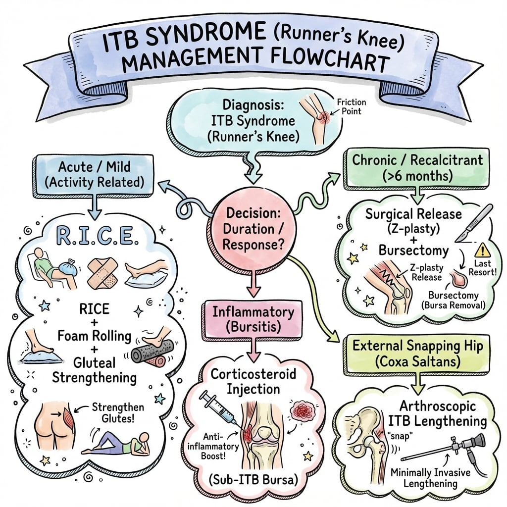
More than 90% of ITB syndrome cases respond to non-operative management. Surgery should only be considered after 6-12 months of comprehensive conservative treatment failure. The key is addressing the biomechanical cause (hip weakness), not just symptomatic treatment.
Conservative Treatment Algorithm
Early Stage Management
Goal: Reduce inflammation, continue running with modifications
Treatment Steps
- Reduce running mileage by 25-50%
- Avoid provocative activities: Downhill running, track (same direction), steep hills
- Cross-training: Swimming, pool running (maintains fitness without impact)
- Surface change: Treadmill (flat), grass, trails (varied terrain)
- Ice: 15-20 minutes after running, 3-4 times daily
- NSAIDs: Ibuprofen 400mg TDS or Naproxen 500mg BD for 1-2 weeks
- Topical NSAIDs: Diclofenac gel applied to lateral knee
- Standing cross-leg stretch: Cross affected leg behind, lean away from affected side
- Side-lying ITB stretch: Bottom leg straight, top leg crosses over, rotate trunk
- Foam rolling: 30 seconds along ITB (may be very painful initially)
- Hold stretches: 30-60 seconds, repeat 3-5 times daily
- Side-lying hip abduction: 3 sets of 15 repetitions
- Clamshells: With resistance band, 3 sets of 15
- Single-leg bridge: Progress to single-leg, 3 sets of 10
- Single-leg squat: Progress difficulty as tolerated
Eccentric strengthening of hip abductors is more effective than concentric. Slow lowering phase (3-5 seconds) during exercises creates greater strength gains.
- Gait analysis: Running store or sports physiotherapist
- Footwear assessment: Replace shoes if over 500-800km
- Orthotics consideration: If significant pronation or supination
- Running form coaching: Increase cadence (reduce stride length), avoid crossover gait
Return to Running Criteria (must meet all before progressing):
Pain-free with daily activities / Negative Noble test / Hip abduction strength 90% of opposite side / Pain-free single-leg squat
Complications and Management
Conservative Treatment Complications
- Incidence
- 30-40% if rehab incomplete
- Prevention/Management
- Complete 6-8 week return protocol, maintain hip strengthening
- Incidence
- 5-10%
- Prevention/Management
- Ensure compliance, exclude other pathology (MRI), consider surgery
- Incidence
- Common
- Prevention/Management
- Cross-training (swimming, cycling), maintain cardiovascular fitness
Surgical Complications
- Incidence
- 10-20%
- Prevention/Management
- Comprehensive post-op rehab, biomechanical correction
- Incidence
- Less than 5%
- Prevention/Management
- Sterile technique, prophylactic antibiotics
- Incidence
- Rare (excessive release)
- Prevention/Management
- Conservative Z-lengthening, avoid excessive ITB excision
- Incidence
- Rare (less than 2%)
- Prevention/Management
- Careful dissection, avoid deep dissection near fibular head
Surgical Management (Rarely Indicated)
Indications for Surgery
Surgery for ITB syndrome is indicated in less than 10% of cases and only after:
- 6-12 months of failed comprehensive conservative treatment
- Confirmed compliance with physiotherapy and activity modification
- Exclusion of other pathology (MRI scan)
- Documented biomechanical correction attempt
- Impact on quality of life (unable to work, exercise, daily activities)
Surgical Options
- Technique
- Z-plasty incision in ITB at friction zone to lengthen
- Rationale
- Reduces tension over lateral epicondyle
- Evidence
- Most common, 80-90% good results in selected cases
- Technique
- Excision of posterior 2cm of ITB over epicondyle ± bursa
- Rationale
- Removes impinging tissue
- Evidence
- Good results but some reports of weakness
- Technique
- Remove ellipse of ITB (4×2cm) over friction zone
- Rationale
- Decompression and reduces friction
- Evidence
- Variable results, theoretical weakness concern
ITB Z-Lengthening (Most Common)
Operative Steps
- Position: Supine, affected leg free-draped or lateral decubitus
- Tourniquet: Optional (improves visualization)
- Landmarks: Mark lateral femoral epicondyle (palpate), Gerdy tubercle
- Incision: 5-7cm longitudinal over lateral femoral epicondyle
- Dissection: Through subcutaneous tissue, identify ITB
- Bursa: Excise if present (usually not)
- Proximal limb: Incise ITB in line with fibers, 2cm proximal to epicondyle, from anterior edge posteriorly (leave 1cm intact posteriorly)
- Distal limb: 2cm distal to epicondyle, from posterior edge anteriorly (leave 1cm intact anteriorly)
- Result: Z-shaped cut allows lengthening
- Tension: Gently separate the two limbs to lengthen ITB by 1-2cm
- Do not repair Z-plasty (allow lengthening to persist)
- Close subcutaneous tissue and skin
- No drain typically needed
Surgical Outcomes
Success rates (selected patients):
- 80-90% return to pre-injury activity level
- Higher success in those who failed conservative care due to anatomical factors (vs poor compliance)
- Failures often due to persistent biomechanical issues (hip weakness not addressed)
Critical concept: Surgery addresses the local anatomical issue (tight ITB) but does not correct hip weakness or running biomechanics. Post-surgical rehabilitation MUST include the same hip strengthening and gait retraining as conservative care, or symptoms recur.
Gait Retraining
The management repeatedly advises to "increase cadence, reduce stride length, avoid crossover gait", and the Brown study supports "gait re-training to decrease stance-phase hip adduction", but the specific evidence-based modifications are never gathered into a coherent programme.
- The target. ITB load peaks with stance-phase hip adduction and contralateral pelvic drop (the mechanics that hip-abductor weakness produces). Gait retraining aims to reduce peak hip adduction - so much so that ITB-syndrome runners who fatigue actually self-reduce hip adduction to offload the band (Brown).
- Increase step (cadence) rate. Raising cadence by about 5-10% at the same speed shortens the stride, reduces the vertical loading rate and typically lowers peak hip adduction and knee-joint load - one of the best-supported single changes.
- Widen step width / stop crossover. A narrow base or crossover gait (foot landing across the midline) increases hip adduction and ITB strain; cueing a slightly wider step width reduces it.
- Cue the pelvis and trunk. "Run tall, don't let the hip drop", or a small contralateral trunk lean, reduces pelvic drop and hip adduction; mirror or real-time (video/wearable) feedback on hip adduction and pelvic drop makes the change stick.
- Load and terrain. Avoiding downhill and cambered surfaces early and progressing load by the 10% rule protects the retrained pattern while capacity rebuilds.
- The caveat. The evidence is mostly small biomechanical studies; gait retraining is an adjunct to hip strengthening and load management, not a replacement, and the change must be practised long enough to become automatic.
Q: What gait-retraining changes reduce ITB load, and why? A: The target is stance-phase hip adduction (and pelvic drop). Increasing cadence by about 5-10% (a shorter stride), widening step width / stopping a crossover gait, and cueing the runner to not let the pelvis drop (with mirror or real-time feedback) all reduce peak hip adduction and ITB strain. It is an adjunct to hip-abductor strengthening and load management, and must be practised until automatic.
Postoperative Care and Rehabilitation
Note: This section applies to the less than 10% of patients who undergo surgical intervention (ITB Z-lengthening or release).
Post-Surgical Rehabilitation Protocol
Goals: Wound healing, pain control, prevent stiffness
Weight-bearing: Full weight-bearing as tolerated with crutches for comfort
ROM: Immediate knee range of motion exercises (avoid stiffness)
Exercises: Gentle ankle pumps, quadriceps sets, passive knee flexion/extension
Pain management: Paracetamol, NSAIDs, ice elevation
Wound care: Keep dressing dry, remove sutures at 10-14 days
Goals: Restore full ROM, begin strengthening, wean crutches
Mobility: Wean crutches as comfortable (usually by week 3-4)
ROM: Active and active-assisted knee ROM (goal: full ROM by week 6)
Strengthening: Begin gentle hip abductor strengthening
Hip exercises: Side-lying hip abduction (light resistance), clamshells, bridges
Cycling: Stationary bike (start week 3-4) for ROM and cardiovascular fitness
Avoid: Running, jumping, impact activities
Goals: Build strength, prepare for running
Strengthening: Intensive hip and knee strengthening program
Exercises: Single-leg squats, single-leg deadlifts, step-ups, resistance band work
Proprioception: Single-leg balance, perturbation exercises
Cardiovascular: Continue cycling, add elliptical if pain-free
Functional testing: Pain-free single-leg hop, single-leg squat (10 repetitions)
Goals: Safe return to running
Prerequisites: Must meet all criteria before starting running:
Pain-free daily activities for 2+ weeks
Full knee ROM
Hip abduction strength equal to or greater than 90% of contralateral
Pain-free single-leg hop (within 90% of opposite)
Running protocol: Follow the same 6-8 week graded return protocol (see Management section, Return to Running tab)
Start: Walk-run intervals on flat, soft surface
Goals: Return to pre-injury activity level
Expected: Full return to running by 3-6 months post-surgery
Maintenance: Lifelong hip strengthening 2 times per week minimum
Prevention: Proper running biomechanics, appropriate mileage progression (10% rule), avoid training errors
Critical concept: Surgical Z-lengthening or release addresses the tight ITB but does not correct hip abductor weakness or running biomechanics. Post-surgical rehabilitation MUST include the same hip strengthening and gait retraining as conservative treatment. Failure to address biomechanics leads to recurrence.
Return to Running Criteria (Post-Surgery)
Must achieve ALL before starting graded running protocol:
- Criteria
- Pain-free daily activities 2+ weeks
- Test
- Stairs, prolonged walking, single-leg stance
- Criteria
- Full active knee ROM
- Test
- 0-135 degrees minimum, symmetrical
- Criteria
- Hip abduction 90%+ of opposite
- Test
- Manual muscle testing or dynamometry
- Criteria
- Pain-free single-leg squat (10 reps)
- Test
- Controlled descent, no pain
- Criteria
- Single-leg hop distance 90%+ of opposite
- Test
- Within 10% limb symmetry
Outcomes and Prognosis
Conservative Treatment Outcomes
- Stage 1-2: 4-6 weeks average
- Stage 3-4: 8-12 weeks average
- Refractory: Consider surgery after 6-12 months
- Early presentation (Stage 1-2)
- Good compliance with rehabilitation
- Correction of biomechanical factors (hip strengthening)
- Appropriate return to running protocol
- Late presentation (chronic symptoms more than 6 months)
- Poor compliance or continued running despite pain
- Persistent hip weakness
- Rapid return to high mileage
Surgical Outcomes
Success rates (return to pre-injury activity):
- 80-90% in selected patients
- Better outcomes in patients with:
- Clear mechanical cause (tight ITB on examination)
- Failed appropriate conservative trial
- Absence of other knee pathology
- Commitment to post-operative rehabilitation
Failures typically due to:
- Persistent biomechanical issues not addressed
- Other unrecognized knee pathology
- Inadequate post-operative rehabilitation
Prevention Strategies
Primary Prevention (For All Runners)
- 10% rule: Increase weekly mileage by no more than 10%
- Vary surfaces: Mix road, trail, grass, treadmill
- Avoid camber: Running same side of cambered road increases ITB tension
- Track direction: Alternate clockwise/counterclockwise on track
- Hill training: Gradual introduction, avoid excessive downhill
- Hip abductor strengthening: 2-3 times per week
- Gluteus medius focus: Side-lying abduction, clamshells, single-leg work
- Core strengthening: Plank variations, rotational exercises
- Running-specific: Single-leg squats, single-leg deadlifts
- Replace shoes: Every 500-800km (300-500 miles)
- Gait analysis: Professional running store assessment
- Orthotics: If biomechanical abnormalities (pronation, leg length discrepancy)
- Bike fit: For cyclists, proper saddle height and cleat position
- ITB stretching: Daily after running
- Foam rolling: ITB, quadriceps, hip flexors
- Rest days: Include in training schedule (not every day running)
- Listen to body: Early lateral knee discomfort = reduce training immediately
Secondary Prevention (Preventing Recurrence)
After ITB syndrome episode:
- Lifelong hip strengthening: Maintenance program 2 times per week minimum
- Running form: Higher cadence (reduce stride length), avoid crossover gait
- Gradual progressions: Never rapid increases in mileage or intensity
- Early intervention: At first sign of lateral knee pain, modify training immediately
30-40% recurrence rate if runners return to previous training patterns without addressing biomechanical causes. The key to preventing recurrence is lifelong hip abductor strengthening - this is not just for rehabilitation, it's a permanent addition to training.
Guidelines, Registries & Global Practice
Global Epidemiology
- Figure
- 5-14%
- Source
- van der Worp systematic review (Sports Med 2012)
- Figure
- ~7.9%
- Source
- Kakouris systematic review (J Sport Health Sci 2021)
- Figure
- Yes
- Source
- Multiple series
- Figure
- Cyclists, rowers, military recruits, court-sport athletes
- Source
- Global cohorts
- ITB syndrome is culturally universal - reported wherever distance running, cycling and military training occur. There is no meaningful geographic variation in the underlying biology; differences are in access to gait analysis and rehabilitation rather than disease itself.
Guidelines and Society Positions (Side by Side)
There is no dedicated national-society clinical guideline specific to ITB syndrome (it is a benign overuse condition managed within broader running-injury pathways). The consistent message across consensus statements and major sports-medicine texts is conservative, load- and biomechanics-focused care.
- Emphasis
- Load management, hip strengthening, gait retraining
- Position on Imaging
- Clinical diagnosis; MRI only for atypical or refractory cases
- Position on Surgery
- Reserved for refractory cases after prolonged rehab
- Emphasis
- Relative rest, progressive loading, address training error
- Position on Imaging
- Imaging not routine; ultrasound/MRI selectively
- Position on Surgery
- Rare; small uncontrolled evidence base
- Emphasis
- Training-load monitoring and prevention
- Position on Imaging
- Diagnosis primarily clinical
- Position on Surgery
- Last resort
- Emphasis
- Evidence is low quality; multimodal conservative care
- Position on Imaging
- No high-level data favouring routine imaging
- Position on Surgery
- No RCT evidence for any surgical technique
Registry and Evidence Notes
- No implant or arthroplasty registry applies to ITB syndrome (it is a soft-tissue overuse condition, not an implant procedure). Quality evidence therefore comes from systematic reviews and small RCTs/cohorts, not joint registries.
- The highest-level evidence is conservative-care systematic review data (van der Worp 2012; Ellis 2007), which is uniformly graded as low to moderate quality - a recurring exam point about the weak evidence base.
High- vs Limited-Resource Practice Variation
- Access to 3D gait analysis, video running assessment and sports physiotherapy
- Ultrasound-guided injection and MRI readily available for refractory cases
- Structured return-to-running programs and load-monitoring technology (GPS/wearables)
- Diagnosis remains fully clinical (history plus Noble test) - no imaging needed
- Core treatment is free or low-cost: activity modification, simple hip-abductor exercises, education on training load
- The condition is self-limiting in most, so outcomes remain good without advanced technology
Across every health system, first-line management is identical: reduce load, correct training error, strengthen hip abductors, retrain gait. Imaging and surgery are exceptions, not the rule. This makes ITB syndrome a model "clinical diagnosis, conservative management" topic for exams worldwide.
Controversies and Areas of Uncertainty
The traditional "friction syndrome" model (ITB rubbing back and forth over the lateral femoral epicondyle) has been challenged by anatomical and MRI evidence. Examiners increasingly expect candidates to acknowledge this nuance rather than recite the old friction dogma uncritically.
Cadaveric dissection and MRI (Fairclough 2006) showed the distal ITB is firmly anchored to the supracondylar femur by fibrous strands and is part of the fascia lata - it cannot truly slide antero-posteriorly. The perception of movement is an illusion from shifting tension in anterior and posterior fibres. The lesion is compression of a richly innervated fat pad deep to the ITB, not a true friction bursitis.
Whether friction or compression, the clinical and rehabilitative endpoint is the same: reduce the load transmitted through the ITB at roughly 30 degrees of flexion. This still means addressing hip abductor function, gait mechanics and training load. The terminology debate does not change first-line management.
A discrete "ITB bursa" is frequently absent on cadaveric and MRI study (Muhle 1999; Fairclough 2006). What is often labelled a bursa may be the lateral synovial recess of the knee or oedematous fat. This undermines historical "bursectomy" surgical rationale.
Fredericson (2000) found hip abductor weakness in the injured limb only, raising the question of whether weakness is the cause or a consequence of pain-related inhibition. The 2012 systematic review (van der Worp) concluded the evidence that hip abductor weakness drives ITB syndrome is limited and conflicting. Strengthening still helps, but a single unifying cause is not proven.
- Stretching/foam rolling: widely prescribed but the ITB is largely inextensible fascia; meaningful lengthening is questionable and benefit may relate to local soft-tissue/neural effects rather than true elongation.
- Corticosteroid injection: only short-term benefit demonstrated (Gunter 2004), and repeated injections risk collagen weakening.
- Surgery: all surgical series are small, retrospective and uncontrolled - there is no high-level evidence comparing surgical techniques.
- MRI grading (Fredericson classification): not validated against outcomes and rarely alters management.
MCQ Practice Points
Q: At what angle of knee flexion does ITB friction occur over the lateral femoral epicondyle? A: 30 degrees of flexion. At this angle, the ITB transitions from anterior to posterior to the knee's axis of rotation. This is when compression and friction are maximal. This is why the Noble compression test is positive at 30 degrees.
Q: What is the primary biomechanical cause of ITB syndrome in runners? A: Gluteus medius (hip abductor) weakness. Weak hip abductors cannot stabilize the pelvis during single-leg stance, leading to contralateral pelvic drop (Trendelenburg), increased hip adduction on the stance leg, and increased ITB tension over the lateral femoral epicondyle.
Q: Describe the Noble compression test and its significance in ITB syndrome. A: Apply thumb pressure 2cm proximal to the lateral femoral epicondyle while passively extending the knee from 90 degrees flexion. Positive test = pain at 30 degrees of flexion. This is pathognomonic (highly specific, more than 90%) for ITB syndrome as it reproduces the exact friction zone pain.
Q: What percentage of running injuries are caused by ITB syndrome? A: Approximately 12% of all running injuries, making it the second most common running injury after patellofemoral pain syndrome (17%). It is particularly common in distance runners (marathons, ultramarathons).
Q: What percentage of ITB syndrome cases respond to conservative management? A: More than 90% respond to conservative treatment with activity modification, hip abductor strengthening, and biomechanical correction. Less than 10% require surgical intervention.
Q: What are the indications for surgical management of ITB syndrome? A: Surgery is indicated only after 6-12 months of failed comprehensive conservative treatment including documented physiotherapy compliance, activity modification, biomechanical correction, and consideration of corticosteroid injection. The patient must have significant functional impairment affecting quality of life or career.
Exam Viva Scenarios
Practise clinical reasoning and management decisions out loud
“A 32-year-old female recreational marathon runner presents with 3 weeks of lateral knee pain. She is training for her first marathon and increased her weekly mileage from 40km to 70km over the past month. The pain starts after about 5km of running and worsens on downhill sections. It resolves with rest but returns with the next run. On examination, she has point tenderness 2cm proximal to the lateral femoral epicondyle. Noble compression test is positive at 30 degrees. What is your assessment and management?”
“The examiner asks: Explain the biomechanics of ITB syndrome. Why does friction occur at 30 degrees of flexion? What is the role of hip abductor weakness?”
“A 28-year-old male competitive runner presents after 10 months of ITB syndrome symptoms. He has failed comprehensive conservative treatment including physiotherapy (3 months of hip strengthening documented), activity modification, two corticosteroid injections, and biomechanical assessment with orthotics. MRI shows thickening of the ITB with T2 hyperintensity deep to the band at the lateral epicondyle. He is unable to run more than 2km without severe lateral knee pain and this is affecting his career as a professional athlete. He asks about surgery. How would you manage this?”
“A 45-year-old recreational runner presents with lateral knee pain. She thinks she has ITB syndrome because her friend had it. However, the pain is more localized to the lateral joint line, she describes occasional catching, and there was an acute onset after a twisting injury playing tennis 2 weeks ago. Noble compression test is negative. What is your assessment?”
Key Anatomy
- ITB = fascial band from iliac crest to Gerdy tubercle (lateral tibia)
- Receives 75% fibers from gluteus maximus, 25% from tensor fasciae latae
- Friction zone = 2cm proximal to lateral femoral epicondyle
- At 30° flexion, ITB transitions anterior to posterior (friction occurs)
- ITB glides over epicondyle (no anatomical attachment)
Clinical Diagnosis
- 12% of running injuries (second most common after patellofemoral pain)
- Lateral knee pain 2cm proximal to joint line, worse during running
- Noble test: pain at 30° with compression = pathognomonic
- Ober test: ITB tightness (hip abduction contracture)
- Trendelenburg test: hip abductor weakness (primary cause)
Biomechanical Cascade
- Gluteus medius weakness → pelvic drop → hip adduction
- Hip adduction → increased ITB tension → friction at epicondyle
- 1,000 foot strikes per mile = 1,000 friction cycles
- Risk factors: training error, downhill running, genu varum, leg length discrepancy
Conservative Treatment (90% Success)
- Activity modification: reduce mileage 50%, avoid downhill/track
- Hip abductor strengthening: gluteus medius exercises (cornerstone)
- ITB stretching and foam rolling
- NSAIDs for 1-2 weeks, ice after running
- Return to running: graded 6-8 week protocol, 10% rule
- Cortisone injection: short-term benefit (4-8 weeks) to facilitate rehab
Surgical Management (Less Than 10%)
- Indications: failed 6-12 months comprehensive conservative care
- ITB Z-lengthening preferred technique
- Success rate: 80-90% in selected patients
- Recovery: 3-6 months to return to competitive running
- Post-op rehab MUST include hip strengthening (surgery doesn't fix biomechanics)
Key Numbers
- 30° = friction zone knee flexion angle
- 2cm = proximal to lateral femoral epicondyle (friction site)
- 12% = percentage of running injuries
- 90% = conservative treatment success rate
- 10% = maximum who need surgery
- 10% rule = maximum weekly mileage increase
- 30-40% = recurrence rate if incomplete rehab
Evidence Base and Key Studies
- Classic descriptive series of 100 consecutive knees in long-distance runners (age 19-48, mean 31)
- Pain over the lateral femoral epicondyle, worse running downhill and with excessive striding
- On initial regimen (single steroid injection plus training reduction), 30 of 73 followed-up resolved; 21 needed a second and 8 a third injection
- 14 underwent total rest for 4-6 weeks; only 5 ultimately required surgery
- Case series: 24 distance runners with ITB syndrome vs 30 healthy distance-runner controls
- Injured limb had significantly weaker hip abductors than the uninjured limb and controls (eg injured females 7.82 vs 9.82 %BWh; p less than 0.05)
- After a 6-week gluteus medius strengthening program, abductor torque rose 34.9% (females) and 51.4% (males)
- 22 of 24 became pain-free and returned to running, with no recurrence at 6 months
- Double-blind RCT: 18 runners with recent-onset (under 2 weeks) grade 2+ ITB syndrome
- Methylprednisolone acetate 40mg plus local anaesthetic versus local anaesthetic alone
- Greater reduction in total pain during a treadmill running test in the steroid group from day 0 to day 14 (p = 0.01)
- Benefit was confined to the early treatment window (first two weeks)
- 17 MRI studies in 16 patients plus MR arthrography of 6 cadaveric knees
- Signal abnormality / fluid lay in a compartment-like space bounded laterally by the iliotibial tract and medially by the epicondyle and capsule
- No primary bursa was found between the ITB and lateral femoral epicondyle in any cadaver
- No interference of the lateral synovial recess with the epicondyle at 0, 30 or 60 degrees
- Gross and microscopic dissection of 15 cadavers plus MRI of 6 volunteers and 2 acute ITB syndrome patients
- Distal ITB is anchored to the femur by fibrous strands and is part of the fascia lata - it cannot roll antero-posteriorly over the epicondyle
- No bursa identified; a richly innervated, vascularised fat layer lies deep to the tract
- MRI signal change in patients was located in this deep fat, with the ITB compressed at ~30 degrees flexion
- Critical anatomical review challenging the friction-syndrome paradigm
- ITB is a thickened part of the fascia lata, tethered to the linea aspera and supracondylar femur - not a discrete sliding band
- Apparent movement over the epicondyle is an illusion from changing anterior/posterior fibre tension
- Proposes compression of vascularised fat and emphasises impaired hip muscle function as the key driver
- Systematic review of aetiology, diagnosis and treatment of ITB syndrome in adult runners
- Estimated incidence of 5-14% of running injuries; the commonest lateral knee injury in runners
- Evidence that hip abductor weakness has a major causal role was judged limited and conflicting
- Hip/knee coordination and running style emerged as key, but overall methodological quality was poor
- Systematic review of running-related injury incidence and prevalence by site and pathology
- Overall injury prevalence 44.6% of runners; the knee was the most frequently affected region
- ITB syndrome had a prevalence proportion of 7.9%, among the highest of named pathologies
- Patellofemoral pain (16.7%) was the single most prevalent specific diagnosis
- 12 symptomatic female ITB syndrome runners vs 20 uninjured controls, run-to-fatigue protocol
- Fatigue reduced peak hip adduction in injured runners, suggesting they self-modify gait to offload the ITB
- Hip abductor/external-rotator moments and frontal-sagittal coupling were not differentially affected by fatigue
- Reducing hip adduction may reduce ITB strain and pain