Nightstick Fracture | Direct Blow | Nonoperative vs ORIF
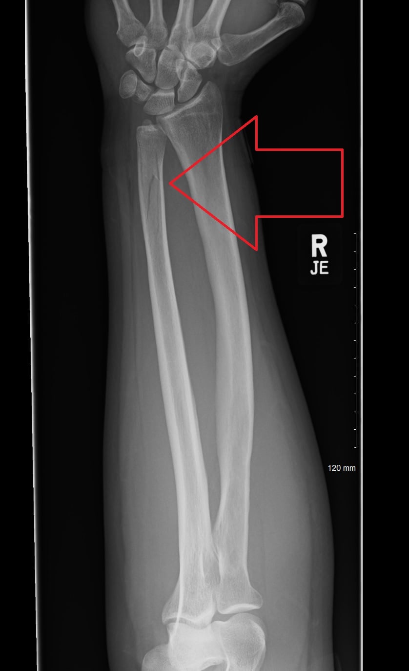
- Nightstick fracture = isolated ulna shaft from direct blow (defensive mechanism)
- EXCLUDE Monteggia (check radial head position on all views)
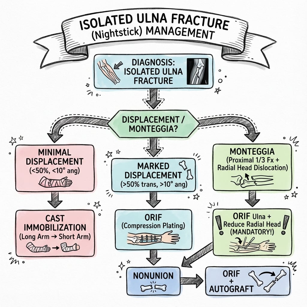
- Nonoperative threshold: less than 50% displacement, less than 10° angulation
- ORIF with 3.5mm DCP plate - 6+ cortices each side
- High union rate both operative and nonoperative
- “ALWAYS check PRUJ - rule out Monteggia lesion
- “Functional bracing allows early motion
- “Plate fixation is gold standard for displaced
- “Refracture risk higher with plate removal
ALWAYS check radial head. An isolated ulna fracture is NOT Monteggia. Get proper views including elbow. A line through radial neck should bisect capitellum on ALL views (radiocapitellar line).
Classic mechanism. Patient raises arm to defend against blow (assault, fall onto object). The subcutaneous position of the ulna makes it vulnerable. Ask about mechanism carefully.
Strict criteria. Less than 50% displacement, less than 10° angulation in proximal 2/3 or less than 15° in distal 1/3. Middle/distal third location. Functional brace allows early motion.
Greater than 50% displacement or significant angulation. Use 3.5mm DCP plate with 6+ cortices each side. Compression plating standard technique.
- Angulation
- Less than 10°
- Location
- Middle/distal
- Treatment
- Functional brace
- Angulation
- 10-15°
- Location
- Any
- Treatment
- Borderline - consider ORIF
- Angulation
- Greater than 15°
- Location
- Proximal
- Treatment
- ORIF recommended
- Angulation
- Any
- Location
- Open fracture
- Treatment
- ORIF + debridement
NIGHTNightstick Fracture Features
Hook:NIGHT stick fracture from a NIGHT time assault!
PLATEORIF Indications
Hook:PLATE the ulna when criteria met!
SAFENonoperative Criteria
Hook:SAFE for functional bracing!
Overview and Epidemiology
Historical name. Called nightstick fracture because the mechanism is typically a direct blow to the raised forearm, as when defending against an assault with a nightstick (baton). The subcutaneous ulna border is vulnerable to direct impact.
- 2-5% of forearm fractures
- Bimodal: young males (assault), elderly (falls)
- More common in males
- Middle to distal third most common
- Direct blow to subcutaneous ulna border
- Defensive arm position (guard)
- Fall onto hard edge
- Sports (hockey stick, bat)
Anatomy and Biomechanics
Ulna Shaft
The posterior border is subcutaneous throughout its length, making it vulnerable to direct trauma.
Triangular proximally, becomes more rounded distally.
Connects ulna to radius. Important for load transfer and forearm stability. Disruption creates longitudinal instability (Essex-Lopresti).
Classification
OTA/AO Classification
Simple fracture of ulna only.
Wedge fracture of ulna only.
Complex fracture of ulna only.
The classification guides complexity but treatment is primarily based on displacement and angulation.
Clinical Assessment
- Mechanism (direct blow vs fall)
- Assault or accident
- Location and timing of impact
- Previous forearm injury
- Hand dominance
- Inspect for deformity, swelling
- Palpate entire ulna and radius
- Check DRUJ and PRUJ
- Test forearm rotation
- Neurovascular exam
An isolated ulna fracture is NOT a Monteggia lesion. Always check the proximal radioulnar joint (PRUJ). The radiocapitellar line (line through radial shaft/neck) should bisect the capitellum on ALL views. If the radial head is dislocated, it is a Monteggia fracture-dislocation, not an isolated ulna fracture.
Differential Diagnosis
- Key Distinguishing Feature
- Single ulna fracture, radiocapitellar line intact, DRUJ congruent
- Pitfall if Missed
- —
- Key Distinguishing Feature
- Ulna fracture PLUS radial head dislocation (radiocapitellar line broken)
- Pitfall if Missed
- Missed radial head dislocation leads to chronic instability and poor function
- Key Distinguishing Feature
- Radius AND ulna both fractured
- Pitfall if Missed
- Treating as isolated underestimates instability
- Key Distinguishing Feature
- Radial shaft fracture with DRUJ disruption (not ulnar shaft)
- Pitfall if Missed
- Different injury; DRUJ must be reduced
- Key Distinguishing Feature
- Radial head fracture, IOM disruption, DRUJ instability (longitudinal)
- Pitfall if Missed
- Missed longitudinal forearm instability
- Key Distinguishing Feature
- Low-energy mechanism, lytic/sclerotic lesion on imaging
- Pitfall if Missed
- Fixing without addressing underlying lesion
Investigations
Essential Views
Both bones, full length.
Essential to assess radiocapitellar alignment and exclude Monteggia.
Assess DRUJ for longitudinal instability.
Displacement as percentage of bone width, angulation in degrees.
Reading the Radiocapitellar Line: Excluding Monteggia
"Exclude Monteggia" is this topic's single most-repeated safety rule — but calling a fracture "isolated" depends entirely on correctly reading the radiocapitellar line, so here is how to do it.
- The line. A line drawn down the long axis of the radial neck/shaft should pass through the centre of the capitellum — on every projection (AP, lateral and oblique), because the radial head can dislocate in any direction. Checking only the lateral is not enough.
- Why all views. A radial-head dislocation missed on one view may be obvious on another; a normal lateral does not exclude an anterior, posterior or lateral Monteggia. Any angulated ulnar shaft fracture should raise suspicion, because the ulnar deformity and the radial dislocation are mechanically linked.
- Get dedicated elbow (and wrist) films. The commonest reason a Monteggia is missed is inadequate imaging — a forearm film that does not include a true elbow. Always image the joint above and below the fracture.
- The paediatric trap. In children the ulna may show only plastic (bowing) deformation or a greenstick fracture alongside a frankly dislocated radial head — an easily missed Monteggia-equivalent. A bowed but apparently "unbroken" ulna with a displaced radiocapitellar line is still a Monteggia. (Bado types and paediatric Monteggia detail are in the Monteggia topics.)
- Bottom line: only once the radiocapitellar line is confirmed intact on all views, with a congruent DRUJ, can the injury be safely labelled an isolated ulnar shaft fracture and treated on the displacement/angulation criteria.
Q: How do you use the radiocapitellar line to be sure an ulnar shaft fracture is not a Monteggia? A: Draw a line through the radial neck/shaft axis — it must bisect the capitellum on AP, lateral AND oblique views (dislocation can be in any plane), so obtain dedicated elbow films, not just a forearm view. In children, beware plastic bowing or a greenstick ulna hiding a dislocated radial head. Only with the line intact on all views and a congruent DRUJ is it a true isolated ulnar shaft fracture.
Management
Functional Bracing
Less than 50% displacement, less than 10° angulation (proximal/middle), less than 15° angulation (distal), intact radiocapitellar joint.
Initial long-arm splint 1-2 weeks. Convert to functional brace. Allow elbow and wrist motion. Serial X-rays at 2, 4, 6 weeks.
Union in 8-12 weeks. Good functional results if criteria met.
If displacement increases or patient non-compliant, convert to ORIF.
Why functional bracing works: The interosseous membrane and surrounding soft tissues provide stability. Early motion prevents stiffness while allowing fracture healing through micromotion. The ulna is primarily a stabilizer (not weight-bearing like radius), so moderate displacement is tolerated.
- Avoids surgery complications
- Lower infection risk
- Early joint motion
- Cost-effective
- No hardware removal needed
- Patient compliance critical
- Regular follow-up essential
- Serial radiographs needed
- Accept some residual deformity
- Longer time to union
Refracture after plate removal is a recognized complication (up to 20% in some series). Counsel patients about this risk. If removing plate, protect arm for 6-12 weeks after removal.
Surgical Technique
Posterior Approach to Ulna
Supine with arm across chest, or lateral with arm on table.
Direct posterior over subcutaneous ulna border.
Between ECU (posterior interosseous) and FCU (ulnar nerve). The ulna is subcutaneous - minimal dissection needed.
Ulnar nerve is anterior and does not need to be identified for shaft fractures.
Complications
- Incidence
- 5-10% (nonoperative)
- Management
- ORIF with bone graft
- Incidence
- Variable
- Management
- Osteotomy if symptomatic
- Incidence
- Up to 20%
- Management
- Protect arm, consider leaving plate
- Incidence
- 1-2% (operative)
- Management
- Antibiotics, debridement
- Incidence
- Common
- Management
- Plate removal after union
Postoperative Care
Rehabilitation Protocol
Splint or brace. Wound care. Finger and shoulder ROM.
Begin elbow and wrist ROM. Gentle forearm rotation. Sling for comfort.
Full ROM goal. Light strengthening. X-ray to confirm healing.
Progressive strengthening. Return to work based on healing. Sports at 4-6 months.
Outcomes and Prognosis
Outcome Factors
Distal third, minimal displacement, good compliance, anatomic reduction.
Proximal third, comminution, delayed treatment, smoking.
Guidelines, Registries & Global Practice
- Isolated ulnar shaft fractures are uncommon (a small fraction of forearm fractures) and remain under-studied
- Bimodal: young men (assault, the classic "nightstick" mechanism) and older adults (low-energy falls)
- High-energy mechanism predominated (85.7%) in one Level I trauma-centre series of 70 cases (Coulibaly 2015)
- Mechanism varies by region: interpersonal violence in urban trauma centres, falls in ageing populations, sport elsewhere
- No dedicated arthroplasty-style registry exists for diaphyseal forearm fractures
- Evidence base is small retrospective series and case-control studies, not RCTs
- No high-level consensus on the exact operative threshold for moderately displaced fractures
- This makes individualised, shared decision-making essential
- Position on Isolated Ulnar Shaft Fractures
- Classify as 2U2 (ulna diaphysis); plate compression for simple, bridge plating for comminuted; minimum 6 cortices each side
- Position on Isolated Ulnar Shaft Fractures
- Nonoperative bracing for minimally displaced (under 50% displacement, under 10° angulation, radial head reduced); ORIF for displaced or proximal-third
- Position on Isolated Ulnar Shaft Fractures
- Lower angulation tolerance suggested (8° or more linked to worse function); favour fixation when displacement near threshold
- Position on Isolated Ulnar Shaft Fractures
- Exclude Monteggia in every case (radiocapitellar line on dedicated elbow views) before labelling 'isolated'
- Prefabricated functional braces readily available for nonoperative pathway
- Locking plates (LCP) stocked for osteoporotic bone
- Early supervised hand therapy and serial imaging routine
- Day-case ORIF feasible with image intensifier
- Custom moulded casts/braces substitute for prefabricated braces
- Standard (non-locking) DCP plating remains effective and lower cost
- Reliable follow-up for serial radiographs may favour definitive fixation when displacement is borderline
- Implant removal may be deferred indefinitely to conserve theatre resources and avoid refracture risk
Safeguarding remains global. In a child or in any patient where the history does not match the injury, an isolated ulnar fracture should prompt consideration of non-accidental injury and appropriate safeguarding pathways, irrespective of healthcare system.
Why the Proximal Third Is Different: Deforming Muscle Forces
The topic recommends a lower ORIF threshold for proximal-third fractures and cites "deforming muscle forces", and Viva 3 asks directly which muscles attach to the proximal ulna — here they are and how they pull.
- Triceps inserts on the olecranon — pulling the proximal fragment posteriorly and into extension.
- Brachialis inserts on the coronoid / ulnar tuberosity — flexing and pulling the proximal fragment anteriorly.
- Anconeus (lateral proximal ulna) plus the origins of the deep forearm muscles — flexor digitorum profundus, flexor carpi ulnaris and extensor carpi ulnaris take origin from the volar and subcutaneous ulnar shaft, adding rotational and angular deforming pull.
- Supinator and pronator forces act across the interosseous space, so a proximal-shaft fracture sits between opposing rotational muscle vectors.
Consequences for management:
- These powerful, opposing muscle vectors make a proximal-third fracture harder to reduce and to hold in a brace, and Sarmiento's own series showed the greatest loss of pronation in proximal-third fractures — the biomechanical basis for the lower operative threshold proximally.
- The same forces mean a "borderline" proximal fracture (even one below the 50% rule) is more likely to displace secondarily, which the controversies data link to malunion and nonunion.
Q: Why does a proximal-third isolated ulna fracture warrant a lower threshold for ORIF? A: Powerful opposing muscle attachments deform it — triceps (olecranon, extension/posterior), brachialis (coronoid, flexion/anterior), and anconeus plus the FDP/FCU/ECU origins and supinator/pronator rotational pull. These make brace-held reduction unreliable, and Sarmiento found the greatest pronation loss proximally — so proximal fractures displace and malunite more readily.
Controversies and Areas of Uncertainty
The classic nonoperative criteria (under 50% displacement, under 10° angulation) come from Sarmiento-era series. Newer data (Coulibaly 2015) link angulation of 8° or more and any secondary displacement to malunion, nonunion and failure to return to prior activity, suggesting the traditional limits may be too permissive for active patients.
Fractures near the 50% threshold are genuinely contested. Ali (2019) reported 5 of 10 nonoperative cases failed, yet operative treatment adds surgical and hardware risk. No RCT resolves this; decisions are individualised by age, demand, compliance and follow-up reliability.
Anderson's historical recommendation to graft minor comminution is no longer supported: Ring (2005) found bone grafting did not reduce nonunion in comminuted forearm fractures. Bridge plating with biological technique is now preferred.
Whether to remove a subcutaneous, prominent ulnar plate is debated. Removal relieves symptoms but carries a refracture risk concentrated at the plate ends; many surgeons leave asymptomatic plates in situ and, if removing, protect the limb afterwards.
MCQ Practice Points
Q: What is the displacement threshold for nonoperative treatment of isolated ulna fractures? A: Less than 50% of bone width. Beyond this, ORIF is recommended.
Q: What angulation is acceptable for nonoperative treatment? A: Less than 10 degrees in proximal/middle third, up to 15 degrees in distal third.
Q: What must be confirmed before diagnosing an isolated ulna fracture? A: Radial head is located. Check radiocapitellar line on all views to exclude Monteggia lesion.
Q: What is the minimum fixation required for ulna shaft ORIF? A: 6 cortices (3 screws) on each side of the fracture with a 3.5mm DCP or LCP.
Q: What is a significant risk after plate removal from the ulna? A: Refracture (up to 20%). Recommend waiting 18-24 months before removal if indicated.
Q: What is the mechanism of a nightstick fracture? A: Direct blow to the subcutaneous ulna border, typically when arm is raised in defense.
Exam Viva Scenarios
Practise clinical reasoning and management decisions out loud
“A 28-year-old man presents after an altercation where he raised his arm to defend himself. X-rays show an isolated mid-shaft ulna fracture with 30% displacement and 5 degrees of angulation. How would you manage this?”
“A 35-year-old woman falls onto a metal railing, striking her forearm. X-rays show an isolated ulna fracture with 75% displacement and 20 degrees of angulation. How would you treat this?”
“An isolated ulna fracture in the proximal third with 40% displacement. The radial head is confirmed located. How would your management differ from a mid-shaft fracture?”
“A 42-year-old motorcyclist presents with a Gustilo grade II open isolated ulna fracture. The radial head is confirmed located. How would you manage this injury?”
Key Features
- Nightstick = direct blow mechanism
- Must exclude Monteggia (check PRUJ)
- Subcutaneous position vulnerable
- Middle/distal third most common
Nonoperative Criteria
- Less than 50% displacement
- Less than 10° angulation prox/mid
- Less than 15° angulation distal
- Radial head located
ORIF Indications
- Greater than 50% displacement
- Greater than 15° angulation
- Proximal third (lower threshold)
- Open fractures
Operative Technique
- Posterior approach to ulna
- 3.5mm DCP or LCP
- 6+ cortices each side
- Lag screw if oblique
Outcomes
- 95%+ union rate (ORIF)
- 90% union rate (nonoperative)
- Refracture risk with plate removal
- Prox third higher complications
Evidence Base and Key Studies
Functional Bracing - Landmark Series (Sarmiento)
- 444 isolated ulnar shaft fractures braced; 287 (65%) followed up
- Union in 99% of fractures; mean shortening only 1.1 mm
- Mean final radial and dorsal angulation 5° each
- Good-to-excellent function in more than 96% (greatest pronation loss in proximal-third)
Compression Plating of Forearm Diaphysis (Anderson)
- 330 acute diaphyseal radius/ulna fractures plated; 137 isolated/combined ulna fractures
- Union rate 96.3% for the ulna and 97.9% for the radius
- Established ASIF compression plating as the standard for forearm diaphyseal fractures
- Iliac bone graft used for severely comminuted patterns (later questioned)
Bone Graft Not Required for Comminution (Ring)
- 41 comminuted both-bone forearm fractures plated with 3.5/4.5 mm DCP (6+ holes)
- Nonunion in 12% (5 patients)
- Bone grafting NOT associated with lower nonunion (OR 0.98, 95% CI 0.15-6.42)
- Open fracture, multiple injury, ipsilateral injury also not significant
Operative vs Nonoperative Nightstick Outcomes (Ali)
- 52 isolated ulnar shaft fractures; 42 ORIF (incl. 6 open) vs 10 nonoperative
- 5 of 10 nonoperative cases failed and required more follow-up visits
- ORIF gave satisfactory outcome with early non-load-bearing mobilisation
- Fractures with less than 50% displacement individualised by age, function, compliance
Displacement Drives Complications (Coulibaly)
- 70 isolated ulnar shaft fractures; 33 nonoperative vs 37 ORIF
- 14 nonunions and 17 malunions overall; nonoperative significantly associated with both
- Angulation of 8° or more linked to failure to return to prior activity level
- Secondary displacement greater than 2 mm contributed to malunion and nonunion
Refracture After Plate Removal (Lindgren)
- 349 surgically fixed forearm fractures; subsequent fracture rate 5-11%
- Plate refractures 10.9% vs flexible-nail 5.1%
- 90% of plate refractures occurred at the proximal or distal plate edge
- 90% of plate refractures required revision surgery