Orthopedic Emergency | Multiligamentous Injury | Vascular Catastrophe
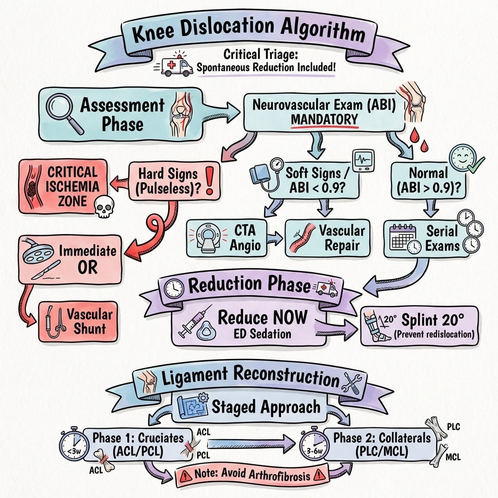
- VASCULAR EMERGENCY - Rule out popliteal artery injury in ALL cases with ABI and CTA
- Immediate reduction reduces vascular compromise - perform in ED under sedation
- Serial neurovascular exams - Document before and after reduction, every 2 hours
- Multiligamentous repair - Staged approach: ACL/PCL first, then collaterals at 3-6 weeks
- 20% missed initially - High suspicion if spontaneous reduction before arrival
- “Popliteal artery injury occurs in 30-40% - Normal pulses do NOT exclude intimal tear
- “ABI less than 0.9 = Mandatory CTA - Sensitivity 95% for arterial injury
- “Peroneal nerve injury (25%) - Check foot dorsiflexion and eversion before/after reduction
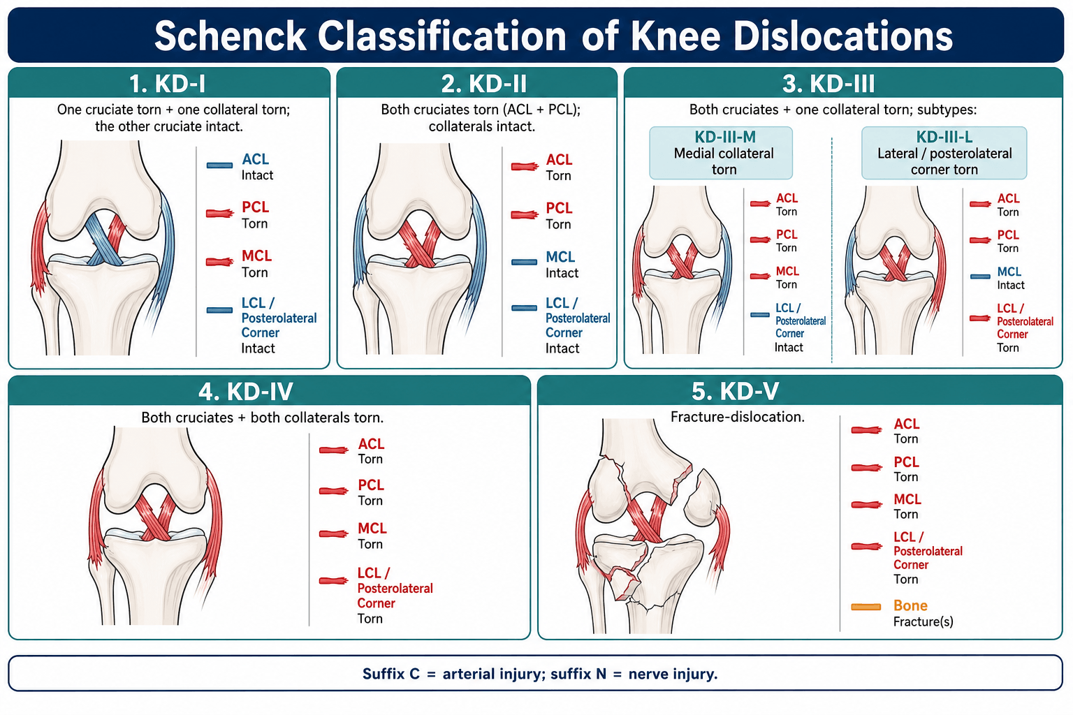
- “Schenck Classification (KD I-V) grades injury severity based on ligaments torn
MANDATORY workup. Normal pulses do NOT exclude arterial injury. Intimal tears can present with normal distal pulses initially then thrombose at 6-24 hours. ABI less than 0.9 requires immediate CTA. Vascular surgery consult if any abnormality.
Reduce immediately. Do NOT delay for imaging if neurovascular compromise present. Longitudinal traction with counter-traction. Reverse mechanism of injury. Reassess pulses post-reduction. Splint in 15-20 degrees flexion to prevent re-dislocation.
KD I-V system. KD I (one cruciate), KD II (both cruciates), KD III (M or L) (cruciate + collateral), KD IV (M or L) (both cruciates + collateral), KD V (fracture-dislocation). Higher grades = worse outcomes.
Staged repair approach. Acute: Reduce and splint, repair vascular/nerve injuries. Early (less than 3 weeks): ACL/PCL reconstruction. Delayed (3-6 weeks): Collateral ligament repair. Prevents arthrofibrosis with early total repair.
- Vascular Status
- Normal pulses, ABI 1.0
- Management
- Serial neurovascular exams, CTA if change
- Key Pearl
- Intimal tears can thrombose at 6-24h
- Vascular Status
- Diminished pulses
- Management
- URGENT CTA + vascular surgery consult
- Key Pearl
- 20% progress to thrombosis
- Vascular Status
- Absent pulses, cold foot
- Management
- IMMEDIATE reduction + vascular surgery OR
- Key Pearl
- 6-hour ischemia window - Amputation risk 86%
PANICStructures at Risk in Knee Dislocation
Hook:Knee dislocation causes PANIC - Check Popliteal artery, ACL/PCL, Nerve, Intima, Collaterals!
Overview and Epidemiology
Knee dislocation is an orthopedic emergency with potential for limb-threatening vascular injury. Despite being rare (0.02% of knee injuries), the consequences of missed diagnosis are catastrophic: amputation rates of 86% if ischemia exceeds 6 hours. The key challenge is that 20% of dislocations spontaneously reduce before arrival, making diagnosis difficult unless high clinical suspicion is maintained.
- High-energy trauma (60%): Motor vehicle collision, fall from height
- Low-energy in obese (40%): Simple fall, hyperextension
- Sports injuries: Dashboard injury, contact sports (football, rugby)
- Ultra-low velocity: Morbidly obese patients (BMI greater than 40)
- Popliteal artery injury: 30-40% (intimal tear most common)
- Peroneal nerve palsy: 25-30% (lateral dislocations highest risk)
- Compartment syndrome: 10-15% (especially after vascular repair)
- Meniscal tears: 50% (often peripheral detachment)
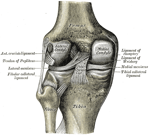
Anatomy
- ACL: Anteromedial and posterolateral bundles
- PCL: Anterolateral and posteromedial bundles
- MCL: Superficial (tibial attachment) and deep (meniscal)
- LCL: Fibular attachment, part of posterolateral corner
- LCL: Primary lateral stabilizer
- Popliteus tendon: Dynamic posterolateral stabilizer
- Popliteofibular ligament: Resists external rotation
- Lateral capsule: Secondary restraint
Pathophysiology
The popliteal artery is tethered proximally at the adductor hiatus and distally at the soleus arch, making it vulnerable to injury during knee dislocation. Intimal tears can occur WITHOUT complete disruption, presenting initially with normal pulses but thrombosing 6-24 hours later. This is why normal pulses do NOT exclude arterial injury.
- Anatomic Feature
- Tethered at adductor hiatus and soleus arch
- Injury Mechanism
- Stretching during anterior dislocation
- Clinical Significance
- Intimal tear common, delayed thrombosis
- Anatomic Feature
- Wraps around fibular neck
- Injury Mechanism
- Traction injury in lateral/posterolateral dislocation
- Clinical Significance
- 25-30% injury rate, poor recovery
- Anatomic Feature
- Intracapsular, minimal blood supply
- Injury Mechanism
- Torn in 80% of dislocations
- Clinical Significance
- Both require reconstruction for stability
Injury Mechanism and Cascade
Pathological sequence in knee dislocation:
- High-energy force - MVA, sports collision, fall from height
- Ligamentous failure - ACL/PCL rupture, capsular disruption
- Joint subluxation/dislocation - Tibiofemoral joint displaced
- Vascular tethering - Popliteal artery stretched over fixed points
- Intimal injury - Dissection, thrombosis, or complete transection
- Nerve traction - Peroneal nerve stretched around fibular head
- Spontaneous reduction - May occur, masking severity of injury
Classification Systems
Schenck Classification (Most Widely Used)
- Ligaments Injured
- Single cruciate (ACL or PCL)
- Frequency
- 5-10%
- Prognosis
- Good with reconstruction
- Ligaments Injured
- Both cruciates (ACL + PCL)
- Frequency
- 15-20%
- Prognosis
- Moderate, requires both repairs
- Ligaments Injured
- Cruciates + MCL
- Frequency
- 25%
- Prognosis
- Fair, staged repair needed
- Ligaments Injured
- Cruciates + LCL/PLC
- Frequency
- 30%
- Prognosis
- Fair, PLC critical for rotatory stability
- Ligaments Injured
- All four major ligaments
- Frequency
- 20%
- Prognosis
- Poor, high stiffness risk
- Ligaments Injured
- Periarticular fracture + ligament
- Frequency
- 10%
- Prognosis
- Variable, fracture healing affects timing
Think I - II - III - IV - V as increasing severity: I = one cruciate, II = two cruciates, III = three ligaments (add one collateral), IV = four ligaments (all), V = five problems (ligaments plus fracture).
CLIMBSchenck Classification (KD I-V)
Hook:CLIMB the knee dislocation severity ladder from KD I to KD V!
Clinical Assessment
- Mechanism: High-energy trauma, dashboard injury, hyperextension
- Reduced in field: 20% spontaneously reduce - Ask if knee "popped out"
- Pain and swelling: Immediate hemarthrosis, inability to bear weight
- Paresthesias: Foot numbness suggests nerve or vascular injury
- Look: Gross deformity (if not reduced), swelling, ecchymosis
- Neurovascular: MANDATORY before and after reduction - ABI, pulses, peroneal nerve
- Stability testing: DO NOT stress test acutely - Risk re-dislocation
- Compartments: Palpate for tightness, especially post-vascular repair


20% of knee dislocations reduce spontaneously before medical evaluation. High clinical suspicion is required if history suggests transient dislocation. Key clues: High-energy mechanism, severe instability on exam, inability to bear weight despite normal X-rays. Obtain MRI to assess multiligamentous injury.
Differential Diagnosis
The spontaneously reduced dislocation and the acutely swollen, unstable knee can be confused with lesser injuries. The distinction matters because only the true (occult) dislocation mandates the full vascular pathway.
- Discriminating Features
- Multidirectional gross instability, 2 or more ligaments torn, high-energy or ultra-low-velocity in obese
- Vascular/Nerve Risk
- High - popliteal artery and peroneal nerve at risk
- Key Investigation
- ABI plus serial exam; MRI for ligament pattern
- Discriminating Features
- Single-plane anterior laxity, positive Lachman, intact PCL and collaterals
- Vascular/Nerve Risk
- Negligible
- Key Investigation
- MRI; vascular workup not required
- Discriminating Features
- Patella displaced (usually lateral), tibiofemoral joint congruent, apprehension sign
- Vascular/Nerve Risk
- Negligible
- Key Investigation
- Skyline/axial radiograph; MRI for MPFL
- Discriminating Features
- Bony deformity plus instability, articular step-off on imaging
- Vascular/Nerve Risk
- High - treat as a dislocation
- Key Investigation
- CT for fracture; ABI plus CTA
- Discriminating Features
- Lateral fibular head prominence, peroneal symptoms, tibiofemoral joint reduced
- Vascular/Nerve Risk
- Peroneal nerve at risk; artery usually spared
- Key Investigation
- AP/oblique radiograph, comparison views
- Normal Value
- Greater than 0.9
- Abnormal Finding
- Less than 0.9
- Action Required
- IMMEDIATE CTA + vascular surgery consult
- Normal Value
- 2+ bilateral
- Abnormal Finding
- Diminished or absent
- Action Required
- Urgent CTA (do NOT rely on pulses alone)
- Normal Value
- Less than 2 seconds
- Abnormal Finding
- Greater than 3 seconds
- Action Required
- Immediate reduction, reassess post-reduction
- Normal Value
- Stable over 24 hours
- Abnormal Finding
- Deterioration at any point
- Action Required
- Urgent CTA - Delayed thrombosis
Investigations
Imaging Protocol
Before reduction: Document dislocation direction, identify fractures (KD V). After reduction: Confirm concentric reduction, assess for occult fractures (tibial plateau, femoral condyle). Stress views should NOT be performed acutely.
MANDATORY in all cases. ABI less than 0.9 has 95% sensitivity for arterial injury. Perform before and after reduction, then serially every 2 hours for 24 hours. Normal ABI does NOT exclude intimal tear.
Gold standard for arterial injury. Sensitivity 95%, specificity 99% for popliteal artery injury. Detects intimal tears, pseudoaneurysms, complete disruption. URGENT vascular surgery consult if any abnormality.
Once vascular status secure. Typically performed at 5-7 days post-injury to assess ligament injury pattern and plan staged reconstruction. T2-weighted sequences show all ligament tears, meniscal injuries, and chondral damage.
If obvious dislocation with neurovascular compromise, reduce IMMEDIATELY in the emergency department under procedural sedation. Do NOT wait for X-rays or CT. Reduction improves vascular flow and reduces compartment pressure. Image AFTER reduction to confirm concentric alignment.
Management Algorithm
Acute Management (First 6 Hours)
Goal: Restore vascular flow, prevent limb loss, document injuries.
ED Protocol
- Identify knee dislocation (obvious or history of reduction)
- Document neurovascular status (ABI, pulses, peroneal nerve)
- Obtain AP/lateral X-rays if time permits
- Procedural sedation (propofol or ketamine)
- Longitudinal traction with counter-traction at thigh
- Reverse mechanism of injury (flex for anterior, extend for posterior)
- Reassess neurovascular status post-reduction
- Repeat ABI and pulses (document improvement or deterioration)
- Confirm concentric reduction on X-ray
- Splint knee in 15-20 degrees flexion (prevents re-dislocation)
- Vascular surgery consult if ABI less than 0.9
- Neurovascular checks every 2 hours for 24 hours
- Watch for compartment syndrome (especially post-vascular repair)
- MRI at 5-7 days to plan ligament reconstruction
For anterior dislocation (most common): Apply longitudinal traction to tibia while assistant provides counter-traction at thigh. Gently extend knee while applying posterior pressure to proximal tibia. For posterior dislocation: Flex hip to 90 degrees, apply traction, then extend knee while lifting tibia anteriorly.
The algorithm above moves from closed reduction/splinting to staged ligament reconstruction, but a viva will expect the intermediate option for the unstable or complex knee dislocation: a knee-spanning (femur-to-tibia) external fixator.
Indications:
- Gross instability where a splint or brace cannot hold the reduction - classically the large/obese limb after an ultra-low-velocity dislocation, where soft tissue simply will not maintain alignment.
- To protect a vascular repair - after popliteal artery repair or bypass, an external fixator immobilises the joint so the fresh anastomosis is not stressed by knee motion.
- Open dislocation, severe soft-tissue compromise, or polytrauma/damage control where definitive ligament surgery must be delayed.
- KD-V fracture-dislocation as part of staged skeletal stabilisation.
Technique principles: apply with the knee in slight flexion (about 15 to 20 degrees), place pins well away from planned ligament tunnel sites and any future surgical zones, and keep it on until soft tissues and vascular status allow (commonly around 6 weeks) before converting to a hinged brace and proceeding to staged reconstruction.
Trade-off: pin-site infection and knee stiffness if left on too long - it is a temporising stabiliser, not a definitive treatment.
Exam point: between the splint and definitive ligament surgery sits the knee-spanning external fixator - use it for the unstable/obese limb, to protect a vascular repair, in open or polytrauma cases, and in KD-V - applied in slight flexion with pins clear of future tunnels, then converted to bracing and staged reconstruction.
REDUCEAcute Management Priorities
Hook:REDUCE is the goal - systematic approach prevents complications!
Surgical Technique
Cruciate Ligament Reconstruction
Surgical Steps (ACL + PCL)
Supine on standard operating table. Lateral post at thigh. Foot of bed dropped for knee flexion. Tourniquet thigh (usually NOT inflated due to vascular concerns). Prepare for arthroscopy and open if needed.
Standard portals (anterolateral, anteromedial). Assess cruciate tears, meniscal injuries, chondral damage. Document with photos. Perform limited debridement of cruciate remnants (preserve tibial footprint).
Bone-patellar tendon-bone (BTB) autograft preferred for ACL (bone blocks aid fixation). Achilles allograft often used for PCL (larger diameter, less donor morbidity). Prepare grafts on back table with whipstitch sutures.
ACL: Femoral tunnel at 10:30 (right knee) or 1:30 (left knee), tibial tunnel at ACL footprint. PCL: Femoral tunnel at 2:00 (right) or 10:00 (left), tibial tunnel via posteromedial portal. Ensure tunnels avoid convergence.
ACL first: Pass graft, fix femur (interference screw or button), tension at 20 degrees flexion, fix tibia. PCL second: Pass via posteromedial portal, fix femur, tension at 90 degrees flexion (posterior drawer reduced), fix tibia.
Check stability: Lachman (ACL), posterior drawer (PCL). Assess ROM (should achieve 0-130 degrees). Document with fluoroscopy. Ensure no graft impingement. Close portals, apply hinged knee brace locked 0-90 degrees.
When reconstructing both ACL and PCL, femoral tunnels can converge (ACL at 10:30/1:30, PCL at 2:00/10:00). Use 3D planning on CT or intraoperative fluoroscopy to ensure adequate bone bridge. If concern, stage PCL reconstruction 6 weeks later.
Complications
- Incidence
- 5-10% overall, 86% if ischemia greater than 6h
- Risk Factors
- Delayed vascular repair, compartment syndrome
- Management
- Prevention: Immediate reduction and vascular surgery consult
- Incidence
- 20-50% (50% if early total repair)
- Risk Factors
- Early total ligament repair, inadequate ROM
- Management
- Prevention: Staged repair. Treatment: Manipulation or arthroscopic lysis
- Incidence
- 15-30%
- Risk Factors
- Missed PLC injury, graft failure
- Management
- Revision reconstruction with attention to PLC
- Incidence
- 10-20% (25-30% have initial injury)
- Risk Factors
- Traction injury, compartment syndrome
- Management
- Ankle-foot orthosis, tendon transfer if no recovery at 12 months
- Incidence
- 10-15%
- Risk Factors
- Vascular repair, immobilization
- Management
- Prophylactic anticoagulation, early mobilization
Compartment syndrome occurs in 10-15% of patients after popliteal artery repair due to reperfusion injury. Maintain HIGH clinical suspicion. Perform 4-compartment fasciotomy liberally if any concern (pain out of proportion, tense compartments). Delayed fasciotomy (greater than 6-8 hours) leads to permanent muscle and nerve damage.
Postoperative Care and Rehabilitation
ACL/PCL Reconstruction Protocol
- Hinged knee brace locked 0-90 degrees
- Weight-bearing as tolerated with crutches
- ROM exercises: Passive extension to 0 degrees, flexion to 90 degrees
- Quad sets, ankle pumps, SLR (avoid hamstring contraction)
- Unlock brace, progress ROM to 0-120 degrees
- Weight-bearing as tolerated, wean crutches by week 4
- Closed-chain exercises (wall sits, mini squats)
- Avoid open-chain hamstring exercises (protect PCL)
- Full ROM expected (0-130 degrees)
- Progress strengthening (leg press, step-ups)
- Proprioception and balance training
- Stationary bike, swimming (no breaststroke)
- Jogging at 4-6 months if quad strength greater than 70%
- Sport-specific training at 6-9 months
- Return to sport at 12 months (MINIMUM)
- Functional testing before clearance
Outcomes and Prognosis
- Stability Outcome
- Good stability 70-80%
- ROM Outcome
- Stiffness 20%, full ROM 60%
- Notes
- Current gold standard approach
- Stability Outcome
- Good stability 60-70%
- ROM Outcome
- Stiffness 50%, full ROM 30%
- Notes
- Historical approach, high stiffness rate
- Stability Outcome
- Variable stability 50-70%
- ROM Outcome
- Better ROM (low stiffness)
- Notes
- Scar tissue makes reconstruction difficult
Poor outcomes are associated with: (1) Vascular injury requiring repair (higher complication rate), (2) KD IV or KD V injuries (all ligaments torn), (3) Peroneal nerve palsy (10-20% permanent), (4) Delayed reconstruction (greater than 6 months), and (5) High-energy mechanism (polytrauma, associated injuries).
Guidelines, Registries & Global Practice
Knee dislocation accounts for under 0.02% of orthopaedic injuries, but true incidence is under-reported because up to 50% reduce spontaneously before assessment. The epidemiological pattern is shifting worldwide from young high-energy trauma (road traffic, falls from height) toward ultra-low-velocity dislocations in obese and morbidly obese patients during everyday activity - a trend tracking rising global obesity. There is no dedicated international knee-dislocation registry; evidence comes from trauma-centre series and systematic reviews.
- Vascular Assessment
- Selective angiography driven by ABI and serial exam; ABI under 0.9 triggers CTA
- Surgical Stance
- Operative reconstruction favoured; reconstruct PLC
- Distinctive Point
- Drove the move away from routine arteriography
- Vascular Assessment
- Immediate neurovascular assessment, documented serial exams, urgent vascular input
- Surgical Stance
- Manage in or refer to a unit with combined vascular and ligament expertise
- Distinctive Point
- Embeds dislocation in major-trauma network pathways
- Vascular Assessment
- Reduce and stabilise first; spanning external fixator if unstable or vascular repair done
- Surgical Stance
- Restore bony anatomy, then staged ligament surgery
- Distinctive Point
- External fixation for the unstable or large (obese) limb
- Vascular Assessment
- Selective imaging plus 24-48h observation, longer vigilance for KD-IV
- Surgical Stance
- Early reconstruction within 3 weeks where soft tissues allow
- Distinctive Point
- Emphasises staged approach to limit arthrofibrosis
- CT angiography and MRI readily available; selective (exam/ABI-driven) vascular pathway is standard
- On-site vascular surgery enables revascularisation within the ischaemia window
- Staged arthroscopic reconstruction with allograft/autograft and hinged bracing
- 24-48h inpatient neurovascular observation routine
- Reliance on clinical exam and ABI where CTA is unavailable; low threshold to transfer
- Prompt closed reduction and a spanning external fixator stabilises the limb for transfer
- Delays to vascular care raise amputation risk - early recognition is the key modifiable factor
- Definitive ligament reconstruction may be deferred or unavailable; functional bracing used
Across every guideline and resource setting, the non-negotiable standard is a documented neurovascular assessment (pulses, ABI, peroneal nerve) before and after reduction with serial re-examination. A knee dislocation discharged on "normal pulses" alone, without ABI or an observation period, can return with a cold, pulseless limb and delayed thrombosis - the single most catastrophic and avoidable outcome of this injury.
Controversies and Areas of Uncertainty
Classic teaching quotes 30-40% popliteal artery injury and 86% amputation if ischaemia exceeds 6 hours - figures from older, selected single-centre series. The largest pooled meta-analysis (Medina 2014, 862 patients) found 18% vascular injury and 12% amputation among those injured. Know both: examiners often expect the classic numbers, but the modern, more accurate figures are lower.
Historic practice was routine arteriography in every dislocation. Prospective work (Stannard 2004; Klineberg 2004) showed a normal vascular exam plus ABI reliably excludes a limb-threatening injury, supporting a SELECTIVE pathway. CT angiography has now largely replaced catheter arteriography where available.
Acute primary repair of the posterolateral corner is attractive (single early operation) but fails far more often than reconstruction (37% vs 9% in Levy 2009). Most contemporary surgeons reconstruct, or augment repair, rather than repair alone - especially for the lateral side.
Early surgery (within 3 weeks) gives better functional scores than delayed surgery, but single-stage all-ligament reconstruction must be balanced against arthrofibrosis risk. There is no randomised evidence; decisions remain individualised by soft-tissue condition, vascular status and surgeon experience.
Whether to explore, neurolyse, graft or simply observe a complete peroneal palsy remains unresolved. There is no high-level evidence that acute exploration improves recovery, and many palsies are managed expectantly with an ankle-foot orthosis, reserving tendon transfer (posterior tibial tendon) for those without recovery by 12 months. Counsel that complete palsies recover poorly.
MCQ Practice Points
Q: What percentage of knee dislocations have associated popliteal artery injury? A: 30-40% - This high rate is why ABI measurement is MANDATORY in all knee dislocations. Normal pulses do NOT exclude intimal tear, which can thrombose 6-24 hours later.
Q: A knee dislocation with complete ACL, PCL, MCL, and PLC tears is classified as: A: Schenck KD IV - All four major ligament complexes torn. KD I (one cruciate), KD II (both cruciates), KD III (cruciate + one collateral), KD IV (all ligaments), KD V (fracture-dislocation).
Q: What is the advantage of staged ligament reconstruction over early total repair? A: Reduced arthrofibrosis rate (20% vs 50%) - Staged approach reconstructs cruciates early (2-3 weeks) then collaterals delayed (3-6 weeks). Early total repair has 50% stiffness rate with similar stability outcomes.
Q: What is the amputation rate if warm ischemia time exceeds 6 hours in knee dislocation with popliteal artery injury? A: 86% - This is why immediate reduction and vascular repair are critical. Time is limb in this injury.
Q: Common peroneal nerve injury occurs in what percentage of knee dislocations, and which dislocation direction has the highest risk? A: 25-30% overall, highest in lateral and posterolateral dislocations - Nerve wraps around fibular neck and is stretched during lateral displacement. Complete palsy has only 10-20% recovery rate.
Exam Viva Scenarios
Practise clinical reasoning and management decisions out loud
“A 35-year-old male presents to ED after motor vehicle collision. Paramedics report his knee was dislocated and they reduced it in the field. On arrival, knee is swollen but reduced. Pedal pulses are present but diminished. What is your assessment and management?”
“The patient from Scenario 1 has secure vascular status (ABI 1.0). MRI at 7 days shows complete ACL, PCL, MCL, and posterolateral corner (PLC) tears (Schenck KD IV-M/L). Walk me through your surgical plan.”
“The patient from Scenario 1 was admitted for observation. Initial ABI was 1.0. At 18 hours post-injury, nurse reports foot is cooler and pedal pulses are diminished. What is your management?”
Key Anatomy
- Popliteal artery = Tethered at adductor hiatus and soleus arch (vulnerable to injury)
- Common peroneal nerve = Wraps around fibular neck (25-30% injury rate)
- ACL + PCL = Both torn in 80% of dislocations
- PLC (LCL, popliteus, popliteofibular) = Critical for rotatory stability
Classification
- Schenck KD I = Single cruciate (rare)
- Schenck KD II = Both cruciates
- Schenck KD III-M/L = Cruciates + one collateral
- Schenck KD IV-M/L = All four ligaments
- Schenck KD V = Fracture-dislocation
Treatment Algorithm
- ED: Immediate reduction, ABI measurement, CTA if ABI less than 0.9
- Vascular injury: URGENT repair within 6 hours (amputation rate 86% if delayed)
- Staged repair: Cruciates at 2-3 weeks, collaterals at 3-6 weeks
- Serial exams: Every 2 hours for 24 hours (detect delayed thrombosis)
Surgical Pearls
- Staged approach reduces stiffness from 50% to 20%
- ACL/PCL tunnels: Avoid convergence (use fluoroscopy)
- PLC reconstruction: Protect common peroneal nerve throughout
- Collateral healing: Lock brace in extension 6 weeks
Complications
- Amputation: 5-10% overall, 86% if ischemia greater than 6h
- Arthrofibrosis: 50% if early total repair, 20% if staged
- Peroneal palsy: 25-30% initial injury, 10-20% permanent
- Persistent instability: 20-30% (often missed PLC)
Evidence Base and Key Trials
There is NO randomised controlled trial in knee dislocation - the injury is too rare and heterogeneous. The literature is dominated by systematic reviews, meta-analyses, and single-surgeon case series (Level III-IV). Every quoted figure below has been verified against the primary source; be cautious of the often-repeated "30-40% vascular injury" and "86% amputation" figures, which come from older selected series - pooled modern data give lower, more accurate numbers.
Decision Making in the Multiligament-Injured Knee: Evidence-Based Systematic Review
- Systematic review (Levels I-IV) of operative vs nonoperative, repair vs reconstruction, and early vs late surgery
- Surgery outperformed nonoperative care: good/excellent IKDC 58% vs 20%, return to full sport 29% vs 10%
- Posterolateral corner REPAIR failed more often than reconstruction (37% vs 9%)
- Early surgery (within 3 weeks) gave higher Lysholm (90 vs 82) and IKDC scores than delayed surgery
Vascular Injuries in Knee Dislocations: Role of Physical Examination in Determining Need for Arteriography
- Prospective cohort: 126 patients (134 knees) with acute multiligament knee injury at a Level-1 trauma centre
- Flow-limiting popliteal artery injury in 9 patients (7% prevalence) - lower than older quoted figures
- Serial physical examination had a single false positive and no missed injury (selective, not routine, arteriography)
- All 9 vascular injuries occurred in KD-III, KD-IV or KD-V; KD-IV warrants serial exams for at least 48 hours
The Role of Arteriography in Assessing Popliteal Artery Injury in Knee Dislocations
- Retrospective review of 55 patients (57 knees) with traumatic knee dislocation over 7 years
- Vascular exam (foot pulses plus ABI of 0.80 or greater) normal in 32 knees, abnormal in 25
- NO knee with a normal vascular examination had an injury requiring treatment
- Of 25 abnormal exams, 12 had vascular injury on angiography and 7 needed reverse saphenous vein grafting
Vascular and Nerve Injury After Knee Dislocation: A Systematic Review
- Meta-analysis of 862 knee dislocations - the largest pooled vascular/nerve dataset
- Weighted vascular injury frequency 18% and nerve injury 25% (lower than classic 30-40% teaching)
- 80% of vascular injuries were repaired; 12% of vascular injuries ended in amputation
- Highest vascular prevalence in KD-IIIL (ACL/PCL/lateral, 32%) and in posterior dislocations (25%)
Incidence of Concurrent Peroneal Nerve Injury in Multiligament Knee Injuries and Outcomes
- Retrospective cohort of 357 surgically treated multiligament knee injuries (mean follow-up 35 months)
- Concurrent peroneal nerve injury in 68 patients (19%)
- Nerve-injured patients had significantly lower final ROM (121 vs 127 degrees) and trended to lower return to work
- Pain (VAS), Lysholm and IKDC scores did NOT differ significantly with or without nerve injury
Arthroscopically Assisted Combined ACL/PCL Reconstruction in the Multiple Ligament Injured Knee: 2- to 10-Year Follow-up
- Case series of 35 combined ACL/PCL reconstructions (19 acute, 16 chronic) followed 2-10 years
- Significant improvement in Lysholm (mean 91), Tegner and HSS scores and in KT-1000 side-to-side laxity
- Normal Lachman/pivot-shift in 33 of 35 (94%); restored posterolateral stability less reliable
- Conclusion: reconstructed knees are functionally stable but NOT normal
Outcomes After Multiligament Knee Injury Worsen Over Time: Systematic Review and Meta-Analysis
- Meta-analysis of 79 studies and 3571 surgically treated multiligament knee injuries (mean age 35.6 years)
- Mean Lysholm 86 and IKDC 81 at 2 years, retaining roughly 80-85% of knee function
- Knee function DETERIORATES yearly (IKDC about -2.0 points/year), not a stable plateau
- PCL-based injuries had significantly worse IKDC (75 vs 84) and Lysholm (84 vs 91) than non-PCL injuries
Knee Dislocations in the Morbidly Obese Patient (Ultra-Low-Velocity Dislocation)
- Review of the increasingly common ultra-low-velocity knee dislocation in obese, morbidly obese and super-obese patients
- These injuries occur during everyday activities yet can be as severe or worse than high-velocity dislocations
- Frequently associated with neurovascular injury - early reduction and vascular assessment are critical to avoid amputation
- Limb size usually mandates external fixation to maintain reduction; reconstruction still improves outcomes