Sports Medicine | Isolated Usually Non-Operative | Combined Injuries Need Surgery | PLC Assessment Critical
- LCL is primary varus stabilizer - especially at 30° flexion when cruciate contribution minimal
- Isolated LCL injuries are RARE - always assess for PLC injury (popliteus, popliteofibular ligament)
- Varus stress test at 0° and 30° - opening at 0° indicates combined cruciate injury
- Peroneal nerve at risk - courses around fibular neck, assess with every lateral knee injury
- Combined injuries require surgery - isolated Grade III may heal non-operatively, combined do not
- “LCL injury with varus opening at 0° = combined PCL/ACL injury until proven otherwise
- “Always document peroneal nerve function BEFORE any intervention
- “MRI essential to assess PLC structures - isolated LCL rare, combined common
- “Fibular head avulsion is pathognomonic of LCL injury - check X-ray carefully
Truly isolated LCL injuries are rare - the great majority of lateral-sided injuries involve the posterolateral corner. When you see lateral instability, always examine for posterolateral corner (PLC) injury. The triad of LCL + popliteus + popliteofibular ligament = complete PLC injury requiring reconstruction.
Common peroneal nerve wraps around fibular neck - directly adjacent to LCL insertion. Document motor (ankle dorsiflexion, toe extension) and sensory (first web space) function. Peroneal nerve injury occurs in around a quarter of knee dislocations with posterolateral disruption (25%, Niall et al, 2005).
At full extension, cruciates provide secondary varus restraint. If varus opens at 0°, you have combined LCL + cruciate injury (usually PCL). This CANNOT be managed non-operatively - needs surgical reconstruction.
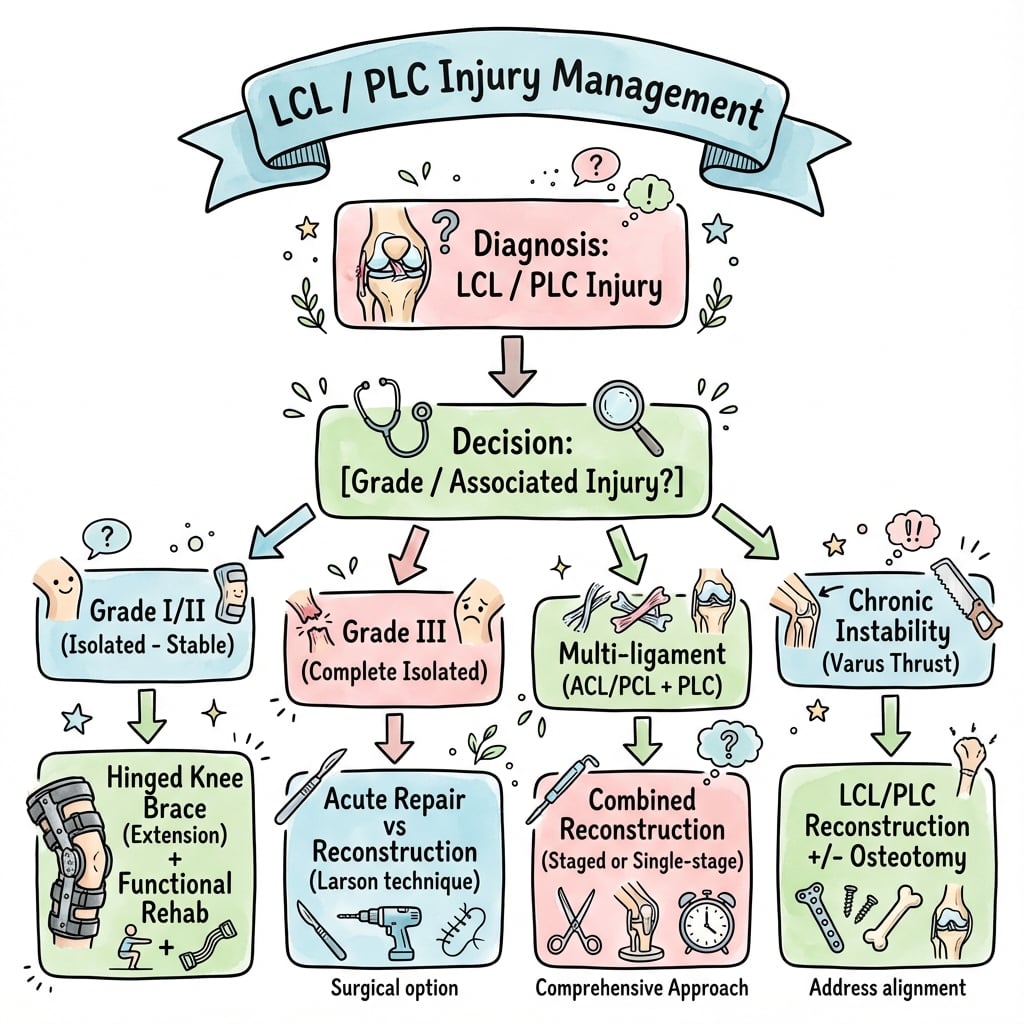
Isolated Grade I-II: Conservative (hinged brace, PT). Isolated Grade III: May heal non-operatively with bracing. Combined LCL + PLC or cruciate: Surgical reconstruction required for functional stability.
- Clinical Finding
- Lateral tenderness, no laxity
- Stability Testing
- Under 5mm varus opening at 30°, firm endpoint
- Treatment
- Functional brace 1-2 weeks, early ROM, PRICE
- Clinical Finding
- Moderate pain, mild laxity
- Stability Testing
- 5-10mm varus opening at 30°, endpoint present
- Treatment
- Hinged brace 4-6 weeks, protected WB, PT
- Clinical Finding
- Severe pain, significant laxity
- Stability Testing
- Over 10mm opening at 30° only, no endpoint
- Treatment
- Bracing 6-8 weeks may suffice - assess healing at 6 weeks
- Clinical Finding
- Posterolateral rotatory instability
- Stability Testing
- Varus + external rotation asymmetry, + dial test
- Treatment
- Surgical reconstruction (LCL + PLC)
- Clinical Finding
- Multi-ligament knee injury
- Stability Testing
- Varus opening at 0° AND 30°
- Treatment
- Staged or simultaneous multi-ligament reconstruction
FIBLCL Anatomy - 'FIB'
Hook:FIB = Fibula is where LCL ends. Think of a fib (lie) - isolated LCL injury is a 'fib', usually combined!
LPPPLC Structures - 'LPP'
Hook:LPP = All three make up the posterolateral corner. Like saying 'LP' (long play) Plus - you need all three!
ZERO-THIRTYVarus Stress Test - 'ZERO THIRTY'
Hook:Remember: TEST AT 30° to isolate LCL, TEST AT 0° to detect combined injuries
DANCEPeroneal Nerve Exam - 'DANCE'
Hook:Do the DANCE exam - if patient can't dance (foot drop), peroneal nerve is injured!
Overview and Epidemiology
Lateral Collateral Ligament (LCL) injuries are uncommon in isolation but frequently occur as part of complex multi-ligament knee injuries. The LCL is the primary static stabilizer against varus stress at the knee, working in concert with the posterolateral corner structures.
- Isolated LCL injuries are rare - most lateral-sided injuries occur with PLC and/or cruciate damage
- Sports-related: Contact sports (rugby, AFL, American football), skiing
- Mechanism: Varus force to weight-bearing knee, often with rotation
- Peak incidence: Males aged 20-40 years in contact sports
The LCL cannot be considered in isolation. Injury to the lateral structures almost always involves the posterolateral corner, and recognition of this combined injury pattern is critical for appropriate management. Failure to identify and treat PLC injuries leads to residual instability and reconstruction failure.
Pathophysiology and Mechanisms
- Origin: Lateral femoral epicondyle, anterior and distal to popliteus origin
- Insertion: Lateral aspect of fibular head, conjoint with biceps femoris
- Length: Approximately 60mm
- Width: 5-8mm (round, cord-like structure)
- Course: Extra-articular (unlike MCL, not attached to capsule or meniscus)
The LCL is separated from the joint by the popliteus tendon. The common peroneal nerve passes 10mm posterior to biceps tendon insertion. The lateral inferior genicular artery runs beneath the LCL. Unlike the MCL, the LCL is not attached to the lateral meniscus (the meniscus has popliteomeniscal fascicles instead).


The LCL is extra-articular and not attached to the lateral meniscus - this is why isolated LCL injuries don't cause meniscal damage. Compare to MCL which is intimately related to the medial meniscus.
Classification Systems
LCL Injury Classification
- Pathology
- Microscopic fiber damage, ligament intact
- Varus Stress at 30°
- Under 5mm opening, firm endpoint
- Clinical Features
- Tenderness, no instability, full ROM
- Pathology
- Partial macroscopic tear, some fibers intact
- Varus Stress at 30°
- 5-10mm opening, endpoint present
- Clinical Features
- Pain with varus stress, mild laxity
- Pathology
- Complete tear (midsubstance or avulsion)
- Varus Stress at 30°
- Over 10mm opening, no endpoint
- Clinical Features
- Gross laxity, may be painless (complete disruption)
Grading is based on varus stress testing at 30° flexion to isolate the LCL.
History
- Varus force to weight-bearing knee (tackle from medial side)
- Non-contact hyperextension with varus moment
- Dashboard injury with knee flexed and externally rotated
- Twisting injury with foot planted
- Lateral knee pain (worse with varus stress)
- Feeling of instability, especially pivoting/cutting
- "Knee giving way" with combined injuries
- Pop or snap at time of injury (less common than ACL)
Was it contact or non-contact mechanism? What was the direction of force? Was there immediate swelling (suggests cruciate involvement)? Was the patient able to weight-bear after injury? Any previous knee injuries? What are the sport and activity demands?
Examination
- Lateral ecchymosis (posterolateral suggests PLC)
- Effusion (intra-articular = cruciate involvement)
- Gait assessment (varus thrust in chronic injuries)
- LCL along its course (epicondyle to fibular head)
- Fibular head (avulsion tenderness)
- Lateral joint line
- Peroneal nerve at fibular neck
- Popliteal fossa (popliteus)
1. Varus stress test (most important) - Test at 0° extension AND 30° flexion. Grade opening as: I (under 5mm), II (5-10mm), III (over 10mm). Note endpoint quality (firm vs soft).
2. Dial test (external rotation) - Test at 30° and 90° flexion, compare ER asymmetry to contralateral side. Over 10° asymmetry at 30° only indicates isolated PLC injury. Over 10° at both 30° and 90° indicates combined PLC plus PCL injury.
3. Posterolateral drawer - At 90° flexion, apply posterior force with external rotation. Positive test indicates PLC injury.
4. Reverse pivot shift - Extend knee from flexion with valgus and external rotation. A reduction clunk indicates PLC laxity and posterolateral rotatory instability.
Always document peroneal nerve function (dorsiflexion power, first web space sensation) BEFORE any intervention including bracing, examination under anaesthesia, or surgery. Medicolegal significance is high.
Differential Diagnosis of Lateral Knee Instability/Pain
- Distinguishing Features
- Varus laxity at 30° only, normal rotation, varus stable at 0°
- Key Test/Investigation
- Varus stress at 0° and 30°; MRI confirms isolated FCL
- Distinguishing Features
- Varus laxity plus increased external rotation; posterolateral rotatory instability
- Key Test/Investigation
- Dial test (over 10° asymmetry at 30°), posterolateral drawer, reverse pivot shift
- Distinguishing Features
- Posterior sag, increased ER at BOTH 30° and 90°
- Key Test/Investigation
- Dial test at 30° and 90°; posterior drawer; MRI
- Distinguishing Features
- Gross multidirectional instability, high-energy mechanism, neurovascular risk
- Key Test/Investigation
- ABI, CT angiography, peroneal nerve exam, MRI
- Distinguishing Features
- Lateral pain with activity, no instability, tender over lateral epicondyle
- Key Test/Investigation
- Noble/Ober tests; no varus laxity
- Distinguishing Features
- Joint-line pain, mechanical catching/locking, no varus laxity
- Key Test/Investigation
- McMurray/Thessaly; MRI
- Distinguishing Features
- Focal fibular tenderness, possible peroneal nerve signs
- Key Test/Investigation
- AP/lateral X-ray; assess associated LCL avulsion (arcuate sign)
The special tests above (varus stress, dial, posterolateral drawer, reverse pivot shift) are the core, but a thorough PLC examination - and a strong viva answer - adds the classic recurvatum and gait signs the topic otherwise omits:
- External rotation recurvatum test (Hughston): with the patient supine, lift both great toes/feet off the bed by the toes with the knees extended. In a PLC-deficient knee the tibia drops into hyperextension (recurvatum), varus and external rotation relative to the other side. It is most positive when the PLC injury is combined with an ACL injury, and is a useful screening sign for significant posterolateral and combined instability.
- Standing/gait varus thrust: a dynamic sign of functional PLC deficiency - watch the patient walk; the knee thrusts laterally (into varus) at heel-strike/stance. Its presence (especially in chronic injury) signals that ligament reconstruction alone will fail without addressing alignment.
- Heel-height / prone vs supine dial nuance: perform the dial test prone (easier to read the foot-thigh angle) as well as supine, and remember the external rotation asymmetry is read against the contralateral side - over about ten degrees at thirty degrees only is isolated PLC, at both thirty and ninety degrees is PLC plus PCL (already covered above).
Exam point: do not stop at varus stress and the dial test - the external rotation recurvatum test and the dynamic varus thrust on gait complete the PLC assessment and the latter specifically flags the chronic, malaligned knee that needs an osteotomy.
Investigations
- AP, lateral, skyline views
- Bilateral weight-bearing if chronic
- Stress views if diagnosis uncertain
- Arcuate sign: Fibular styloid avulsion (pathognomonic of PLC injury)
- Segond fracture: Lateral tibial avulsion (anterolateral capsule - suggests ACL injury)
- Lateral capsular avulsion
- Fibular head fracture
- Varus alignment (chronic deficiency)
Varus stress at 20° flexion with side-to-side comparison. A difference over 4mm is significant. Useful for pre-operative planning and documenting degree of instability.

The "arcuate sign" (avulsion of fibular styloid process) on X-ray is pathognomonic of PLC injury. Don't miss this - look carefully at the fibular head on every lateral knee X-ray!
Non-Operative Management
Indications for conservative management:
- Grade I injuries (all)
- Grade II injuries (most)
- Isolated Grade III injuries (selected cases)
- Elderly/low-demand patients
- Significant medical comorbidities
- PRICE protocol (Protection, Rest, Ice, Compression, Elevation)
- Functional hinged brace (optional)
- Weight-bearing as tolerated
- NSAIDs for pain/inflammation
- ROM exercises (aim full ROM by 2 weeks)
- Quadriceps and hamstring strengthening
- Proprioception exercises
- Stationary cycling
- Sport-specific drills
- Functional brace for contact sports initially
- Full return when strength 90% and no pain with stress
Expected outcome: Full recovery, 2-4 weeks typical
Management Algorithm
- Initial Assessment
- Under 5mm varus, firm endpoint
- Management
- Functional treatment, early ROM
- Timeline
- Return 2-4 weeks
- Initial Assessment
- 5-10mm varus, endpoint present
- Management
- Hinged brace 4-6 weeks, PT
- Timeline
- Return 6-12 weeks
- Initial Assessment
- Over 10mm varus at 30° only
- Management
- Trial bracing 6-8 weeks, reassess
- Timeline
- Surgery if persistent laxity
- Initial Assessment
- Varus + external rotation asymmetry
- Management
- Surgical reconstruction (LCL + PLC)
- Timeline
- Surgery within 2-3 weeks ideal
- Initial Assessment
- Varus at 0° AND 30°, + cruciate tests
- Management
- Multi-ligament reconstruction
- Timeline
- Staged or single-stage, surgeon preference
Treatment decisions are based on injury severity, associated structures, and patient demands.

Surgical Management
- Combined LCL + PLC injury (Fanelli B/C)
- Combined LCL + cruciate injury
- Multi-ligament knee injury
- Bony avulsion with displacement (repair/fixation)
- Peroneal nerve injury requiring exploration
- Failed non-operative treatment of isolated Grade III
- High-demand athlete with isolated Grade III
- Persistent symptomatic instability
- Varus thrust gait in chronic injury
Acute injuries (under 3 weeks) allow primary repair or augmented repair. Subacute injuries (3-6 weeks) can be repaired if tissue quality is adequate. Chronic injuries (over 6 weeks) require reconstruction as tissue is not repairable.
The topic correctly says a chronic varus thrust may need an HTO, but the examinable framework that explains why is the Noyes classification of the varus knee, which separates the contributions to varus and dictates the staged plan:
- Primary varus: varus due to tibiofemoral bony/articular alignment alone (constitutional varus +/- lateral compartment cartilage/meniscus loss). The lateral soft tissues are competent.
- Double varus: primary (bony) varus plus separation of the lateral tibiofemoral compartment from deficiency/laxity of the lateral soft-tissue restraints (LCL/PLC stretch) - the limb is more varus on weight-bearing than the bony alignment alone predicts.
- Triple varus: double varus plus posterolateral rotatory instability and varus recurvatum (frank PLC incompetence with hyperextension/external rotation) - this is the knee with a visible varus thrust.
Why it matters: in double and especially triple varus, a ligament reconstruction performed on a malaligned limb is loaded by the uncorrected varus mechanical axis and stretches out/fails. The principle is therefore to correct alignment first (or concurrently) with a valgus-producing high tibial osteotomy, which shifts the weight-bearing axis laterally, abolishes the varus thrust and protects the subsequent (or staged) PLC reconstruction. Some triple-varus knees become asymptomatic after osteotomy alone, deferring or avoiding ligament surgery.
Exam point: "why correct alignment before reconstructing the PLC?" - because in double/triple varus (Noyes) the uncorrected varus axis will fail the graft; HTO unloads the lateral side, eliminates the thrust and is done first or with the reconstruction.
Complications
- Risk Factors
- Grade III injury, posterolateral trauma, fibular fracture
- Prevention/Management
- Document pre-op function, careful dissection, explore if no recovery by 3 months
- Risk Factors
- Missed PLC injury, inadequate reconstruction, non-anatomic repair
- Prevention/Management
- Complete assessment pre-op, anatomic reconstruction technique
- Risk Factors
- Prolonged immobilization, associated intra-articular injury
- Prevention/Management
- Early ROM, avoid over-tensioning graft
- Risk Factors
- Chronic instability, failed treatment
- Prevention/Management
- Correct with reconstruction, may need HTO for varus malalignment
- Risk Factors
- Untreated posterolateral instability
- Prevention/Management
- Always address PLC with cruciate reconstruction
Critical point: Untreated posterolateral corner injury is the most common cause of ACL and PCL reconstruction failure. The lateral structures must be addressed to protect cruciate grafts.
Postoperative Care
Hinged brace locked at 0°, toe-touch weight-bearing, ice and elevation, gentle quad sets, ankle pumps. No active hamstring exercises (protects PLC repair).
Progress ROM in brace (0-90° by week 4, full by week 6), progress to 50% weight-bearing by week 4, stationary cycling, pool exercises, continue quad strengthening.
Full weight-bearing, wean from brace by week 8, closed chain exercises, proprioception training, progress strengthening, avoid pivoting/cutting.
Sport-specific training progression, agility drills (straight-line first, then cutting), plyometrics, functional testing at 6 months.
Full return when passing functional tests (hop tests over 90%, isokinetic strength over 85%), sport-specific brace recommended for first season, ongoing maintenance program.
Avoid active hamstring exercises in early postoperative period - the biceps femoris inserts with the LCL and can stress the reconstruction. Quad-dominant rehabilitation initially.
Outcomes and Prognosis
Non-operative outcomes:
- Grade I: 100% return to sport, no residual laxity
- Grade II: 95%+ return to sport, minimal residual laxity
- Isolated Grade III: 70-80% satisfactory with bracing, 20-30% need delayed surgery
Surgical outcomes:
- Return to Sport
- 85-90%
- Stability Restoration
- 90%+
- Complications
- Under 5%
- Return to Sport
- 75-85%
- Stability Restoration
- 80-90%
- Complications
- 5-10%
- Return to Sport
- 70-80%
- Stability Restoration
- 75-85%
- Complications
- 10-15%
- Return to Sport
- 60-75%
- Stability Restoration
- 70-85%
- Complications
- 15-20%
Factors affecting outcome:
- Positive: Acute surgery, isolated injury, young patient, anatomic technique
- Negative: Chronic injury, multi-ligament, varus alignment, nerve injury
Guidelines, Registries & Global Practice
Global epidemiology. Isolated LCL injury is rare; lateral-sided injuries usually involve the posterolateral corner and are frequently part of multiligament injury or frank knee dislocation. PLC injury is commonly missed at first presentation - Pacheco et al found it was unrecognised in 72% of referred cases, with a mean diagnostic delay of 30 months (J Bone Joint Surg Br, 2011). In knee dislocation, common peroneal nerve palsy complicates around 25% and is associated with posterolateral disruption (Niall et al, 2005). Sport-related cohorts confirm that lateral and bicruciate patterns carry a worse return-to-play prognosis than medial-sided injuries (Bakshi et al, Sports Health, 2018).
Guidance across major bodies. No society has published a high-level (Level I) standalone guideline for isolated LCL/PLC injury; recommendations are consensus-based and consistent internationally.
- Region
- USA
- Core guidance
- Reconstruct (not repair) complete PLC tears; address PLC with concomitant cruciate reconstruction
- Evidence level
- Expert consensus / Level III-V
- Region
- UK
- Core guidance
- Early specialist referral and MRI for suspected PLC; anatomic reconstruction for complete injury
- Evidence level
- Consensus
- Region
- UK
- Core guidance
- No condition-specific guideline; general acute knee soft-tissue injury pathways apply
- Evidence level
- n/a
- Region
- Europe
- Core guidance
- Anatomic reconstruction preferred over repair for chronic/complete PLC; treat associated cruciate injury
- Evidence level
- Consensus / Level III-V
- Region
- Global
- Core guidance
- Fix displaced fibular/arcuate avulsions; reconstruct chronic ligamentous deficiency
- Evidence level
- Expert opinion
Registry evidence. Joint-replacement registries (AOANJRR, NJR, AJRR) do not capture isolated ligament reconstruction, so there is no national LCL/PLC implant-survival dataset; the evidence base remains cohort studies and biomechanical work. Multiligament and dislocation cohorts (including military and professional-sport series) provide the best available outcome data.
Practice variation. Surgeons increasingly favour anatomic two-graft reconstruction (after LaPrade) over older single-graft (Larson) techniques and over primary repair, reflecting Stannard's finding of higher failure with repair. Timing is convergent internationally: acute injuries are best addressed within roughly 2-3 weeks while primary repair/augmented repair remains feasible, with reconstruction reserved for chronic deficiency.
MCQ Practice Points
Q: What is the primary restraint to varus stress at 30 degrees knee flexion? A: The lateral collateral ligament (LCL) is the primary varus stabilizer, providing 69% of varus restraint at 25-30 degrees flexion. At this angle, the cruciates relax making the LCL the isolated primary restraint.
Q: What does varus opening at both 0 and 30 degrees indicate? A: Combined injury to both the LCL AND the cruciate ligaments (particularly PCL). Opening only at 30 degrees suggests isolated LCL injury since the cruciates are taut at 0 degrees and contribute to varus restraint.
Q: What is the dial test and what does asymmetry at 30 degrees only indicate? A: The dial test assesses external rotation of the tibia relative to the femur. Asymmetry greater than 10 degrees at 30 degrees ONLY indicates isolated PLC injury. Asymmetry at BOTH 30 and 90 degrees indicates combined PLC plus PCL injury.
Q: What is the arcuate sign? A: A small avulsion fracture of the fibular styloid on AP knee radiograph. It is pathognomonic of posterolateral corner (PLC) injury and indicates avulsion of the conjoint tendon insertion (LCL plus biceps femoris).
Q: Why is untreated PLC injury important in ACL reconstruction? A: Untreated posterolateral corner instability is the NUMBER ONE cause of ACL graft failure. The abnormal tibial external rotation places excessive stress on the ACL graft, leading to elongation or rupture.
Q: What is the relationship between the common peroneal nerve and the LCL? A: The common peroneal nerve passes approximately 10mm posterior to the biceps femoris tendon at the fibular head level. Peroneal nerve injury complicates around 25% of knee dislocations with posterolateral disruption (Niall et al, 2005).
Clinical Decision Scenarios
Practise clinical reasoning and management decisions out loud
“25-year-old rugby player presents after a tackle from the medial side. Has lateral knee pain and weakness of ankle dorsiflexion. X-ray shows fibular styloid avulsion (arcuate sign). Varus stress at 30° shows over 10mm opening. Varus at 0° is normal.”
“30-year-old AFL player has lateral knee injury. Varus stress positive at 30° (Grade III). Dial test shows 15° external rotation asymmetry at 30° but not at 90°. What is your diagnosis and management?”
“A 28-year-old presents 18 months after ACL reconstruction with recurrent instability. Original MRI showed isolated ACL tear. Examination shows 2+ Lachman and Grade II varus laxity. What happened?”
“45-year-old presents with lateral knee pain and 'knee bowing outward' when walking. History of knee injury 5 years ago, managed conservatively. Standing alignment shows 8° varus. Stress testing shows Grade II varus laxity.”
Key Numbers
- 30° flexion - optimal angle for varus stress testing (isolates LCL)
- Under 5mm opening = Grade I, 5-10mm = Grade II, over 10mm = Grade III
- Dial test: over 10° asymmetry at 30° = PLC injury; both 30° and 90° = PLC + PCL
- Isolated LCL injury is uncommon - most lateral injuries involve the PLC
- Peroneal nerve injury ~25% in knee dislocation with PLC disruption (Niall 2005)
Critical Concepts
- Isolated LCL injury is RARE - always assess PLC
- Varus opening at 0° = combined LCL + cruciate injury
- Peroneal nerve documentation BEFORE any intervention is mandatory
- Untreated PLC is #1 cause of ACL/PCL reconstruction failure
- Acute repair/reconstruction (under 3 weeks) has better outcomes
Must-Know Anatomy
- LCL: Lateral epicondyle to fibular head (extra-articular)
- PLC triad: LCL + popliteus + popliteofibular ligament
- Peroneal nerve: 10mm posterior to biceps tendon at fibular neck
- Arcuate sign: Fibular styloid avulsion = PLC injury
Management Principles
- Grade I-II isolated: Non-operative (brace, PT)
- Grade III isolated: Trial bracing, surgery if fails
- Combined LCL + PLC: Surgical reconstruction required
- Combined + cruciate: Address all structures, PLC protects graft
- Chronic instability + varus: HTO before or with reconstruction
Viva Pearls
- Always examine BOTH 0° and 30° for varus stress
- Dial test at 30° AND 90° differentiates PLC vs PLC+PCL
- Check peroneal nerve before doing ANYTHING
- Failed cruciate reconstruction - think missed PLC injury
- Chronic varus thrust needs alignment correction first
Evidence Base
LaPrade Anatomic Two-Graft PLC Reconstruction (Landmark Technique)
- Cadaveric study (10 specimens): two-graft technique anatomically reconstructs FCL, popliteus tendon and popliteofibular ligament
- Reconstruction significantly improved varus stability versus the cut (Grade III) state at 0°, 30°, 60° and 90° of flexion
- No significant difference in external rotation between intact and reconstructed knees at any flexion angle
- Provided the biomechanical basis for the modern anatomic PLC reconstruction
PLC Repair versus Reconstruction (Stannard)
- Prospective cohort of 64 PLC tears (39 repairs, 25 reconstructions; minimum 24-month follow-up)
- Acute primary repair failed in 13 of 35 (37%) versus 2 of 22 (9%) for reconstruction (statistically significant)
- Reconstruction using the modified two-tailed technique gave significantly better stability than repair
- Authors now favour reconstruction over repair for most high-energy PLC tears
PLC Injuries: A Serious Injury Commonly Missed (Pacheco)
- Retrospective review of 68 referred PLC injuries; injury was not identified at initial presentation in 49 of 68 patients (72%)
- Mean delay to correct diagnosis was 30 months from time of injury
- MRI correctly identified 14 of 15 injuries when performed within 12 weeks, but only 4 of 15 when performed later
- PLC injury was usually recognised only when severe multiligament injury was present
Untreated Grade III PLC Injury Increases ACL Graft Force (LaPrade)
- Cadaveric biomechanical study of ACL-reconstructed knees with sequential sectioning of the FCL, popliteofibular ligament and popliteus tendon
- ACL graft force was significantly higher after FCL sectioning during varus loading at both 0° and 30° of flexion
- Coupled varus and internal rotation moments increased graft force further beyond varus alone
- Supports the clinical observation that untreated Grade III PLC injury contributes to ACL graft failure
Common Peroneal Nerve Palsy After Knee Dislocation (Niall)
- Common peroneal nerve injury in 14 of 55 patients (25%) with knee dislocation; all had posterolateral structure disruption
- Palsy occurred in 14 of 34 (41%) of those with combined bicruciate and posterolateral injury
- Complete recovery in only 3 (21%) and partial useful motor recovery in 4 (29%); no useful recovery in 7 (50%)
- Lesions in continuity under 7 cm long recovered within 6 to 18 months
Posterolateral Attachments of the Knee: Surgical Anatomy (LaPrade)
- Cadaveric morphologic study (10 knees) quantifying attachments of the FCL, popliteus tendon and popliteofibular ligament
- FCL femoral attachment averaged 1.4 mm proximal and 3.1 mm posterior to the lateral epicondyle; fibular attachment 8.2 mm posterior to the anterior fibular head
- Popliteus tendon femoral attachment was consistently anterior to the FCL (mean separation 18.5 mm)
- Popliteofibular ligament had constant anterior and posterior divisions at the fibular styloid
Return to Play After Multiligament Knee Injury in NFL Athletes (Bakshi)
- Retrospective cohort of 50 NFL athletes; overall return-to-play rate 64%
- Athletes with ACL and PCL/LCL injury had a lower RTP rate (55.6%) and longer recovery than ACL/MCL injuries (70.8%)
- Mean time to RTP was 459 days for combined ACL and PCL/LCL injury versus 305 days for ACL/MCL injury
- Lateral-sided and bicruciate injury patterns carry a worse prognosis than medial-sided patterns