The Classic Connective Tissue Disorder
- FBN1 Gene: Fibrillin-1 mutation.
- Aortic Root: Annual echo screening essential.
- Scoliosis: Common, may be progressive.
- Protrusio Acetabuli: Characteristic finding.
- Dural Ectasia: Expand the dural sac.
- “FBN1 mutation
- “Aortic root is main mortality cause
- “Scoliosis screening needed
- “Protrusio is characteristic
Overview and Epidemiology
Marfan syndrome is an autosomal dominant connective tissue disorder caused by mutations in FBN1, the fibrillin-1 gene on chromosome 15. Its incidence is 1 in 5,000-10,000, and the diagnosis is made on the revised Ghent criteria of 2010.
Three systems. The disease affects the cardiovascular system, the eye, where the lens dislocates upwards, and the skeleton. Aortic root dilatation and dissection are the main causes of death.
The orthopaedic problems. Scoliosis affects 30-50% of patients. The others are pectus deformity, protrusio acetabuli, pes planus and ligamentous laxity.
Dural Ectasia: Definition, Measurement and Significance
What it is. Dural ectasia is widening, or ballooning, of the dural sac, most marked in the lumbosacral spine where CSF hydrostatic pressure is greatest. The chronically stretched, fibrillin-deficient dura expands and can scallop the posterior vertebral bodies, widen the neural foramina, and form anterior sacral meningocoeles or nerve-root-sleeve (Tarlov) cysts. It is both a diagnostic feature and an operative hazard.
When it appears. It is present in the large majority of adults with Marfan syndrome and may first appear or progress during adulthood, so a normal scan in childhood does not exclude it later.
How it is measured. Diagnosis is radiological, on MRI, or on CT if MRI is contraindicated. The most-used quantitative measure is the dural sac ratio: the dural sac diameter divided by the vertebral body diameter at the same level. In ectasia the sac is wider relative to the vertebral body, a ratio above the threshold and the opposite of the normal taper.
No agreed cut-off. There is no single agreed threshold, so reported prevalence varies, and so does the weight dural ectasia carries within the Ghent systemic score. One validated proposal raises the S1 dural sac ratio threshold to about 0.64. Supportive qualitative signs include posterior vertebral scalloping, a widened sac and an anterior meningocoele.
Symptoms. It can cause postural (low-pressure) headaches, low back pain and proximal leg pain, and rarely genitourinary symptoms from a large meningocoele.
For the spine surgeon. Dural ectasia is the key reason to image the lumbosacral dura before fusion. The thin, distended dura raises the risk of incidental durotomy and CSF leak. Ectasia also enlarges the canal and thins the posterior elements, and it complicates pedicle-screw purchase where bone is scalloped.
Pulmonary Manifestations and Spontaneous Pneumothorax
The pulmonary involvement changes how these patients tolerate surgery and ventilation.
Blebs and pneumothorax. The same fibrillin-deficient connective tissue weakens the lung parenchyma and produces apical bullae and blebs. Their rupture causes spontaneous pneumothorax, which occurs in a meaningful minority of Marfan patients (far above the general-population rate) and is often recurrent. Recurrent pneumothorax may need pleurodesis.
Restriction. Severe pectus excavatum and scoliosis reduce chest-wall compliance and lung volumes, lowering pulmonary reserve. Obstructive sleep apnoea is more common, from craniofacial and airway laxity.
Under anaesthesia. Positive-pressure ventilation can convert a simple pneumothorax, or rupture a bleb, into a tension pneumothorax. Combined with the chest-wall restriction of pectus and scoliosis, this reduces the ventilatory margin during long prone spinal fusions, so document baseline lung status before major surgery.
Sudden pleuritic chest pain and breathlessness in a Marfan patient is a spontaneous pneumothorax until proven otherwise, and the same symptoms can herald aortic dissection. Both are connective-tissue consequences and both can present identically: exclude each.
Pathophysiology and Mechanisms
Fibrillin and the elastic fibre. Fibrillin is a component of elastic fibres. Defective fibrillin produces abnormal elastic fibres in the aorta, the ligaments and the lens zonules, and increased TGF-β signalling contributes to the aortic disease.
Why scoliosis develops. Ligamentous laxity combines with abnormal connective tissue. The curves may resemble idiopathic scoliosis but can be more severe.
Clinical Assessment
History. Ask about the family history, cardiac symptoms such as palpitations and chest pain, visual symptoms and musculoskeletal symptoms.
Examination. Look for the whole phenotype:
- Habitus: tall and thin, with an arm span greater than height
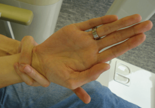
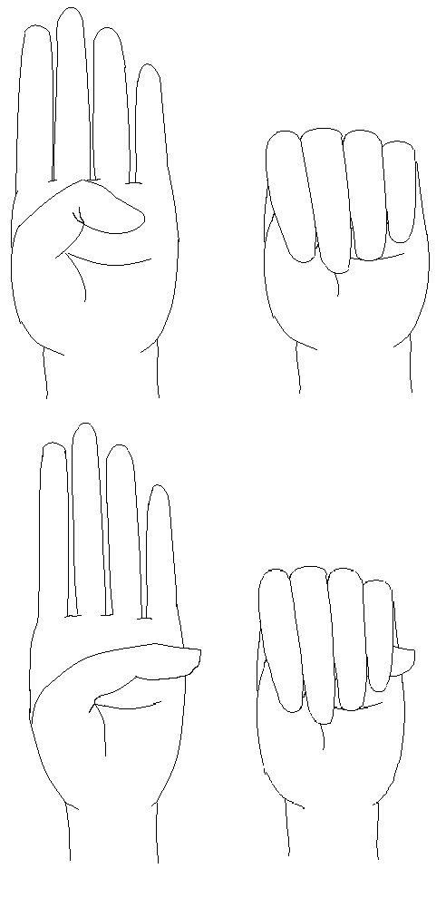
- Hands: arachnodactyly, shown by the wrist (Walker-Murdoch) and thumb (Steinberg) signs below
- Chest: pectus excavatum or carinatum
- Spine: scoliosis
- Hips: protrusio acetabuli, with limited internal rotation
- Feet: pes planus and hindfoot valgus
- Eyes: lens dislocation on slit-lamp examination
Diagnostic Criteria
The revised Ghent criteria (2010) are built from four features:
- Aortic root dilatation (Z-score ≥2) or dissection
- Ectopia lentis (lens dislocation)
- A causative FBN1 mutation
- A systemic score of 7 or more
Making the diagnosis. Without a family history, any one of these pairings makes the diagnosis:
- Aortic root dilatation plus ectopia lentis
- Aortic root dilatation plus an FBN1 mutation
- Aortic root dilatation plus a systemic score of 7 or more
- Ectopia lentis plus an FBN1 mutation known to be associated with aortic disease
With a family history, aortic root dilatation or ectopia lentis is sufficient.
The systemic score. Features are weighted, and a total of 7 or more counts as one of the four building blocks. Dural ectasia also scores (see below).
- Points
- 3
- Points
- 1
- Points
- 2
- Points
- 1
- Points
- 2
- Points
- 1
- Points
- 2
- Points
- 1
- Points
- 1
- Points
- 1
- Points
- 1
- Points
- 1
- Points
- 1
Investigations
An FBN1 mutation confirms the diagnosis; the other investigations measure the aorta and image the spine and pelvis.
- FBN1 genetic testing: confirmatory
- Echocardiography: aortic root measurement
- Spine radiographs: scoliosis
- Pelvic radiograph: protrusio acetabuli
- MRI of the spine: dural ectasia
Differential Diagnosis
Marfan-like Syndromes:
- Gene
- FBN1
- Key Differentiator
- Aortic root dilation, lens UP
- Gene
- TGFBR1/2
- Key Differentiator
- Bifid uvula, hypertelorism
- Gene
- CBS
- Key Differentiator
- Lens DOWN, thrombosis, cognitive issues
- Gene
- COL3A1
- Key Differentiator
- Thin skin, arterial rupture
- Gene
- COL2A1
- Key Differentiator
- Flat face, hearing loss, retinal detachment
- Gene
- FBN2
- Key Differentiator
- Crumpled ears, camptodactyly
Key Distinguishing Features:
- Lens dislocation direction: Marfan UP, Homocystinuria DOWN
- Cognitive function: Normal in Marfan, impaired in Homocystinuria
- Uvula: Normal in Marfan, bifid in Loeys-Dietz
- Gene: FBN1 for Marfan, FBN2 for Beals
Management
Aortic surveillance. The aortic root is measured by echocardiography every year, and this is mandatory. Beta-blockers reduce the rate of aortic root growth, and losartan (a TGF-β blocker) may also help. Avoid contact sports and strenuous activity.
Beta-blocker or losartan. The Pediatric Heart Network trial (Lacro 2014) found no significant difference between losartan and atenolol for aortic-root growth, and subsequent meta-analyses suggest the two are broadly comparable. Whether combination therapy or angiotensin-receptor blockade adds benefit over beta-blockade alone remains unsettled.
Aortic root replacement. The dilated root is replaced. The 5.0 cm threshold is pragmatic, not absolute: a family history of early dissection, rapid growth (over 3 to 5 mm/year) and planned pregnancy all argue for earlier surgery, and the relative merits of valve-sparing versus composite root replacement are debated.
Bracing scoliosis. A brace may be trialled for curves of 25-40 degrees in a growing child. Marfan-related scoliosis progresses faster and resists bracing more than idiopathic scoliosis, which has been attributed to the underlying connective-tissue abnormality rather than to any failure of technique (DOI). An older review of paediatric spinal deformity makes the same point about the syndromic curves as a group.
A shorter leash. Bracing is not abandoned: it can still buy growth in a very young child, and case series document success in early-onset Marfan curves. Set explicit progression thresholds at the outset and refer for surgical opinion early, rather than letting a brace become the reason a fast curve was watched for two more years. Over-bracing, which wastes the growth window in a curve that was never going to respond, is the commoner error in practice.
Operating on scoliosis. Posterior spinal fusion is indicated for progressive curves greater than 40-50 degrees.
The remaining skeletal features are managed as follows.
- Description
- Excavatum or Carinatum
- Management
- Surgical repair if severe
- Description
- Medial protrusion of acetabulum
- Management
- Usually observation
- Description
- Flat feet, hypermobile
- Management
- Orthotics; rarely surgery
Protrusio acetabuli. Older recommendations for prophylactic triradiate closure in growing children are largely abandoned. Sponseller's data show most protrusio is well tolerated, so intervention is now reserved for genuinely symptomatic or rapidly progressive cases, and acetabular closure is rarely needed, in severe cases before skeletal maturity.
Surgical Technique
Posterior spinal fusion. The operation is a standard posterior fusion, but the curves are often longer than idiopathic ones. The shift to posterior-only constructs is clear. Curve length, kyphosis, pelvic obliquity and bone fragility complicate level selection, and distal fusion levels and the role of pelvic fixation remain individualised.
The aorta is the hazard that belongs to this syndrome. The paper cited above reports that "risky" pedicle screws, those lying within 1 to 3 mm of the aorta or other adjacent structures, were found in 5.8 to 15.2% of screws in scoliosis surgery, and argues for careful preoperative aorto-vertebral metrology. That matters more in Marfan than in any other spinal deformity population. The aorta sits closer to the vertebral body than you expect, and the vessel a misplaced screw may abut is a dilated, fragile one in a patient whose connective tissue heals and holds sutures poorly.
- Cardiac clearance and echocardiography, which every candidate says
- CT assessment of the aorta's position relative to the pedicles, which few do
- Recognition of dural ectasia, by imaging the lumbosacral dura
Blood pressure. Avoid hypotension; blood pressure management is critical. Under-recognising the vascular and dural anatomy turns a routine posterior fusion into a catastrophe, and it is the graver of the two errors in Marfan scoliosis.
Complications and Postoperative Care
Aortic dissection is the perioperative risk, managed by careful blood pressure control, and cardiac monitoring continues that control after surgery. Dural ectasia brings the risk of CSF leak, and hardware may fail in weak bone and tissue, so it is monitored. Otherwise the standard spine protocol of mobilisation and physiotherapy applies.
Outcomes and Prognosis
Life expectancy is reduced, historically to a median of 40-50 years, and has improved with aortic monitoring and surgery. Scoliosis surgery outcomes are similar to idiopathic, although a national database found more neurological complications in Marfan patients than in controls (see the Evidence Base).
Guidelines, Registries & Global Practice
Global epidemiology. Prevalence is approximately 1 in 5,000 worldwide with no clear ethnic or geographic predilection. Inheritance is autosomal dominant with high penetrance but markedly variable expressivity; roughly 25 percent of cases arise from de novo FBN1 mutations and so present without a family history.
Side-by-side guidelines:
- Position
- Diagnostic standard worldwide; aortic root and ectopia lentis are cardinal; systemic score of 7 or more
- Position
- Annual imaging of aortic root; elective root replacement at approximately 5.0 cm (lower with family history of dissection, rapid growth, or pregnancy)
- Position
- Similar surveillance; root surgery typically at 5.0 cm, individualised; valve-sparing root replacement favoured in expert centres
- Position
- Posterior spinal fusion is the dominant approach; pre-operative MRI to define dural ectasia; cardiac clearance mandatory
Surgical orthopaedic thresholds (broadly concordant globally). Brace curves of 25 to 40 degrees in the growing child; operate on curves over 40 to 50 degrees, which progress faster and respond less to bracing than idiopathic curves. Observe asymptomatic protrusio acetabuli.
Registry and resource notes. No dedicated Marfan implant registry exists; arthroplasty data are sparse because protrusio rarely requires replacement before late adulthood. National Marfan patient organisations (for example The Marfan Foundation in the US, the Marfan Trust in the UK, and equivalent bodies in many countries) coordinate multidisciplinary clinics.
High- vs limited-resource practice. In well-resourced settings, lifelong echocardiographic (and cross-sectional CT/MRI) aortic surveillance, FBN1 sequencing, valve-sparing root replacement, and neuromonitored spinal fusion are standard. Where genetic testing and advanced imaging are limited, diagnosis leans on the clinical Ghent features and echocardiography; the priority everywhere is recognising aortic risk, restricting high-intensity isometric and contact sport, and arranging cardiac assessment before any major orthopaedic surgery.
MCQ Practice Points
Q: What gene is mutated in Marfan Syndrome? A: FBN1 (Fibrillin-1) on chromosome 15.
Q: What is the main cause of death in Marfan? A: Aortic dissection/rupture.
Q: What hip finding is characteristic of Marfan? A: Protrusio acetabuli.
Q: In which direction does the lens dislocate in Marfan? A: Superiorly (upward). Compare to Homocystinuria where it dislocates inferiorly.
Q: What must be done before scoliosis surgery in Marfan? A: Pre-op cardiac clearance with echo is MANDATORY. Watch for dural ectasia (CSF leak risk), curves often longer than idiopathic, and blood pressure control is critical.
Q: What are the Walker-Murdoch and Steinberg signs? A: Walker-Murdoch: Thumb overlaps fifth finger when grasping wrist. Steinberg: Thumb extends beyond ulnar border of closed fist. Both indicate arachnodactyly.
Self-Assessment Quiz
Additional Quiz Questions
Viva Scenarios
Practise clinical reasoning and management decisions out loud
“15-year-old male with known Marfan Syndrome. Scoliosis of 45 degrees. Aortic root is 4.2cm on recent echo.”
“How do you diagnose Marfan Syndrome?”
“X-ray shows protrusio acetabuli. What is this and how do you manage?”
GENETICS
- FBN1 Mutation
- Chromosome 15
- Autosomal Dominant
- Fibrillin-1 defect
CARDIAC
- Aortic root dilation
- Annual echo
- Beta-blockers
- Main mortality cause
SKELETAL
- Scoliosis 30-50%
- Protrusio acetabuli
- Pectus
- Pes planus
DIAGNOSIS
- Ghent criteria
- Aortic root + FBN1
- Systemic score ≥7
- Ectopia lentis
CLINICAL SIGNS
- Walker-Murdoch sign
- Steinberg sign
- Arm span exceeds height
- Arachnodactyly
SURGERY PEARLS
- Cardiac clearance first
- Dural ectasia risk
- BP control critical
- Curves often longer
Evidence Base
Loeys et al — Revised Ghent Nosology
- International expert panel re-weighted criteria toward cardiovascular disease
- Aortic root aneurysm and ectopia lentis are now the two cardinal features; together they are sufficient for diagnosis when there is no family history
- New weighted systemic score (threshold of 7 or more) and greater emphasis on FBN1 testing; molecular confirmation possible in over 95 percent
Lacro et al (Pediatric Heart Network) — Atenolol vs Losartan
- Randomised trial, 608 children and young adults with aortic-root z-score over 3.0
- No significant difference in rate of aortic-root z-score change between losartan and atenolol over 3 years (P=0.08)
- No difference in aortic-root surgery, dissection, or death between groups
Sponseller et al — Protrusio Acetabuli
- Cross-sectional study, 173 patients (346 hips) with Marfan syndrome
- Prevalence of protrusio 16 to 27 percent depending on the radiographic criterion; rises through the first two decades then plateaus
- Only a slight reduction in hip range of motion and no clinically meaningful loss of Iowa hip score
Böker et al — Dural Ectasia 10-year Follow-up
- Prospective 10-year cohort; 45 of 46 Marfan patients had dural ectasia at follow-up
- Dural ectasia may first appear or worsen during adulthood; anterior sacral meningocoele present in roughly half
- Proposed dural-sac-ratio cut-off at S1 raised to 0.64 for diagnosis
Kurucan et al — National Spinal Fusion Trends
- National database, 314 (1,410 weighted) Marfan patients undergoing spinal fusion 2003 to 2014
- Posterior spinal fusion rose from 67 percent to 92 percent of cases
- Marfan patients had higher neurologic complications than matched non-Marfan controls (2.4 vs 0.79 percent)
Benes et al — Postoperative Opioid Use (MFS vs AIS)
- Matched cohort, 20 Marfan vs 40 adolescent idiopathic scoliosis patients after posterior spinal fusion
- Marfan patients used more inpatient opioid (4.9 vs 2.1 mg/kg morphine-equivalents) despite similar pain scores
- Marfan was the only independent predictor of an outpatient opioid request (odds ratio 4.1)