Fibrocartilage | Load Distribution | Shock Absorption | Knee Stability
- Menisci transmit 50-70% of compressive load in extension, 85% in flexion
- Type I collagen arranged circumferentially provides hoop stress resistance
- Red-red zone (peripheral 3mm) has blood supply from perimeniscal capillary plexus
- Meniscectomy increases contact stress by 235%, accelerating osteoarthritis
- Lateral meniscus more mobile (10-12mm) than medial (5mm) during knee flexion
- “Viva starter: 'Draw the meniscus showing fiber orientation and vascular zones'
- “Key biomechanical function is load transmission and shock absorption
- “Complete meniscectomy → 235% increase in contact stress → OA within 10-20 years
- “Meniscal extrusion (over 3mm) → loss of hoop stress function → degenerative changes
Type I collagen arranged circumferentially. Radial tie fibers prevent longitudinal splitting. This architecture resists hoop stresses generated during load transmission.
Peripheral 10-25% is vascular (red-red zone, perimeniscal capillary plexus). The transitional red-white zone lies central to this, and the central majority is avascular (white-white). Determines healing potential.
Load transmission, shock absorption, joint stability. Transmits 50-70% load in extension, 85% in flexion. Meniscectomy increases contact stress by 235%.
Meniscus extrusion over 3mm → loss of hoop stress function. Total meniscectomy → OA develops in 10-20 years. Preserve tissue whenever possible.
RRWMeniscal Zones and Healing
Hook:Red blood means healing! RRW zones from peripheral (vascular) to central (avascular).
Overview and Introduction
Gross Anatomy
The menisci are crescent-shaped fibrocartilaginous structures interposed between the femoral condyles and tibial plateaus. Each knee contains two menisci:
Medial Meniscus:
- C-shaped, covering approximately 60% of medial tibial plateau
- Anterior horn attaches to tibial plateau anterior to ACL
- Posterior horn attaches posterior to ACL, anterior to PCL
- Peripheral attachment to joint capsule and deep MCL (less mobile)
- Translates approximately 5mm during knee flexion
Lateral Meniscus:
- More circular (covers 80% of lateral plateau)
- Anterior horn attaches anterior to tibial eminence (near ACL)
- Posterior horn attaches posterior to tibial eminence
- No attachment to LCL (popliteus tendon separates them)
- Translates approximately 10-12mm during knee flexion (more mobile)
Microstructure
- Water: 70-75% of wet weight
- Collagen: 15-25% (predominantly Type I)
- Proteoglycans: 1-2% (aggrecan, decorin)
- Cells: Fibrochondrocytes (outer) and chondrocytes (inner)
- Circumferential fibers (Type I): Resist hoop stress
- Radial tie fibers: Prevent longitudinal splitting
- Surface mesh: Random orientation at articular surface
- Organized outer to inner: structured to random
The circumferential arrangement of Type I collagen is the key structural feature. During weight-bearing, axial loads convert to radial displacement of the meniscus. The peripheral attachments prevent extrusion, generating hoop stresses (tensile forces) in the circumferential fibers. This mechanism is lost if the meniscus is excised or extruded.
Vascular Supply
The meniscus has zonal vascularity that profoundly affects tear management:
- Red-Red Zone (peripheral 0-3mm): Vascularized from perimeniscal capillary plexus (superior and inferior geniculate arteries)
- Red-White Zone (middle 3-5mm): Transitional zone with variable blood supply
- White-White Zone (central, over 5mm from periphery): Avascular, relies on synovial fluid diffusion
Only the red-red zone has consistent healing potential. This is why peripheral vertical tears can be repaired, while central horizontal cleavage tears cannot heal and require partial meniscectomy.
In children, the entire meniscus is vascular. By skeletal maturity only the peripheral 10-25% retains vascularity (Arnoczky & Warren), and the central two-thirds is avascular, relying on synovial-fluid diffusion.
Meniscofemoral Ligaments (Humphry and Wrisberg)
The root-tear section and the Bao finite-element card both invoke the "meniscofemoral ligaments" as the reason a lateral root tear is not equivalent to meniscectomy - but the ligaments themselves are never described. They are a discrete, high-yield piece of meniscal anatomy.
The two ligaments run from the posterior horn of the LATERAL meniscus to the lateral aspect of the medial femoral condyle, straddling the posterior cruciate ligament:
- Position relative to PCL
- Passes ANTERIOR to the PCL
- Mnemonic
- H comes before W = Humphry is in front (anterior)
- Position relative to PCL
- Passes POSTERIOR to the PCL
- Mnemonic
- Wrisberg = posterior; usually the larger/more constant of the two
Why they matter (structure → function):
- They are present in most knees (at least one in the large majority; both in a minority) and arise only from the lateral meniscus - a key asymmetry between the two menisci.
- Functionally they tether the posterior horn of the lateral meniscus and restrain its radial extrusion under load. This is precisely why a lateral posterior root tear is not biomechanically equivalent to lateral meniscectomy (Bao): the meniscofemoral ligaments keep the torn meniscus partly load-bearing, whereas the medial side has no such backup (hence the Allaire "medial root tear = meniscectomy" finding).
- On MRI or arthroscopy a meniscofemoral ligament can be mistaken for a tear fragment of the posterior horn or for the PCL.
The meniscofemoral ligaments run from the posterior horn of the lateral meniscus to the medial femoral condyle, one on each side of the PCL: Humphry anterior, Wrisberg posterior (alphabetical = anterior-to-posterior). They restrain lateral-meniscus extrusion, which is the structural reason a lateral root tear behaves better than a medial one.
Concepts and Biomechanical Principles
Core Functional Concepts
Circumferential Fiber Architecture:
The meniscus is predominantly composed of Type I collagen arranged in circumferential bundles. This orientation is critical for resisting hoop stresses generated during axial loading. Radial tie fibers interconnect the circumferential bundles, preventing longitudinal splitting.
Hoop Stress Mechanism:
When axial load is applied across the knee, the wedge-shaped meniscus is squeezed radially outward. The circumferential collagen fibers convert this compressive force into circumferential (hoop) tension, distributing load over a larger contact area and reducing peak stresses on articular cartilage.
Clinical Implication: Meniscal extrusion (over 3mm) disrupts hoop stress function → loss of load distribution → accelerated cartilage degeneration
Biomechanical Functions
Load Transmission
The primary function of the meniscus is load transmission across the knee joint:
- In extension: Menisci transmit 50-70% of compressive load
- In flexion: Menisci transmit 85% of compressive load
- After total meniscectomy: Contact stress increases by 235%
- After partial meniscectomy: Contact stress increases proportional to amount removed
- Contact Area
- 100% (baseline)
- Contact Stress
- 100% (baseline)
- Clinical Consequence
- Normal joint mechanics
- Contact Area
- Reduced 20-50%
- Contact Stress
- Increased 100-200%
- Clinical Consequence
- Accelerated cartilage wear
- Contact Area
- Reduced 50-70%
- Contact Stress
- Increased 235%
- Clinical Consequence
- OA in 10-20 years
Shock Absorption
The meniscus deforms under load, absorbing and dissipating energy. The viscoelastic properties of the fibrocartilage allow it to:
- Deform under cyclic loading (time-dependent behavior)
- Recover shape after load removal
- Dissipate energy through internal friction
Peak impact forces during gait are reduced by approximately 20% by intact menisci.
Joint Stability
The menisci contribute to knee stability by:
- Deepening the tibial plateaus (increases concavity)
- Resisting anterior-posterior translation (wedge effect)
- Secondary restraint to anterior tibial translation in ACL-deficient knee (especially posterior horn of medial meniscus)
The medial meniscus becomes a primary stabilizer after ACL rupture. This is why combined ACL and medial meniscus injury has particularly poor prognosis.
Lubrication and Nutrition
- Articular cartilage lubrication: Meniscus spreads synovial fluid across cartilage surface
- Proprioception: Mechanoreceptors in meniscal tissue (especially anterior and posterior horns) contribute to joint position sense
LASSMeniscal Functions
Hook:Think of meniscus as the knee's shock-absorbing LASS-o that distributes LOAD!
Viscoelastic and Biphasic Material Behaviour
The topic repeatedly calls the meniscus "viscoelastic" and "time-dependent" and the figure above shows its creep curve, but the underlying material science is never explained. It is the deep "function" answer an examiner is fishing for when they ask how a meniscus absorbs shock.
The meniscus is a biphasic (poroelastic) material. It has two phases:
- a solid phase - the collagen-proteoglycan matrix, and
- a fluid phase - interstitial water (~70-75% of wet weight), held by the negative charge of the proteoglycans.
Under load, the matrix has low permeability, so the water cannot escape instantly. The pressurised interstitial fluid carries most of the load in the first moments (fluid load support), and only flows out slowly through the matrix. This fluid flow is the source of the time-dependent (viscoelastic) behaviour:
- What is held constant
- A constant LOAD is applied
- What happens over time
- The tissue keeps DEFORMING (strain rises) as fluid is slowly squeezed out - exactly the curve in the figure
- Meaning
- Why a sustained load gradually flattens the meniscus
- What is held constant
- A constant DEFORMATION is held
- What happens over time
- The internal STRESS falls over time as fluid redistributes
- Meaning
- Why peak stress on cartilage drops after the initial impact
How this produces shock absorption: energy is dissipated through fluid flow and internal friction (and through the viscoelastic matrix) rather than being transmitted intact to the cartilage. On unloading, the proteoglycans osmotically re-imbibe water and the meniscus recovers its shape, ready for the next cycle. This is why the meniscus damps impact loads and why rapid (impulsive) loading is more dangerous than slow loading - there is no time for fluid to redistribute, so peak stresses are higher. (The general biphasic theory of articular cartilage is developed in articular-cartilage-structure; here it is applied to the meniscus.)
The meniscus is biphasic: a low-permeability collagen-proteoglycan solid plus interstitial water. Under load the pressurised fluid carries most of the load and exudes slowly, giving creep (deforms under constant load) and stress-relaxation (stress falls under constant deformation). Shock absorption is energy dissipated by this fluid flow - which is why fast impact loading, leaving no time for fluid redistribution, is the most damaging.
Clinical Relevance and Applications
Meniscal Tears and Treatment Implications
Vascular Zone Determines Treatment
- Blood Supply
- Excellent
- Healing Potential
- 90% healing rate
- Treatment Options
- Repair preferred
- Blood Supply
- Moderate
- Healing Potential
- 50-70% healing rate
- Treatment Options
- Repair with augmentation
- Blood Supply
- None
- Healing Potential
- Under 10% healing rate
- Treatment Options
- Resection/meniscectomy
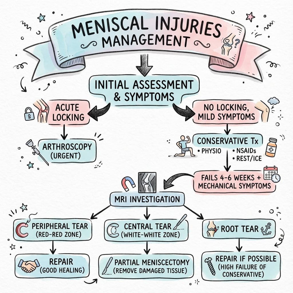
Q: What determines whether a meniscal tear should be repaired or resected?
A: Location relative to vascular zones is the primary determinant. Red-red zone tears (peripheral 3mm) should be repaired due to excellent healing potential (90%). White-white zone tears (central avascular) should be resected as they cannot heal (under 10%). Red-white zone (transitional) may be repaired with augmentation techniques (fibrin clot, PRP) with 50-70% success.
Meniscal Root Tears
The meniscal root attachments (anterior and posterior horns) anchor the circumferential fibres and are essential for hoop-stress generation. A root avulsion lets the meniscus extrude under load, abolishing its biomechanical function:
- Posterior medial root tear: Most common; often degenerative in middle-aged knees
- Biomechanical consequence (medial): A posterior medial root tear raises peak medial contact pressure by ~25% and is not significantly different from total medial meniscectomy (Allaire et al.). Anatomic repair restores contact pressure and kinematics to normal.
- Lateral side is different: Finite-element work (Bao et al.) suggests a lateral root tear is not fully equivalent to lateral meniscectomy because the meniscofemoral ligaments restrain radial extrusion - one reason lateral root tears are repaired aggressively, especially alongside ACL reconstruction.
- MRI signs: "Ghost meniscus" (meniscus seen on sagittal but absent on coronal), radial linear defect at the root, and meniscal extrusion over 3mm on coronal images
- Treatment: Transtibial pull-out or suture-anchor root repair to restore hoop stress
Differential Diagnosis of the Painful, "Meniscus-Like" Knee
Several entities mimic a symptomatic meniscal tear or extrusion and are common viva discriminators.
- Typical patient / history
- Young, twisting injury, locking/catching
- Key examination / imaging
- Joint-line tenderness, positive Thessaly/McMurray; MRI line reaching surface
- Distinguishing feature
- Mechanical symptoms; vertical/bucket-handle pattern
- Typical patient / history
- Over 45, insidious, activity-related ache
- Key examination / imaging
- Often coexisting cartilage change; horizontal cleavage on MRI
- Distinguishing feature
- Frequently incidental; responds to physiotherapy (ESCAPE)
- Typical patient / history
- Middle-aged, sudden pop squatting, rapid pain
- Key examination / imaging
- Coronal extrusion over 3mm, ghost meniscus
- Distinguishing feature
- Behaves biomechanically like meniscectomy
- Typical patient / history
- Older, sudden severe pain, often after root tear
- Key examination / imaging
- Subchondral oedema and crescent on MRI
- Distinguishing feature
- Pain out of proportion; bone - not meniscal - source
- Typical patient / history
- Child/adolescent, audible snapping knee
- Key examination / imaging
- Wide meniscus over 3 consecutive sagittal slices ('bow-tie' sign)
- Distinguishing feature
- Congenital morphology, lateral symptoms
- Typical patient / history
- Lateral more than medial, localised swelling
- Key examination / imaging
- Para-meniscal fluid collection on MRI, linked to horizontal tear
- Distinguishing feature
- Palpable mass at joint line
Controversies and Areas of Uncertainty
Multiple RCTs (including the ESCAPE trial) show no clinically meaningful benefit of arthroscopic partial meniscectomy over structured exercise for degenerative tears without locking. Debate persists over the minority with true mechanical symptoms who may still benefit.
The "root tear = total meniscectomy" rule is robust for the medial posterior root (Allaire) but is less absolute laterally, where meniscofemoral ligaments preserve some load transfer (Bao). Side matters when counselling on urgency of repair.
Contact stress rises broadly in proportion to tissue removed, but no precise threshold defines a "safe" resection volume. The pragmatic principle is to preserve every fibre possible and protect the peripheral rim that carries hoop stress.
Fibrin clot, PRP, marrow venting and scaffolds aim to extend repair into the red-white zone, but high-level evidence remains limited and outcomes are heterogeneous. Meniscal allograft transplantation has a clear but narrow indication (symptomatic meniscus-deficient compartment, aligned, stable, non-arthritic knee).
Guidelines, Registries & Global Practice
Global Epidemiology
- Meniscal injury is among the most common knee pathologies, with surgical incidence reported around 60-70 per 100,000 person-years in high-income populations; arthroscopic meniscus procedures remain among the most frequently performed orthopaedic operations worldwide.
- Bimodal pattern: traumatic tears in young, active people (often sport- and pivot-related, frequently with ACL injury) versus degenerative tears in those over 45, where prevalence rises with age and obesity and tears are commonly incidental on MRI.
- Medial more than lateral in degenerative disease; lateral and root/discoid pathology feature more in younger and paediatric cohorts.
Side-by-Side Guideline Positions
- Core position
- Preserve meniscal tissue; limited role for arthroscopic surgery in degenerative tears with osteoarthritis
- Practical implication
- Favour repair over resection; non-operative first for degenerative knees
- Core position
- Arthroscopic lavage and debridement not recommended for osteoarthritis; surgery reserved for true mechanical locking
- Practical implication
- Structured exercise and weight management first-line
- Core position
- Consensus: degenerative tear is a feature of early OA - exercise-based therapy first; surgery only after failed rehab or with clear mechanical symptoms
- Practical implication
- Shared decision-making; avoid 'incidental tear' surgery
- Core position
- No clinically meaningful benefit of APM over physiotherapy/sham for degenerative tears
- Practical implication
- Underpins all of the above guidance
Registry and Practice Variation
- Large national datasets (e.g. UK and Scandinavian arthroscopy/knee registries) have tracked a decline in arthroscopic meniscectomy for degenerative tears following the landmark RCTs, alongside a rising share of meniscal repair - a measurable shift toward preservation.
- Resource-rich settings: ready access to MRI, arthroscopic repair implants, and allograft transplantation; greater use of root repair and biologic augmentation.
- Resource-limited settings: MRI and arthroscopy access may be constrained, so diagnosis leans on clinical tests (joint-line tenderness, McMurray, Thessaly) and treatment emphasises rehabilitation; when surgery is needed, repair capability and allograft availability are often limited, favouring tissue-sparing techniques and conservative care.
MCQ Practice Points
Q: What is the predominant collagen type in the meniscus and how is it arranged?
A: Type I collagen arranged circumferentially with radial tie fibers. This architecture allows the meniscus to resist hoop stresses generated during weight-bearing, which is the mechanical basis for load transmission.
Q: What percentage of load is transmitted through the menisci in knee extension versus flexion?
A: 50-70% in extension, 85% in flexion. This increased load transmission in flexion explains why meniscal tears are more symptomatic with activities involving knee flexion under load (squatting, pivoting).
Q: A vertical longitudinal tear is identified 2mm from the peripheral edge. What is the expected healing potential with repair?
A: Excellent (approximately 90%). This tear is in the red-red zone (0-3mm from periphery) which has vascular supply from the perimeniscal capillary plexus and demonstrates excellent healing after repair.
Q: By how much does total meniscectomy increase peak contact stress in the knee?
A: 235% (Ahmed and Burke, 1983). This dramatic increase in contact stress explains the predictable development of osteoarthritis within 10-20 years after total meniscectomy.
Q: What is meniscal extrusion and why is it biomechanically significant?
A: Displacement of the meniscus over 3mm beyond the tibial plateau edge. This prevents the meniscus from generating hoop stress, making it biomechanically equivalent to total meniscectomy despite the tissue being physically present.

Exam Viva Scenarios
Practise clinical reasoning and management decisions out loud
“You are shown a diagram of the knee in cross-section. The examiner asks: 'Please describe the structure and function of the meniscus.'”
“A patient has undergone total medial meniscectomy 15 years ago and now presents with medial knee pain and early osteoarthritis. The examiner asks: 'Explain the biomechanical basis for this patient's degenerative changes.'”
“A 54-year-old presents with 3 months of medial knee ache, no locking, and an MRI showing a horizontal cleavage tear of the posterior medial meniscus with mild chondral wear. They ask whether they need keyhole surgery. The examiner asks: 'How would you counsel and manage this patient, and what is your evidence?'”
Key Microstructure
- Type I collagen arranged circumferentially = resists hoop stress
- Radial tie fibers prevent longitudinal splitting
- Fibrochondrocytes (outer) → chondrocytes (inner)
- 70-75% water, 15-25% collagen, 1-2% proteoglycans
Vascular Zones
- Red-Red (0-3mm) = vascular = 90% healing with repair
- Red-White (3-5mm) = transitional = 50-70% healing
- White-White (over 5mm) = avascular = under 10% healing
- Vascularity decreases with age (full in children, peripheral 20-30% in adults)
Biomechanical Functions
- Load transmission: 50-70% (extension), 85% (flexion)
- Shock absorption via viscoelastic deformation
- Stability: Deepens plateau, resists translation
- Secondary ACL stabilizer (medial posterior horn)
Meniscectomy Consequences
- Total meniscectomy → 235% increase in contact stress
- Contact area reduced by 50-70%
- Fairbank changes: Flattening, ridge, space narrowing
- OA develops in 10-20 years
Clinical Correlations
- Meniscal extrusion over 3mm = loss of hoop stress function
- Root tear = functionally equivalent to meniscectomy
- Lateral meniscus more mobile (10-12mm vs 5mm medial)
- Preserve tissue whenever possible
Evidence Base
Static Pressure Distribution on the Tibial Surface and the Effect of Meniscectomy
- Cadaveric pressure-mapping in 18 knees (8 also after medial meniscectomy) across flexion angles
- A significant fraction of the joint compressive load is transmitted through the menisci
- Total meniscectomy caused a drastic alteration of tibial-surface pressure distribution
- Provided the mechanical basis for post-meniscectomy degenerative change
Microvasculature of the Human Meniscus (Vascular Zones)
- 20 cadaver knees studied by histology and Spalteholz tissue clearing
- Perimeniscal capillary plexus (medial, lateral, middle genicular arteries) supplies only the peripheral 10-25%
- Posterolateral lateral meniscus (adjacent to popliteus) is avascular - no penetrating vessels
- Anterior and posterior horn attachments have a good blood supply
Biomechanical Consequences of a Posterior Root Tear of the Medial Meniscus: Similar to Total Meniscectomy
- Nine cadaver knees, 1000 N axial load at 0-90 degrees flexion (Fuji film + robotic kinematics)
- Posterior medial root tear raised peak medial contact pressure by 25% versus intact
- Root-tear contact pressure was not different from total medial meniscectomy
- Anatomic root repair restored contact pressure and kinematics to normal
Arthroscopic Partial Meniscectomy vs Physical Therapy for Degenerative Tears (ESCAPE RCT)
- Multicentre noninferiority RCT, 321 patients aged 45-70 with MRI-confirmed degenerative tear
- Both meniscectomy and a supervised exercise programme produced clinically meaningful improvement
- Between-group difference (Patient-Specific Functional Scale) was below the minimal important change
- No clinically meaningful advantage for surgery at any time point to 24 months
Finite-Element Analysis of Lateral Meniscus Posterior Root Tear
- Finite-element knee model under 1000 N comparing intact, lateral root tear, root tear + MFL deficiency, and lateral meniscectomy
- Lateral root tear reduced contact area and raised lateral contact pressure but less than total meniscectomy
- The meniscofemoral ligament restrained radial extrusion and preserved some load transfer
- Concluded a lateral root tear is NOT functionally equivalent to total meniscectomy
Knee Joint Changes After Meniscectomy (Fairbank Changes)
- Original description of radiographic changes after open meniscectomy
- Anterposterior ridge formation, femoral condyle flattening, joint space narrowing
- Established that meniscectomy is not a benign procedure
- These features are now eponymously termed Fairbank changes