Symptomatic shoulder instability in 2+ directions without significant trauma
- MDI is clinical diagnosis: symptomatic instability in 2+ directions without significant trauma
- Rehabilitation is first-line: minimum 6 months structured physiotherapy before surgery
- Sulcus sign pathognomonic: measure in adduction and external rotation positions
- Beighton score ≥4/9 indicates generalized hypermobility affecting treatment decisions
- Inferior capsular shift gold standard open procedure: T-capsulorrhaphy or lateral shift
- Arthroscopic capsulorrhaphy success rates 70-85%: lower than unidirectional instability
- Thermal capsulorrhaphy abandoned: high failure rates and chondrolysis risk
- Burkhead-Rockwood protocol: structured 6-12 month rehabilitation program
- “Examine BOTH shoulders: bilateral involvement in 50-80% of MDI patients
- “Sulcus sign graded with shoulder in neutral rotation AND external rotation
- “Positive sulcus in ER suggests rotator interval pathology requiring specific treatment
- “Load-and-shift test: compare to contralateral shoulder, grade on 3-point scale
- “Hyperabduction test: humeral head translation with arm overhead indicates inferior laxity
- “Apprehension test often NEGATIVE in MDI: distinguishes from traumatic instability
- “Document generalized hypermobility: affects surgical planning and expectations
Missing bilateral examination (50-80% bilateral involvement), confusing MDI with unidirectional instability (completely different algorithms), failing to assess sulcus sign in external rotation (rotator interval indicator), not documenting generalized hypermobility (Beighton score affects decisions), overlooking voluntary component (psychiatric assessment may be needed), and premature surgical intervention (rehabilitation is first-line with 80-90% success). MDI is a clinical diagnosis requiring comprehensive bilateral examination.
Inadequate rehabilitation trial (minimum 6 months required), wrong surgical procedure (thermal capsulorrhaphy abandoned), excessive capsular shift (overtightening causes stiffness), missing rotator interval closure (persistent sulcus in ER), inadequate postoperative protection (early mobilization causes recurrent stretching), and unrealistic expectations (MDI surgery has lower success than unidirectional). Conservative management succeeds in 80-90% with proper compliance.
Improper sulcus sign technique (test in adduction, neutral, and ER), inadequate load-and-shift grading (compare to contralateral), missing hyperabduction test (specific for inferior laxity), failing to examine under anesthesia (reveals true laxity), not assessing voluntary control (distinguishes structural from non-structural), and overlooking associated pathology (labral tears can coexist). Systematic bilateral examination is essential.
Arthroscopic capsulorrhaphy in severe MDI (open shift has better outcomes), inadequate capsular volume reduction (inferior pouch must be addressed), missing rotator interval closure (when sulcus positive in ER), over-tensioning capsular shift (balance stability vs motion), insufficient rehabilitation (4-6 months for capsular healing), and operating on voluntary dislocators (psychological issues first). Patient selection is critical.
Quick Decision Guide: MDI vs Unidirectional Instability
AMBRIIAMBRII Criteria for MDI
Hook:Remember AMBRII as the complete MDI story from diagnosis to treatment: starts Atraumatic and Multidirectional, often Bilateral, treat with Rehabilitation first, then Inferior capsular shift with Interval closure if needed.
SULCUSSulcus Sign Grading
Hook:Think SULCUS as the systematic approach to examining inferior instability: Standard position first, apply Unload force, Look for depression, Compare sides, Use grading, and crucial Second test in external rotation to assess rotator interval.
Overview and Epidemiology
Definition
Multidirectional instability (MDI) is defined as symptomatic glenohumeral instability in two or more directions (anterior, posterior, and/or inferior) occurring without significant trauma. The hallmark is inferior laxity demonstrated by a positive sulcus sign, distinguishing it from unidirectional instability patterns.
MDI represents a spectrum of capsular redundancy and muscular insufficiency, ranging from generalized connective tissue disorders to acquired capsular stretch. The key diagnostic criteria include atraumatic onset, instability in multiple directions, and characteristic examination findings of inferior translation.
The critical distinction between MDI and unidirectional instability is not just the number of directions of instability, but the underlying pathology: MDI involves global capsular redundancy and often generalized ligamentous laxity, whereas unidirectional instability typically results from specific traumatic labral and capsular injury. This fundamental difference drives completely different treatment algorithms.
Epidemiology
MDI predominantly affects adolescents and young adults, with peak incidence between 15 and 25 years of age. There is a female predominance with a 2:1 ratio compared to males, likely related to higher baseline ligamentous laxity and hormonal influences on connective tissue.
Bilateral involvement occurs in 50-80% of patients, reflecting the systemic nature of capsular laxity in many cases. Approximately 30-40% of MDI patients demonstrate generalized joint hypermobility with Beighton scores of 4 or greater, indicating underlying connective tissue disorder.
Sport participation, particularly overhead activities (swimming, gymnastics, volleyball), is common in the history. However, unlike traumatic instability, there is no identifiable injury event. Symptoms develop insidiously with repetitive overhead use causing progressive capsular stretch.
Risk Factors
Intrinsic factors include generalized joint hypermobility (Beighton score ≥4), connective tissue disorders (Ehlers-Danlos syndrome, Marfan syndrome), family history of hypermobility, and female gender. These constitutional factors predispose to global capsular laxity.
Extrinsic factors include repetitive overhead activities (swimming, gymnastics, throwing sports), improper training techniques leading to capsular stretch, delayed muscle maturation in adolescents, and poor scapular mechanics causing secondary glenohumeral instability.
Screen all adolescent patients with shoulder pain for signs of MDI, even without instability symptoms. Early identification allows preventive rehabilitation to strengthen dynamic stabilizers before symptomatic instability develops. Bilateral examination is mandatory as contralateral involvement may be subclinical.
The combination of constitutional laxity and repetitive overhead loading creates a cumulative capsular stretch pattern. Athletes with generalized hypermobility are particularly vulnerable to developing symptomatic MDI when participating in overhead sports without adequate rotator cuff and scapular strengthening.
Pathophysiology and Mechanisms
Glenohumeral Joint Anatomy
The glenohumeral joint is inherently unstable, relying on both static and dynamic stabilizers to maintain congruity. The bony anatomy provides minimal constraint, with the humeral head being three times larger than the shallow glenoid fossa. This design allows extensive range of motion but depends on soft tissue integrity.
Static stabilizers include the glenoid labrum (deepens socket by 50%), glenohumeral ligaments (anterior band of IGHL most important), joint capsule (provides volume constraint), and negative intra-articular pressure. The inferior glenohumeral ligament complex (IGHL) is the primary restraint to anterior and posterior translation with the arm in abduction and external rotation.
Dynamic stabilizers include the rotator cuff muscles (compressive force creates concavity-compression effect), long head of biceps (superior stability), scapular stabilizers (maintain glenoid position), and proprioceptive neuromuscular control. The rotator cuff provides approximately 50% of shoulder stability through concavity-compression mechanism.
The rotator interval is the space between the supraspinatus and subscapularis tendons, bounded by the coracoid process medially and the bicipital groove laterally. This interval contains the coracohumeral ligament and superior glenohumeral ligament, which are primary restraints to inferior translation, particularly with the arm in adduction and external rotation.
Pathophysiology of MDI
MDI develops when capsular volume exceeds the normal capacity to constrain the humeral head within the glenoid. The primary pathology is global capsular redundancy, particularly affecting the inferior capsular pouch and rotator interval. This allows excessive translation in multiple directions.
The inferior glenohumeral ligament complex becomes attenuated and stretched, losing its normal restraint function. The capsular volume increase is most pronounced in the inferior and posterior aspects, explaining the characteristic positive sulcus sign and posterior instability component in MDI.
Rotator interval pathology is present in most MDI cases, manifested by persistent positive sulcus sign even when the arm is held in external rotation. The coracohumeral ligament and superior glenohumeral ligament become elongated, allowing excessive inferior translation that is not constrained by rotation.
The glenoid labrum in MDI is typically attenuated or absent rather than torn, reflecting the congenital or developmental nature rather than traumatic etiology. There is no discrete Bankart lesion as seen in traumatic anterior instability, although secondary labral fraying may occur with chronic instability.
The key pathoanatomic difference between MDI and traumatic instability: MDI involves global capsular volume increase with attenuated labrum, while traumatic instability has specific focal capsulolabral injury (Bankart lesion) with normal capsular volume elsewhere. This explains why Bankart repair alone fails in MDI - the underlying capsular redundancy remains unaddressed.
Biomechanical Consequences
The enlarged capsular volume disrupts the normal concavity-compression mechanism. With excessive laxity, the humeral head cannot maintain centered position in the glenoid despite rotator cuff contraction. Translation occurs despite intact dynamic stabilizers, leading to symptomatic instability.
Inferior capsular redundancy creates a "hammock effect" where the inferior pouch allows the humeral head to sag inferiorly with the arm at the side. This manifests as the positive sulcus sign and explains inferior subluxation symptoms. The stretched capsule cannot be tensioned by muscle contraction alone.
Scapular dyskinesis commonly develops secondary to MDI as the scapular stabilizers attempt to compensate for glenohumeral instability. The scapula protracts and tilts anteriorly, further compromising the glenoid position and exacerbating the instability pattern. This creates a vicious cycle of progressive dysfunction.
Proprioceptive deficits occur as mechanoreceptors in the stretched capsule fail to provide accurate position sense. Patients lose normal neuromuscular control patterns, leading to aberrant muscle activation sequences. This contributes to persistent symptoms even when capsular volume is surgically reduced.
Stanmore Triangle Concept
The Stanmore classification recognizes three distinct MDI phenotypes based on underlying pathology:
Type I (Traumatic, Structural) occurs when significant trauma causes capsular disruption in a previously normal shoulder. The capsular injury creates structural laxity allowing multidirectional instability. These patients have clear injury history and structural findings on imaging.
Type II (Atraumatic, Structural) represents the classic MDI patient with constitutional capsular laxity, often associated with generalized hypermobility. There is no trauma history, and symptoms develop insidiously. These patients have global capsular redundancy and often bilateral involvement.
Type III (Atraumatic, Non-Structural, Muscle Patterning) involves psychological factors and voluntary muscle inhibition causing instability. The capsule may be normal volume, but aberrant neuromuscular control allows symptomatic translation. These patients often have voluntary control of their instability.
Identifying the Stanmore type is critical for treatment planning. Type I and II (structural) may benefit from capsular shift if rehabilitation fails. Type III (non-structural, muscle patterning) rarely benefits from surgery and may require psychiatric evaluation. Operating on Type III patients leads to poor outcomes and potential psychiatric complications.
Understanding these distinct pathophysiologic mechanisms guides appropriate treatment selection and prognostication in MDI patients.
Classification Systems
The Stanmore Triangle classification, developed by Lewis et al., categorizes shoulder instability based on etiology and underlying pathology. This classification is particularly valuable for MDI as it distinguishes structural from non-structural causes, which have completely different treatment implications.
Type I: Traumatic, Structural
- Significant traumatic event causing capsular injury
- Structural damage on imaging (capsular tears, labral injury)
- Unidirectional or multidirectional depending on injury pattern
- Apprehension positive in direction of trauma
- Normal generalized joint laxity (normal Beighton score)
These patients may require surgery if conservative management fails. Structural repair of the specific capsulolabral injury is appropriate.
Type II: Atraumatic, Structural
- No significant trauma history
- Constitutional capsular laxity and redundancy
- Often generalized hypermobility (Beighton ≥4)
- Bilateral involvement in 50-80%
- Positive sulcus sign pathognomonic
- Insidious onset of symptoms
Rehabilitation first-line with 80-90% success. Surgery (inferior capsular shift) only after minimum 6 months failed conservative treatment.
Type III: Atraumatic, Non-Structural, Muscle Patterning
- No structural pathology on examination or imaging
- Voluntary component to instability
- Psychological factors often present
- May have secondary gain from symptoms
- Normal capsular volume on examination under anesthesia
- Aberrant neuromuscular control patterns
Physiotherapy focusing on motor control and neuromuscular retraining. Psychiatric evaluation may be required. Surgery contraindicated as underlying pathology is not structural.
The Stanmore classification is essential for surgical decision-making. Type I and II have structural pathology amenable to surgical correction if rehabilitation fails. Type III patients have normal anatomy with muscle patterning dysfunction - surgery will fail and may worsen psychological issues. Always document which Stanmore type when assessing MDI patients.
This classification emphasizes that "MDI" is not a single entity but encompasses different pathologies requiring individualized treatment approaches based on underlying mechanism.
Clinical Assessment
History
Presenting Symptoms
Instability symptoms are the hallmark complaint. Patients describe shoulder "slipping," "going out," or "feeling loose" with specific activities. Unlike traumatic instability, there is typically no fear or apprehension, but rather a sensation of looseness or inability to control the shoulder position.
Symptoms occur with overhead activities (reaching, throwing, swimming) and arm positions that stress the capsule (abduction and external rotation, forward flexion). Patients may report multiple direction symptoms: anterior with external rotation, posterior with internal rotation and horizontal adduction, inferior with traction or carrying loads.
Pain is often present but secondary to instability rather than primary complaint. Pain location is typically diffuse shoulder region rather than focal. It occurs during or after activities involving instability episodes. Some patients describe a "dead arm" sensation with overhead activities.
Functional limitations include difficulty with overhead sports, inability to carry heavy objects, trouble sleeping on affected shoulder, and avoidance of positions that provoke instability. Swimming is classically difficult due to repetitive overhead loading in multiple planes.
Key History Questions
"Did you have a specific injury?" (MDI is atraumatic). "When did symptoms start?" (insidious onset over months). "Which activities make it worse?" (overhead, carrying loads).
"In what positions does your shoulder feel loose?" "Can you demonstrate the positions that cause problems?" "Does it slip forward, backward, or downward?" (document all directions).
"Can you make your shoulder slip out on purpose?" "Do you ever demonstrate this to others?" (positive suggests Stanmore Type III, non-structural).
"Does your other shoulder have similar problems?" "Do you have looseness in other joints?" (elbows, knees, fingers).
"Have you tried physiotherapy?" "For how long and how many sessions?" "What exercises were prescribed?" (document adequacy of conservative trial).
"What activities can you no longer do?" "How does this affect your work, sport, daily life?" (functional impact assessment).
Red Flags in History
Voluntary dislocation with secondary gain suggests psychological component (Stanmore Type III). These patients demonstrate instability to others, may have psychiatric comorbidity, and rarely benefit from surgery.
Acute traumatic onset suggests unidirectional traumatic instability rather than true MDI. Presence of significant trauma should prompt search for labral injury and reconsideration of MDI diagnosis.
Unilateral symptoms only in patient without generalized laxity raises question of true MDI versus microtraumatic unidirectional instability. Bilateral examination may reveal subclinical contralateral involvement.
Always screen for generalized hypermobility syndromes (Ehlers-Danlos, Marfan) by asking about joint hyperextensibility, skin hyperelasticity, easy bruising, and family history. These connective tissue disorders have systemic implications beyond shoulder instability and require specialized management.
A thorough history establishes the atraumatic multidirectional nature of symptoms and excludes voluntary component before proceeding with examination and imaging.
Physical Examination
Inspection
Observe scapular position from behind. Look for winging, protraction, or asymmetry suggesting scapular dyskinesis. MDI patients commonly demonstrate inferior angle prominence and protracted scapular posture.
Note any atrophy of rotator cuff (supraspinatus, infraspinatus fossae) or deltoid suggesting chronic dysfunction or neurologic involvement. Generalized muscle underdevelopment common in adolescent MDI patients.
Compare heights of shoulders. Inferior subluxation may cause affected shoulder to appear lower. Measure with patient standing relaxed, arms at sides.
Observe for genu recurvatum (knee hyperextension), elbow hyperextension, or other stigmata of hypermobility while patient standing.
Palpation
Palpate AC joint (exclude AC pathology), coracoid process (evaluate for tenderness), and humeral head position (may be subluxed inferiorly even at rest).
Palpate greater tuberosity and supraspinatus/infraspinatus tendons. Tenderness suggests secondary rotator cuff pathology from chronic instability.
Palpate rhomboids, serratus anterior, trapezius for tenderness or trigger points. Scapular dyskinesis creates muscular overload patterns.
Range of Motion
Assess forward flexion, abduction, internal/external rotation in neutral and 90-degree abduction. MDI patients typically have full or even excessive ROM.
Often exceeds normal limits due to capsular laxity. May have greater than 90 degrees external rotation at 90 degrees abduction. Compare bilaterally.
External rotation greater than 110 degrees suggests anterior capsular laxity. Internal rotation with thumb reaching contralateral scapula suggests posterior laxity.
Specific MDI Tests
MANDATORY test for MDI diagnosis. Perform with arm in neutral rotation and again in external rotation. Grade I less than 1cm, Grade II 1-2cm, Grade III greater than 2cm. Document whether sulcus persists in external rotation (indicates rotator interval incompetence).
Patient supine, shoulder at table edge. Stabilize scapula, load humeral head into glenoid, then apply anterior and posterior translation force. Grade 0-3 scale (see classification section). Test in multiple abduction angles.
Passively abduct arm overhead. Normal maximum abduction approximately 180 degrees. MDI patients may achieve greater than 180 degrees with inferior subluxation of humeral head.
Usually NEGATIVE in pure MDI (distinguishes from traumatic anterior instability). Position arm in 90 degrees abduction and progressive external rotation. No fear response typical.
Forward flex shoulder to 90 degrees, internally rotate, and apply posterior directed force on elbow. Posterior subluxation occurs in MDI but without apprehension.
Usually negative in MDI as no anterior apprehension to relieve. Positive relocation suggests anterior labral pathology and traumatic component.
Beighton Hypermobility Score
Systematically assess all 9 Beighton criteria. Score of 4 or greater indicates generalized joint hypermobility. Document score in all MDI patients as it affects prognosis and surgical outcomes.
The "push-pull test" is highly specific for MDI: stabilize scapula with one hand, grasp proximal humerus with other hand, and alternately push posterior then pull anterior while feeling for excessive translation. Grade 2-3 translation in BOTH directions confirms multidirectional laxity. Compare to contralateral shoulder.
Complete bilateral examination is mandatory. Subclinical contralateral involvement common and affects diagnosis of bilateral MDI requiring modified treatment approach.
Examination Under Anesthesia
Examination under anesthesia (EUA) is routinely performed immediately before surgical intervention for MDI to quantify the true degree of instability without muscle guarding and to confirm the diagnosis.
Indications for EUA
Performed in operating room immediately before arthroscopy or open stabilization to confirm MDI diagnosis and plan surgical strategy.
Eliminate voluntary muscle control and guarding to reveal true capsular laxity. Distinguish structural laxity from muscle patterning dysfunction.
EUA of both shoulders (even if only one symptomatic) provides true baseline for comparison and may reveal subclinical bilateral involvement.
Degree of laxity on EUA determines extent of capsular shift required and need for rotator interval closure.
EUA Technique
Patient supine with shoulder at table edge. Complete muscle relaxation confirmed with anesthesiologist.
Perform load-and-shift in anterior and posterior directions at 0, 45, and 90 degrees of abduction. Grade translation on 0-3 scale at each position.
Apply inferior traction with arm in neutral rotation, then repeat in external rotation. Measure depth of sulcus and document whether it persists in external rotation.
Passively abduct arm and note degree of hyperabduction possible before reaching bony block. Excessive hyperabduction (greater than 180 degrees) indicates inferior capsular redundancy.
Measure maximum external rotation (normal approximately 90 degrees, MDI often greater than 110 degrees) and internal rotation.
Documentation Requirements
Document Grade 0-3 for anterior, posterior, and inferior directions. MDI diagnosis requires Grade 2 or 3 laxity in at least two directions.
Always examine contralateral shoulder. Some patients have bilateral Grade 2 laxity but only unilateral symptoms - this remains MDI requiring bilateral awareness.
Specifically document whether sulcus persists in external rotation, indicating rotator interval incompetence requiring surgical closure.
Note whether instability is easily reducible or whether humeral head locks out over glenoid (latter suggests more severe pathology).
Clinical Significance
EUA findings guide surgical decision-making. Grade 3 laxity in multiple directions indicates need for extensive capsular shift. Persistent sulcus in external rotation mandates rotator interval closure. Bilateral Grade 2 laxity may indicate need for eventual contralateral surgery.
If EUA reveals only Grade 1 laxity or laxity in single direction only, reconsider MDI diagnosis. Patient may have Stanmore Type III (muscle patterning) disorder where muscle relaxation eliminates the instability, or may have unidirectional instability misdiagnosed as MDI. EUA that does not confirm clinical findings should prompt abandoning planned surgery and reassessment.
EUA provides objective quantification of instability severity, confirms MDI diagnosis, and is essential for appropriate surgical planning when conservative management has failed.
Special Tests Summary
High-Yield Examination Maneuvers
- Pathognomonic when Grade II-III
- Must test in neutral AND external rotation
- Persistent sulcus in ER indicates rotator interval pathology
- Always compare bilaterally
- Grade 2-3 in BOTH anterior and posterior confirms multidirectional
- Test at multiple abduction angles
- Must stabilize scapula and apply axial load first
- Grade 3 (dislocation) highly specific for severe MDI
- Passive abduction greater than 180 degrees abnormal
- Indicates inferior capsular pouch redundancy
- Correlates with sulcus sign severity
- Compare to contralateral side
- Positive apprehension suggests traumatic component
- Helps distinguish MDI from traumatic anterior instability
- Absence of apprehension despite laxity characteristic of MDI
- If positive, consider hybrid pathology
- Measure passive abduction with scapula stabilized
- Normal less than 105 degrees
- Greater than 105 degrees indicates inferior capsular laxity
- Correlates with sulcus sign and need for inferior capsular shift
Examination Findings That Distinguish MDI
- Sulcus sign Grade II-III
- Load-and-shift Grade 2-3 in multiple directions
- Negative anterior apprehension despite laxity
- Bilateral involvement
- Beighton score 4 or greater
- Excessive rotational ROM
- Positive apprehension test (traumatic anterior instability)
- Unidirectional laxity only (TUBS pattern)
- Acute trauma history (traumatic instability)
- Focal labral tenderness (labral tear)
- Voluntary demonstration with secondary gain (Stanmore Type III)
- Normal capsular laxity on EUA despite clinical symptoms (muscle patterning)
The "MDI triad" of examination findings: (1) Positive sulcus sign Grade II-III, (2) Load-and-shift Grade 2-3 in at least two directions, (3) Negative anterior apprehension test. Presence of all three findings is highly specific for MDI and distinguishes from unidirectional traumatic instability.
Systematic bilateral examination using these validated maneuvers establishes MDI diagnosis with high confidence and guides treatment planning.
Differential Diagnosis of Suspected MDI
Investigations
Radiographic Assessment
AP in internal and external rotation, axillary lateral, scapular Y view. Radiographs in MDI are typically NORMAL, which helps distinguish from traumatic instability with bony injury.
Hill-Sachs lesion (suggests traumatic anterior dislocation), bony Bankart (anterior glenoid fracture), reverse Hill-Sachs and posterior glenoid fracture (posterior dislocation), os acromiale, degenerative changes.
Not routinely performed for MDI. Weighted radiographs showing inferior subluxation are of historical interest only and not required for diagnosis.
Advanced Imaging Indications
Not routinely required for MDI diagnosis (clinical diagnosis) but useful to exclude labral pathology if traumatic component suspected or if considering surgery.
May show capsular redundancy and patulous capsule, but findings subtle. Cannot reliably quantify capsular volume. Useful to exclude rotator cuff pathology if clinical concern.
No role in MDI assessment unless bony pathology suspected. Does not assess capsular volume adequately.
MDI is a CLINICAL diagnosis based on history and examination. Imaging is primarily to EXCLUDE other pathology rather than to confirm MDI. Normal radiographs and MRI in a patient with positive sulcus sign and multidirectional laxity on examination confirms MDI diagnosis. Do not over-image these patients.
The role of imaging in MDI is limited compared to traumatic instability where imaging defines the structural pathology requiring surgical repair.
Management Algorithm
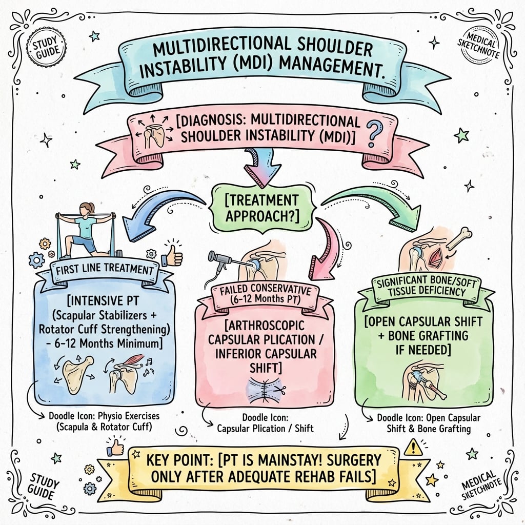
- 1Initial Assessment
Clinical diagnosis with positive sulcus sign, multidirectional laxity, atraumatic history. Classify per Stanmore triangle.
Type I/II proceed to rehab; Type III needs psychiatric evaluation
- 2Structured Rehabilitation
Burkhead-Rockwood protocol: rotator cuff and scapular strengthening, proprioception training for 6-12 months
80-90% success rate with full compliance
- 3Surgical Candidacy
Assess after failed 6-month rehabilitation trial with documented compliance and realistic expectations
Type I/II structural MDI only; exclude voluntary dislocators
- 4Examination Under Anesthesia
Quantify true laxity without muscle guarding in all directions
Grade 2-3 confirms structural laxity; Grade 1 suggests muscle patterning
- 5Technique Selection
Arthroscopic for low-demand Grade 2; Open capsular shift for high-demand or Grade 3
Arthroscopic 70-85% success; Open 85-90% success
- 6Postoperative Protocol
6 weeks strict immobilization, progressive ROM weeks 6-16, strengthening from week 16
Return to sport 6-9 months minimum
Conservative Management Details
The Burkhead-Rockwood rehabilitation protocol is the gold standard for MDI conservative treatment, with 80-90% success rate when properly executed with patient compliance.
Phase 1 (Weeks 0-6): Rotator cuff strengthening
- Internal rotation strengthening (subscapularis)
- External rotation strengthening (infraspinatus, teres minor)
- Deltoid strengthening (anterior, middle, posterior)
- Avoid provocative positions during strengthening
- Low resistance, high repetition protocol
Phase 2 (Weeks 6-12): Scapular stabilization
- Serratus anterior strengthening (wall slides, protraction exercises)
- Rhomboid and middle trapezius (rows, scapular retraction)
- Lower trapezius strengthening (prone Y exercises)
- Scapular control exercises with shoulder motion
- Integration of scapular and rotator cuff activation
Phase 3 (Weeks 12-24): Proprioception and neuromuscular control
- Closed chain exercises (wall push-ups, quadruped exercises)
- Proprioceptive training (unstable surfaces, perturbation)
- Plyometric exercises progression
- Dynamic stabilization drills
- Sport-specific movement patterns
Phase 4 (Weeks 24+): Return to activity
- Gradual return to overhead activities
- Sport-specific training and conditioning
- Maintenance program development
- Ongoing strengthening and proprioception
- Lifelong compliance required
The critical success factors for rehabilitation in MDI are: (1) Minimum 6 months duration before declaring failure, (2) Proper progression through all four phases without skipping steps, (3) Patient compliance with home exercise program, (4) Avoidance of provocative positions during strengthening phase, and (5) Lifelong maintenance program after return to activity. Rehabilitation "failure" is often inadequate trial rather than true biological failure.
The Watson MDI Programme (Motor-Control Approach)
The Burkhead-Rockwood programme is the classic strengthening protocol, but the highest-level evidence in MDI (the Warby/Watson randomised controlled trial in the Evidence Base) showed that a motor-control and scapular-focused programme - the Watson MDI programme - was superior to the Rockwood strengthening programme on validated instability scores (WOSI, Melbourne Instability Shoulder Score) and pain at 24 weeks. Knowing what this programme actually involves is high-yield.
MDI is driven not only by capsular redundancy but by altered scapular and humeral-head neuromuscular control, so the programme retrains control and positioning first, before loading - in contrast to the strengthening-first Rockwood approach.
- Stage 1 - Scapular control: restore correct resting scapular position and dynamic scapular setting (correcting downward rotation, anterior tilt and protraction) as the stable base for the glenohumeral joint
- Stage 2 - Humeral-head control: retrain the deltoid and rotator cuff to centre the humeral head, progressing through increasing range and functional positions while maintaining scapular control
- Stage 3 - Progressive loading and speed: add resistance, then functional and sport-specific patterns once control is established
- Stage 4 - Return to sport and maintenance: sport-specific progression and a lifelong maintenance programme
it reframes MDI rehabilitation from "strengthen the cuff" to "restore neuromuscular control of the scapula and humeral head," and it is the programme with Level-II RCT support. Both programmes still require a prolonged (minimum 6-month) compliant trial before surgery is considered. (Scapular dyskinesis itself is developed in the scapular-dyskinesis topic.)
Managing Muscle-Patterning (Stanmore Type III) Instability
Stanmore Type III (atraumatic, non-structural) instability is repeatedly flagged as a surgical contraindication, but its management deserves explicit treatment, because operating on it fails whereas the right non-operative pathway can succeed.
- Positional non-structural: involuntary subluxation occurring only in certain arm positions, driven by abnormal muscle recruitment (inappropriate activation of pectoralis major or latissimus, or inhibition of the cuff) rather than capsular deficiency
- Habitual / willful (voluntary): the patient can demonstrate the instability on demand, and there may be secondary gain or psychological factors
- The unifying feature is normal capsular volume (Grade 0-1) on examination under anaesthesia despite symptomatic instability when awake, with an abnormal muscle-recruitment (EMG) pattern
- EMG biofeedback-based neuromuscular retraining (the Stanmore biofeedback approach) is the cornerstone: surface EMG shows the patient their abnormal recruitment, then normal firing sequences are retrained and the aberrant muscle activation that produces the subluxation is suppressed
- Address psychological factors and secondary gain: screen for and manage psychiatric comorbidity, with clinical-psychology input where there is a willful component
- No stabilisation surgery: capsular shift will not correct a control problem and tends to fail; surgery is reserved only for a genuinely coexisting structural lesion
- EUA is the gatekeeper: if laxity is normal under anaesthesia, abandon any planned stabilisation and commit to the retraining pathway
motivated patients with positional muscle patterning often improve substantially with biofeedback retraining; outcomes are poorer where there is entrenched willful behaviour or unaddressed secondary gain.
This completes the conservative management protocol.
Surgical Technique
Arthroscopic capsular plication has become increasingly popular for MDI treatment, offering reduced morbidity compared to open techniques. However, outcomes are inferior to open inferior capsular shift, particularly in severe MDI.
Indications
- Young patients with lower functional demands
- Grade 2 laxity (Grade 3 better suited to open)
- Failed appropriate rehabilitation trial
- Stanmore Type II (atraumatic structural)
- Realistic expectations about success rates
- Grade 3 laxity in multiple directions
- High-level athletes requiring return to contact sport
- Revision surgery after failed previous stabilization
- Generalized hypermobility with Beighton greater than 6
- Significant voluntary component
Patient Positioning
30-45 degrees upright, head secured in neutral position. Affected arm free to move through full ROM. Table articulation allows position changes during procedure.
Alternative positioning with arm in traction (10 pounds). Provides better visualization of inferior capsule but less physiologic for assessment.
Portal Placement
Standard viewing portal, 2cm inferior and 1cm medial to posterolateral acromion. Establishes first for orientation.
Just anterior to biceps tendon at rotator interval. Working portal for superior and rotator interval plication.
Through rotator interval or just superior to subscapularis. Working portal for anterior capsular plication.
5:30 position (right shoulder) at inferior glenoid margin. Allows access to inferior capsular pouch.
Posterolateral, just lateral to standard posterior portal. Allows better access to posterior capsule for plication.
Arthroscopic Assessment
Systematic evaluation:
- Drive-through sign: excessive capsular laxity allows scope to pass from posterior to anterior without resistance
- Inferior pouch redundancy: visualize from anterior portal, note excessive volume
- Rotator interval patulous: widened space between supraspinatus and subscapularis
- Labral appearance: typically attenuated or absent, not torn
- Cartilage evaluation: exclude chondral injury
Capsular Plication Technique
- Anterior inferior portal for suture passage
- Multiple horizontal mattress sutures (PDS or FiberWire)
- Plicate inferior capsule in anterior-to-posterior direction
- 3-5 sutures typically required
- Reduce inferior pouch volume significantly
- Avoid over-tensioning (loss of motion)
- Wilmington portal for suture management
- Plicate posterior capsule from inferior to superior
- Address posterior laxity component
- 2-3 sutures typically sufficient
- Check external rotation ROM after plication
- MANDATORY if sulcus persists in external rotation on EUA
- Close interval between supraspinatus and subscapularis
- Suture coracohumeral ligament to superior glenohumeral ligament
- Obliterate the interval space
- Eliminates inferior translation in external rotation
Key Technical Points
Use suture shuttling techniques to pass sutures through capsule. Multiple devices available (suture lasso, penetrator, bird beak). Ensure adequate tissue purchase with each pass.
Plicate until drive-through sign eliminated but preserve ROM. Check ROM intraoperatively after each plication. Over-tensioning causes stiffness.
Identify axillary nerve inferiorly (risk with inferior plication). Stay anterior to mid-glenoid line. Visualize all sutures before tying.
Arthroscopic knot tying or knotless anchor techniques. Ensure knots well-seated and secure. Typical knots: SMC, Duncan loop.
The axillary nerve is at risk during inferior capsular plication. It courses along the inferior capsule approximately 1-2cm from the inferior glenoid margin. Stay superior to the 6 o'clock position on the glenoid face. Never pass sutures blindly in the inferior capsule. Direct visualization essential to avoid nerve injury.
Intraoperative Assessment
After plication, assess ROM before closing. Should maintain 140 degrees forward flexion, 40 degrees external rotation at side, and 60 degrees external rotation at 90 degrees abduction. Restricted motion requires suture release.
Load-and-shift should demonstrate Grade 0-1 translation. Complete elimination of laxity not goal (causes stiffness). Goal is Grade 0-1 with preserved motion.
Should be eliminated or significantly reduced. Inability to pass scope from posterior to anterior indicates adequate volume reduction.
Outcomes
Success rates for arthroscopic capsulorrhaphy in MDI range from 70-85%, lower than the 85-90% for open inferior capsular shift. Recurrence rates higher in patients with severe laxity, generalized hypermobility, and high-demand activities.
Patient satisfaction correlates with appropriate expectations and understanding that some residual laxity may persist. Return to sport rates approximately 80% at pre-injury level, achieved at 6-9 months postoperatively.
FAILEDIndications for Surgical Intervention in MDI
Hook:Remember patient has FAILED when ready for surgery: proper rehabilitation Failed, patient shows Adequate compliance, has Inability to function, Lifestyle is compromised, Examination confirms structural pathology, and there is Documented objective instability on examination.
Complications
Stiffness is the most common complication after MDI surgery, occurring in 10-20% of patients. The risk is inherent when tightening a lax capsule - balancing stability and motion preservation is challenging.
Risk Factors
- Tendency toward stiffness (previous adhesive capsulitis)
- Diabetes mellitus (higher stiffness risk)
- Female gender (higher baseline stiffness rates)
- Age over 40 years
- Underlying connective tissue disorder
- Over-tensioning capsular shift
- Aggressive rotator interval closure
- Combined anterior and posterior procedures
- Thermal capsulorrhaphy (historical)
- Inadequate intraoperative ROM assessment
- Prolonged immobilization (greater than 6 weeks)
- Non-compliance with rehabilitation
- Delayed initiation of ROM exercises
- Pain limiting rehabilitation participation
Clinical Presentation
Patients report restricted shoulder motion affecting activities of daily living. Forward flexion limited (less than 120 degrees), external rotation limited (less than 30 degrees), internal rotation limited (unable to reach back).
Pain with end-range motion is common. Patients describe feeling "tight" and "restricted." Night pain may occur due to capsular inflammation. Quality of life significantly impacted.
Prevention Strategies
After capsular shift, assess ROM before final closure. Release sutures if motion inadequate (less than 140 degrees elevation, less than 40 degrees ER).
Goal is reducing laxity to Grade 0-1, not complete elimination. Some laxity preservation necessary for motion. Avoid over-tensioning.
Modern protocols use 4-6 weeks immobilization rather than historical 8 weeks. Balance healing with motion preservation.
Initiate gentle passive ROM at 6 weeks. Avoid delay beyond 6 weeks as stiffness risk increases significantly.
Counsel regarding stiffness risk and importance of rehabilitation compliance. Prepare patients for potentially slow ROM recovery.
Treatment
- Intensive physiotherapy with passive stretching
- Heat application before stretching
- Joint mobilization techniques
- NSAIDs for pain and inflammation
- Home exercise program compliance
- Duration: 3-6 months minimum trial
- Indicated if conservative fails after 3-6 months
- Performed at 4-6 months postoperatively (allow healing)
- Gentle manipulation to restore ROM
- Risk of capsular re-injury and recurrent instability
- Followed by intensive physiotherapy
- Reserved for refractory cases failing manipulation
- Selective release of contracted capsule and adhesions
- Risk of destabilizing previous stabilization
- Requires experienced surgeon
- Success rate 70-80% for ROM improvement
The key to preventing stiffness after MDI surgery is intraoperative ROM assessment. Before final closure, test ROM in all planes. Target minimum ROM: 140 degrees forward flexion, 40 degrees ER at side, 60 degrees ER at 90 degrees abduction. If these minimums not achieved, release capsular sutures until adequate motion restored. Accepting some residual laxity preferable to creating stiffness.
Stiffness remains a challenging complication requiring prolonged rehabilitation and potentially additional intervention to restore function.
Rehabilitation and Postoperative Care
Postoperative Protocol
MDI surgery requires prolonged protection compared to traumatic instability repair due to need for capsular healing in tensioned position. Premature mobilization risks capsular stretch-out and recurrent instability.
Phase 1: Protection (Weeks 0-6)
Shoulder immobilized in sling with arm at side in neutral rotation. Remove for hygiene only. Sleep in sling. Strict compliance essential.
Protect capsular repair, allow initial healing, prevent capsular stretch, minimize pain and inflammation.
- Hand, wrist, elbow ROM exercises
- Grip strengthening
- Pendulum exercises (controversial, some surgeons avoid)
- Scapular isometrics without shoulder motion
- Active shoulder ROM
- Passive shoulder ROM
- Resisted shoulder exercises
- Lifting objects
- Reaching or overhead activities
- Sleeping on operative side
Oral analgesics, ice application, NSAIDs (after first 6 weeks to avoid healing impairment).
Phase 2: Early Motion (Weeks 6-12)
Discontinue sling at 6 weeks. Gradual weaning if stiffness concern.
Restore passive ROM, begin active-assisted ROM, protect capsular repair integrity, avoid stretching repaired capsule.
- Forward flexion to 90 degrees (weeks 6-8), progress to 120 degrees (weeks 8-10), then 140 degrees (weeks 10-12)
- External rotation in scapular plane to 20 degrees (weeks 6-8), progress to 30 degrees (weeks 8-10), then 40 degrees (weeks 10-12)
- Internal rotation to neutral (weeks 6-8), progress to 40 degrees (weeks 8-12)
- Therapist-assisted stretching, avoid patient forcing motion
Begin at week 8 with table slides, wand exercises, pulley-assisted elevation. Patient assists with motion but does not force.
- Resisted strengthening
- Aggressive stretching
- Overhead reaching
- Lifting greater than 5 pounds
- Return to sport activities
Phase 3: Strengthening (Weeks 12-24)
Restore full ROM, progressive strengthening, improve neuromuscular control, prepare for functional activities.
Achieve full forward flexion (160-180 degrees), external rotation 50-60 degrees at side, internal rotation to T8-T10 level. Gentle stretching if plateau.
- Weeks 12-16: Isometric rotator cuff, light resistance band (0.5-1 pound)
- Weeks 16-20: Progressive resistance strengthening, increase to 2-3 pounds
- Weeks 20-24: Functional strengthening patterns, sport-specific preparation
Rows, scapular retraction, serratus anterior strengthening (wall slides, protraction), trapezius strengthening (shrugs, Y-T-W exercises).
Closed chain exercises, rhythmic stabilization, perturbation training, unstable surface exercises.
Pain-free ROM, adequate strength (4/5 or better on manual muscle testing), no signs of instability, patient compliance demonstrated.
Phase 4: Return to Activity (Months 6-9)
Return to full activities including sport, maintain strength and ROM, establish maintenance program.
Progressive return to overhead activities. Interval throwing program for throwing athletes. Swimming progression for swimmers. Contact sport progression with protective padding.
Assess before clearing for full return:
- Full pain-free ROM
- Strength 85% of contralateral
- Negative instability examination
- Sport-specific functional tests passed
- Psychological readiness
Lifelong rotator cuff and scapular strengthening essential. MDI patients have inherent capsular laxity requiring ongoing dynamic stabilizer maintenance.
- Non-contact sports: 6 months minimum
- Contact sports: 9 months minimum
- Overhead sports (baseball, volleyball): 9-12 months
- Swimming: 6-9 months with gradual progression
The most common cause of failure after MDI surgery is premature progression through rehabilitation phases. The capsule requires 12-16 weeks to achieve adequate tensile strength after surgical shift. Pushing ROM or starting strengthening too early risks capsular stretch-out and recurrent instability. Patience during rehabilitation is as important as surgical technique for successful outcomes.
Rehabilitation After Arthroscopic vs Open
May allow slightly faster progression due to less tissue disruption and subscapularis preservation. Some protocols advance to active ROM at week 4-6 rather than 6-8.
Requires protection of subscapularis repair. More conservative ROM progression. Emphasize external rotation limits to protect subscapularis tendon healing.
Red Flags During Rehabilitation
Patient reports looseness, subluxation sensation. Stop progression, reassess with surgeon, consider examination under anesthesia.
Failure to regain ROM despite appropriate therapy. Consider more aggressive therapy, possible manipulation if no progress by month 4-6.
Pain should gradually improve. Persistent or worsening pain suggests complication (infection, chondrolysis, nerve injury). Requires surgical reassessment.
After open technique, weakness of internal rotation, positive belly-press test. MRI to assess tendon integrity, may require revision repair.
Close communication between surgeon, therapist, and patient throughout rehabilitation maximizes outcomes and identifies complications early.
Outcomes and Prognosis
Conservative Treatment Outcomes
Structured rehabilitation following the Burkhead-Rockwood protocol achieves 80-90% success rates in appropriately selected MDI patients who are compliant with the program. Success is defined as return to activities without functional limitation from instability.
- Patient age under 25 years (younger patients respond better)
- Excellent compliance with therapy program
- Lower Beighton score (less than 4, localized laxity)
- Unilateral involvement (better than bilateral)
- Stanmore Type II (atraumatic structural)
- Willingness to modify provocative activities
- Poor compliance with exercise program
- Generalized hypermobility (Beighton greater than 6)
- Bilateral severe involvement
- Grade III sulcus sign with severe inferior laxity
- Stanmore Type III (muscle patterning, needs different approach)
- Unrealistic expectations about activity return
The key is adequate trial duration - minimum 6 months with excellent compliance required before declaring rehabilitation failure. Many apparent "failures" are actually inadequate trials with poor compliance.
Surgical Outcomes
Surgical success rates vary significantly based on technique, patient selection, and severity of pathology.
- Success rate: 85-90% good to excellent outcomes
- Recurrent instability: 10-15%
- Return to sport: 80-85% at previous level
- Time to return: 6-9 months minimum
- Patient satisfaction: 85-90%
- Success rate: 70-85% good to excellent outcomes
- Recurrent instability: 15-25%
- Return to sport: 75-80% at previous level
- Time to return: 6-9 months minimum
- Patient satisfaction: 75-85%
- Appropriate patient selection (failed adequate conservative trial)
- Stanmore Type I or II (structural pathology)
- Lower Beighton score (less than 4)
- Compliant patient willing to follow postoperative restrictions
- Realistic expectations about outcomes and activity modification
- Experienced surgeon with MDI surgical expertise
- Severe generalized hypermobility (Beighton greater than 7)
- Stanmore Type III (muscle patterning) - should not operate
- Voluntary component or secondary gain
- Premature return to provocative activities
- Inadequate capsular shift or technical errors
- Missed rotator interval pathology
The critical determinant of outcome in MDI is patient selection. Success rates quoted above apply to properly selected patients - Stanmore Type I or II, failed adequate conservative trial, realistic expectations, compliant with restrictions. Operating on poorly selected patients (Stanmore Type III, inadequate rehabilitation trial, unrealistic expectations) leads to failure regardless of surgical technique quality.
Long-term Outcomes
Long-term studies (5-10 years) after MDI surgery show some deterioration compared to early results. Recurrence rates increase over time as capsular tissue gradually stretches, particularly in hypermobile patients.
- 80-85% maintain good to excellent results
- 15-20% develop recurrent symptoms (versus 10-15% at 2 years)
- Most recurrences are mild and manageable conservatively
- Patient satisfaction remains high (80-85%)
- Stiffness resolves in most patients by 5 years
- 75-80% maintain satisfactory results
- 20-25% have some recurrent laxity
- Many patients adapt and remain functional despite some laxity
- Revision surgery rates low (less than 10%)
- Arthritis rates similar to general population
Functional Outcomes
Return to activity is a primary goal for most MDI patients, particularly young athletes. Realistic expectations are critical.
- 75-80% return to pre-injury level after surgery
- 15-20% return at lower level (recreational versus competitive)
- 5-10% unable to return due to recurrent symptoms or fear
- Average time to return: 9-12 months
- Permanent activity modification often required
- 70-75% return to competitive level
- Higher risk of recurrent injury with contact
- May require protective equipment or bracing
- Average time to return: 9-12 months
- Some positions may be unsuitable (offensive line, etc.)
- 90-95% return to full work duties
- Overhead occupations may require modification
- Heavy manual labor often requires permanent restrictions
- Average time to full duty: 4-6 months
- Occupational therapy may assist with work modifications
Quality of Life Outcomes
Patient-reported outcome measures show significant improvement after both conservative and surgical treatment of MDI, though some residual symptoms common.
- Baseline MDI: average 40-50% of maximum score
- Post-rehabilitation: improvement to 75-85%
- Post-surgery: improvement to 80-90%
- Plateau occurs at 12-24 months
- Baseline MDI: average 50-60 points
- Post-treatment: improvement to 80-90 points
- Correlates with return to activities and satisfaction
Prognostic Factors Summary
- Localized MDI without generalized hypermobility
- Excellent rehabilitation compliance
- Realistic expectations and activity modification acceptance
- Stanmore Type II, younger age
- Open surgical technique if surgery required
- Mild generalized hypermobility (Beighton 4-6)
- Good compliance with structured treatment
- Unilateral involvement
- Willingness to modify high-risk activities
- Severe generalized hypermobility (Beighton greater than 6)
- Bilateral involvement
- High-demand overhead athlete
- Revision surgery scenario
- Arthroscopic technique in severe MDI
- Stanmore Type III (muscle patterning) - surgery should not be performed
- Voluntary dislocation with secondary gain
- Unrealistic expectations refusing activity modification
- Connective tissue disorder (Ehlers-Danlos, Marfan)
- Multiple previous failed surgeries
Understanding these prognostic factors allows appropriate patient counseling and shared decision-making regarding treatment approach and expectations.
Guidelines, Registries & Global Practice
Global Epidemiology
According to PubMed, population-level data come predominantly from all-cause (mostly traumatic) glenohumeral dislocation cohorts, which frame the young, active demographic in whom instability - including atraumatic and multidirectional patterns - concentrates. MDI itself remains a clinical diagnosis without a dedicated registry denominator.
Population-Level Glenohumeral Instability Epidemiology
Guideline & Society Positions
No single high-level clinical practice guideline is dedicated to MDI; guidance is drawn from instability-focused society statements and the highest-quality primary evidence. The consistent global position is rehabilitation-first, with surgery reserved for structural MDI that fails an adequate supervised programme.
Side-by-Side Guidance on MDI Management
Registry Evidence
No national arthroplasty or instability registry (AOANJRR, NJR, AJRR) captures soft-tissue MDI stabilisation as a discrete procedure, so registry-grade survivorship data are not available for capsular shift or arthroscopic capsulorrhaphy. The best population data remain emergency-department surveillance for dislocation incidence (above). This absence of registry tracking is itself an exam-relevant point: MDI outcomes are reported from institutional case series and a small number of trials rather than registries.
Practice Variation
- Rehabilitation programme selection varies internationally. The classical Rockwood/Burkhead strengthening protocol is widely taught, but Level I/II evidence favours a motor-control and scapular-focused programme (Watson), and uptake differs between centres.
- Surgical technique preference is centre-dependent. Open inferior capsular shift (Neer) is favoured for severe Grade III laxity and high-demand athletes, while arthroscopic capsular plication is increasingly used for moderate structural MDI; both are acceptable in appropriately selected patients.
- Access and timing differ by health system. Public systems may have prolonged waits for elective stabilisation, favouring extended supervised rehabilitation; this aligns with the rehabilitation-first evidence and rarely disadvantages structurally appropriate candidates.
- Sporting context shapes presentation. Overhead and aquatic sports (swimming), gymnastics and throwing populations generate a higher proportion of atraumatic MDI presentations worldwide.
MCQ Practice Points
High-Yield Multiple Choice Concepts
- Requires symptomatic instability in TWO or more directions (anterior, posterior, inferior)
- Positive sulcus sign Grade II or III is pathognomonic
- Atraumatic or minimal trauma history distinguishes from TUBS
- Bilateral involvement in 50-80% of cases
- Beighton score ≥4 indicates generalized hypermobility
- AMBRII: Atraumatic, Multidirectional, Bilateral, Rehabilitation, Inferior capsular shift, Interval closure
- TUBS: Traumatic, Unidirectional, Bankart lesion, Surgery
- Classic vignette: young female swimmer with bilateral shoulder looseness = AMBRII
- Classic vignette: rugby player with anterior shoulder dislocation = TUBS
Q: How do you distinguish AMBRII from TUBS in an exam vignette?
A: AMBRII describes MDI pathway - Atraumatic and Multidirectional instability, often Bilateral, treat with Rehabilitation first, then Inferior capsular shift with rotator Interval closure if conservative fails. TUBS describes traumatic anterior instability - Traumatic onset, Unidirectional, Bankart lesion present, requires Surgery. Classic AMBRII vignette: young female swimmer with bilateral shoulder looseness. Classic TUBS vignette: rugby player with anterior dislocation.
- Pathognomonic examination finding for MDI
- Grade I less than 1cm, Grade II 1-2cm, Grade III greater than 2cm
- MUST test in neutral rotation AND external rotation
- Persistent sulcus in external rotation indicates rotator interval incompetence
- Always compare to contralateral shoulder
- Type I: Traumatic, Structural (specific injury causing capsular damage)
- Type II: Atraumatic, Structural (constitutional laxity, classic MDI)
- Type III: Atraumatic, Non-Structural, Muscle Patterning (voluntary, psychological)
- Type III should NOT undergo surgery - will fail
- Identifying Stanmore type critical for treatment decisions
Q: What is the critical examination finding that distinguishes Stanmore Type III from Type I/II MDI?
A: Stanmore Type III patients demonstrate normal capsular laxity (Grade 0-1) on examination under anesthesia despite appearing lax when awake due to muscle patterning or voluntary control. This contrasts with Type I/II who have Grade 2-3 structural laxity under EUA. Surgery is contraindicated in Type III because there is no structural pathology to correct - always perform EUA before MDI surgery and abort if laxity is normal under anesthesia.
- Rehabilitation is ALWAYS first-line for MDI (not surgery)
- Burkhead-Rockwood protocol: 6-12 months structured physiotherapy
- Success rate 80-90% with proper compliance
- Four phases: rotator cuff strengthening, scapular stabilization, proprioception, sport-specific
- Minimum 6 months trial required before declaring failure
- Failed minimum 6 months structured rehabilitation
- Documented compliance with therapy program
- Persistent symptoms affecting function
- Stanmore Type I or II (structural pathology)
- Objective instability on examination (Grade 2-3 laxity)
- Realistic expectations about outcomes
- Open inferior capsular shift: gold standard, 85-90% success rate
- Arthroscopic capsulorrhaphy: 70-85% success rate, less invasive
- Thermal capsulorrhaphy: ABANDONED due to failure and chondrolysis
- Rotator interval closure: required if sulcus positive in external rotation
- T-capsulorrhaphy (Neer technique) is classic open approach
Q: Why is thermal capsulorrhaphy never the correct answer for MDI surgical treatment questions?
A: Thermal capsulorrhaphy was completely abandoned in the early 2000s due to unacceptably high failure rates (50-70%) and devastating complications including glenohumeral chondrolysis. Despite appearing in older literature, this technique should never be selected in modern exam questions. If it appears as an option, it is always wrong regardless of the clinical scenario presented.
- Performed before surgery to confirm structural laxity
- Eliminates muscle guarding and voluntary control
- Grade 0-1 laxity on EUA despite clinical symptoms = Stanmore Type III
- If EUA normal laxity, abandon surgery (muscle patterning, not structural)
- EUA guides surgical technique and extent of capsular shift
- Stiffness: most common (10-20%), prevent with ROM assessment intraoperatively
- Recurrent instability: 10-25% depending on technique and patient factors
- Axillary nerve injury: risk during inferior capsular work, stay above 6 o'clock
- Chondrolysis: devastating complication of thermal capsulorrhaphy (now abandoned)
- Subscapularis failure: after open technique, causes anterior instability
- 6 weeks strict immobilization (longer than TUBS repair)
- Passive ROM weeks 6-12, active ROM weeks 12-16
- Strengthening not before 12 weeks
- Return to sport minimum 6-9 months
- Premature progression leads to capsular stretch and recurrence
- Beighton score ≥6 predicts higher failure rate
- Bilateral involvement common but doesn't predict failure
- Generalized connective tissue disorder (Ehlers-Danlos) poor prognosis
- High-level overhead athletes have lower return to sport rate
- Stanmore Type III should not undergo surgery
- MDI does NOT require trauma history (atraumatic by definition)
- Positive apprehension test suggests traumatic instability, NOT typical for MDI
- Normal radiographs and MRI do not exclude MDI (clinical diagnosis)
- Labral tears uncommon in pure MDI (attenuated labrum, not torn)
- Thermal capsulorrhaphy is NEVER the answer (completely abandoned)
Q: What does a positive anterior apprehension test indicate in a patient with multidirectional laxity?
A: MDI patients typically have NEGATIVE apprehension test despite significant laxity because they lack the traumatic mechanism creating the fear response. A positive apprehension in a patient with multidirectional laxity suggests mixed pathology - traumatic anterior injury superimposed on underlying constitutional laxity - rather than pure MDI. This changes management as the Bankart lesion may need addressing along with capsular redundancy.
Evidence-based answers:
- Burkhead-Rockwood study: 80% success with rehabilitation
- Neer inferior capsular shift: 90% success rate, gold standard open technique
- Gartsman study: arthroscopic 76% success versus open 89% success
- Thermal capsulorrhaphy: abandoned due to 50-70% failure and chondrolysis risk
- Stanmore classification: Type III surgery contraindicated
Q: What is the minimum duration of structured rehabilitation required before considering surgical treatment for MDI?
A: Six months is the minimum rehabilitation trial required before surgery is considered. The Burkhead-Rockwood protocol demonstrated 80% success rate with structured rehabilitation over 6-12 months. Patients who do not complete adequate physiotherapy should not be offered surgery, as premature surgical intervention has higher failure rates and patients miss the opportunity for successful conservative management.
Q: When should rotator interval closure be performed in addition to capsular shift for MDI?
A: Rotator interval closure should be added when the sulcus sign remains positive with the shoulder in external rotation. Normal anatomy closes the rotator interval when the arm is externally rotated, eliminating inferior translation. A persistent sulcus in external rotation indicates pathological rotator interval laxity that must be addressed surgically for successful outcome. Without interval closure in these patients, inferior instability will persist despite adequate capsular shift.
These high-yield points cover the most commonly tested MDI concepts in Orthopaedic examinations and should be memorized for rapid recall during MCQ sections.
BEIGHTONBeighton Hypermobility Score (9 Points Total)
Hook:Remember the Beighton score tests peripheral joints first (fingers, thumbs - 4 points), then major joints (elbows, knees - 4 points), then trunk (1 point). Score of 4 or more means generalized hypermobility affecting treatment.
Clinical Decision Scenarios
Practise clinical reasoning and management decisions out loud
“A 17-year-old female competitive swimmer presents with bilateral shoulder pain and instability. She reports feeling her shoulders 'slip out' with overhead swimming strokes. No specific injury. On examination, she has a positive sulcus sign Grade III bilaterally, load-and-shift Grade 2 in anterior and posterior directions, and a Beighton score of 7/9. How would you manage this patient?”
“A 24-year-old male patient underwent arthroscopic capsulorrhaphy for MDI 18 months ago. He now returns with recurrent instability symptoms. Examination shows Grade II sulcus sign and Grade 2 anterior and inferior laxity. MRI shows intact capsular plication but some capsular stretch. He is frustrated and wants revision surgery. How would you manage this recurrence?”
“You are asked to assess the surgical video from a colleague who performed arthroscopic capsulorrhaphy for MDI. On the video, you see thermal energy being applied to the capsule in a 'shrinkage' pattern rather than suture plication. The patient is now 18 months post-op with recurrent instability and shoulder pain. What are your thoughts and how would you manage?”
Must-Know Facts
- MDI definition: symptomatic instability in ≥2 directions without significant trauma
- AMBRII mnemonic: Atraumatic, Multidirectional, Bilateral, Rehabilitation, Inferior capsular shift, Interval closure
- Sulcus sign pathognomonic: Grade II (1-2cm) or III (over 2cm). Test in neutral AND external rotation
- Stanmore classification critical: Type I/II (structural) versus Type III (muscle patterning) - surgery contraindicated in Type III
- Beighton score ≥4 indicates generalized hypermobility affecting surgical prognosis
- Rehabilitation FIRST-LINE: 80-90% success with Burkhead-Rockwood protocol (6-12 months)
- Examination under anesthesia mandatory before surgery: Grade 0-1 laxity = abandon surgery (not structural)
- Open inferior capsular shift gold standard: 85-90% success versus arthroscopic 70-85%
- Rotator interval closure required if sulcus persists in external rotation
- Thermal capsulorrhaphy ABANDONED: high failure rates and chondrolysis complication
Clinical Pearls
- Bilateral examination mandatory: 50-80% bilateral involvement even if unilateral symptoms
- Negative anterior apprehension test distinguishes MDI from traumatic instability despite laxity
- Intraoperative ROM assessment prevents stiffness: target minimum 140° elevation, 40° ER at side
- Axillary nerve protection: never pass sutures below 6 o'clock on glenoid face
- EUA findings trump clinical findings: normal laxity under anesthesia = muscle patterning, not structural
- Premature rehabilitation progression causes failure: capsule needs 12-16 weeks tensile strength
- Patient selection determines outcome: proper selection more important than surgical technique perfection
- Recurrence often inadequate trial: reassess compliance before declaring rehabilitation failure
Common Pitfalls
- Operating on Stanmore Type III (voluntary, muscle patterning): will fail, may worsen psychological issues
- Inadequate conservative trial (under 6 months): premature surgery when rehabilitation might have succeeded
- Missing rotator interval pathology: persistent sulcus in ER requires interval closure, not just capsular shift
- Over-tensioning capsular shift: causes stiffness worse than residual laxity. Accept Grade 0-1 final laxity
- Thermal capsulorrhaphy: never the answer, completely abandoned technique
- Bilateral simultaneous surgery: high complication rate, address more symptomatic side first
- Early return to sport: minimum 6-9 months required, premature return causes capsular stretch-out
- Ignoring generalized hypermobility: Beighton ≥6 predicts surgical failure, counsel appropriately
Viva Questions
- What is the definition of MDI and how does it differ from unidirectional instability?
- Describe the AMBRII criteria and their clinical significance
- How do you perform and grade the sulcus sign? Why test in external rotation?
- Explain the Stanmore Triangle classification and treatment implications
- What is the Burkhead-Rockwood rehabilitation protocol for MDI?
- What are the indications for surgical intervention in MDI after failed conservative management?
- Compare arthroscopic capsulorrhaphy versus open inferior capsular shift: indications and outcomes
- Describe the inferior capsular shift surgical technique (T-capsulorrhaphy)
- What is rotator interval closure and when is it indicated?
- Why was thermal capsulorrhaphy abandoned? What complications occurred?
- What is the role of examination under anesthesia before MDI surgery?
- How would you manage recurrent instability after previous MDI surgery?
- What factors predict poor outcomes after MDI surgery?
- Describe the postoperative rehabilitation protocol timeline after capsular shift
- How does the Beighton hypermobility score influence MDI treatment and prognosis?
Evidence Base
According to PubMed, the evidence cards below have been verified against the indexed primary sources; key findings reflect what each paper's abstract actually reports.
Burkhead-Rockwood Exercise Programme for Shoulder Instability
- 140 shoulders (115 patients) with anterior, posterior or multidirectional subluxation treated with a structured muscle-strengthening programme
- Good or excellent result in 53 of 66 atraumatic shoulders (80%)
- Good or excellent result in only 12 of 74 traumatic shoulders (16%)
- Atraumatic (MDI-type) instability responded far better to exercise than traumatic instability
- Authors emphasised careful history, examination and radiographs to identify the aetiology before predicting success
Neer Inferior Capsular Shift (Original Description)
- Preliminary report of 36 patients (40 shoulders) with involuntary inferior and multidirectional subluxation/dislocation
- All shoulders treated with an inferior capsular shift - a capsular flap reinforced by overlying tendon to reduce redundancy on all three sides through one incision
- Only one shoulder began to sublux again (within 7 months); no other unsatisfactory results reported at the time
- 17 shoulders followed for more than two years
- Authors stressed meticulous psychiatric appraisal, conservative treatment and repeated examination before operating
Arthroscopic Capsulorrhaphy for Multidirectional Instability
- Prospective case series of 47 patients (26 men, 21 women; mean age 30 years) with MDI confirmed clinically and at arthroscopy
- Good to excellent (Rowe) outcome rose from 0% preoperatively to 94% (44 of 47) at mean 35 months
- ASES improved from 45.4 to 94.7 and Rowe from 14.2 to 93.7 (both P=0.001)
- One patient failed with persistent instability and required a second operation
- 22 of 26 patients (85%) returned to their desired level of sport
Chondrolysis After Thermal Capsulorrhaphy
- Case report of two young patients who developed glenohumeral chondrolysis after arthroscopic thermal capsulorrhaphy for instability
- Both progressed to disabling pain, stiffness and rapid cartilage loss in a previously preserved joint
- Implicated thermal energy as a direct cause of cartilage death
- Contributed to the wider recognition of chondrolysis as a catastrophic complication of thermal techniques
- Helped drive the abandonment of thermal capsulorrhaphy in favour of suture plication
Watson MDI Programme vs Rockwood Programme (Randomised Controlled Trial)
- Randomised controlled trial (Level of evidence 2) of 41 patients with nontraumatic, nonstructural MDI
- Compared the Watson MDI programme with the Rockwood Instability programme over 12 weekly physiotherapy sessions
- Watson programme superior on the WOSI at 24 weeks (effect size 12.6; 95% CI 3.4-21.9; P=0.008)
- Watson programme superior on the Melbourne Instability Shoulder Score at 24 weeks (effect size 15.4; 95% CI 5.9-24.8; P=0.002)
- Watson programme also produced greater pain reduction at 24 weeks (P=0.003)
Aetiology, Classification and Non-Operative Management of MDI
- Narrative review updating the aetiology, classification and rehabilitation of MDI
- Frames MDI aetiology as multifactorial (capsulolabral redundancy, abnormal scapular and humeral neuromuscular control), which complicates classification
- Confirms a structured rehabilitation programme as the recommended initial treatment for MDI
- Notes that available rehabilitation programmes carry varying levels of supporting evidence
- Synthesises the authors' single-group studies and randomised controlled trial into an evidence-based programme
Epidemiology of Shoulder Dislocation (US Population)
- National Electronic Injury Surveillance System analysis of 8,940 shoulder dislocations (2002-2006)
- Overall incidence 23.9 per 100,000 person-years (95% CI 20.8-27.0)
- Male incidence 34.9 vs female lower, incidence rate ratio 2.64; 71.8% of dislocations in males
- Peak incidence 47.8 per 100,000 in the 20-29 year age group; 46.8% of dislocations in those aged 15-29
- 48.3% of injuries occurred during sports or recreation