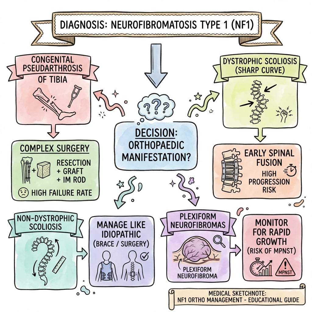
NF1 Orthopaedic Challenges
Orthopaedic Issues
Critical Must-Knows
- Dystrophic Scoliosis: Sharp, angular curve with poor prognosis.
- Tibial Dysplasia: Anterolateral bowing → pseudarthrosis risk.
- Cafe-au-lait Spots: greater than 6 spots greater than 5mm (prepubertal).
- NF1 Gene: Tumor suppressor (neurofibromin).
- Surgery is challenging: High failure rates.
Clinical Pearls
- "Dystrophic vs non-dystrophic scoliosis
- "Anterolateral tibial bowing
- "6 cafe-au-lait spots diagnostic
- "NF1 is a tumor suppressor
Dystrophic Scoliosis
Dystrophic scoliosis in NF1 has a POOR prognosis.
- Features: Short segment, sharp angular curve, vertebral scalloping, rib penciling, dural ectasia.
- Progresses rapidly, even after fusion.
- Early combined (anterior + posterior) fusion may be needed.
- Monitor closely.
Dystrophic vs Non-Dystrophic Scoliosis
| Feature | Dystrophic | Non-Dystrophic |
|---|---|---|
| Short, sharp, angular | Similar to idiopathic | |
| Scalloping, wedging, rotation | Minimal | |
| Poor - rapid progression | Better - like idiopathic | |
| Early combined fusion | Standard scoliosis Rx |
NF1 DNF1 Diagnostic Criteria
| C | Cafe-au-lait ≥6 spots ≥5mm |
| F | Freckling Axillary or inguinal |
| N | Neurofibromas ≥2 or 1 plexiform |
| O | Optic Glioma Tumor |
| L | Lisch Nodules Iris hamartomas |
| B | Bony Dysplasia (sphenoid, tibia) |
| R | Relative First-degree with NF1 |
| C | Cafe-au-lait ≥6 spots ≥5mm | O | Optic Glioma Tumor | R | Relative First-degree with NF1 |
| F | Freckling Axillary or inguinal | L | Lisch Nodules Iris hamartomas | ||
| N | Neurofibromas ≥2 or 1 plexiform | B | Bony Dysplasia (sphenoid, tibia) |
Hook:CFNOLBR - 2 or more criteria.
SSRDDystrophic Scoliosis Features
| S | Sharp Curve Short segment, angular |
| S | Scalloping Vertebral body |
| R | Rib Penciling Thin ribs |
| D | Dural Ectasia Expanded dural sac |
| S | Sharp Curve Short segment, angular | R | Rib Penciling Thin ribs |
| S | Scalloping Vertebral body | D | Dural Ectasia Expanded dural sac |
Hook:SSRD - Sharp, Scalloping, Rib, Dural.
BIVATibial Dysplasia Management
| B | Brace Protect tibia from fracture |
| I | Ilizarov For pseudarthrosis |
| V | Vascularized Fibula Graft option |
| A | Amputation Last resort |
| B | Brace Protect tibia from fracture | V | Vascularized Fibula Graft option |
| I | Ilizarov For pseudarthrosis | A | Amputation Last resort |
Hook:BIVA - Brace, Ilizarov, Vascularized graft, Amputation.
HARDNF1 Surgery Challenges
| H | High failure rate Pseudarthrosis recurs, fusions fail |
| A | Abnormal healing Impaired bone consolidation |
| R | Rapid progression Dystrophic curves progress post-fusion |
| D | Difficult revision Multiple procedures often required |
| H | High failure rate Pseudarthrosis recurs, fusions fail | R | Rapid progression Dystrophic curves progress post-fusion |
| A | Abnormal healing Impaired bone consolidation | D | Difficult revision Multiple procedures often required |
Hook:NF1 surgery is HARD - plan for complications and counsel families accordingly!
Overview/Epidemiology
Neurofibromatosis Type 1 (NF1) is a common genetic disorder.
- Genetics: Autosomal dominant. NF1 gene on chromosome 17 (encodes neurofibromin, a tumor suppressor).
- Incidence: 1 in 3,000.
- 50% new mutations: Often no family history.
- Key Features: Cafe-au-lait spots, neurofibromas, Lisch nodules.
- Malignancy Risk: 8-13% lifetime risk of malignant peripheral nerve sheath tumor (MPNST).
Pathophysiology, Anatomy & Pathomechanics
NF1 Gene Function
- Neurofibromin is a tumor suppressor (Ras-GAP).
- Loss leads to uncontrolled cell proliferation (neurofibromas).
- Affects Schwann cells, melanocytes, bone.
Why Scoliosis in NF1?
- Dystrophic features suggest mesodermal dysplasia.
- Vertebral scalloping from dural ectasia and abnormal bone.
- Non-dystrophic curves likely from neurofibromas affecting paraspinal muscles.
Why Tibial Pseudarthrosis?
- Local mesodermal defect in tibial periosteum/bone.
- Reduced blood supply and poor bone healing.
- Anterolateral bowing progresses to fracture and non-union.
Classification Systems
Crawford Classification (Tibial Dysplasia)
- Type I: Anterolateral bowing with increased cortical density.
- Type II: Anterolateral bowing with sclerotic medullary canal.
- Type III: Anterolateral bowing with cystic lesion.
- Type IV: Anterolateral bowing with frank fracture/pseudarthrosis.
Prognosis: Types III and IV have worst outcomes.
Clinical Assessment
NIH Diagnostic Criteria (≥2):
- ≥6 cafe-au-lait spots (≥5mm prepubertal, ≥15mm postpubertal).
- ≥2 neurofibromas or 1 plexiform neurofibroma.
- Freckling in axillary or inguinal region.
- Optic glioma.
- ≥2 Lisch nodules.
- Bony lesion (sphenoid dysplasia, tibial dysplasia).
- First-degree relative with NF1.
Physical Exam:
- Skin: Cafe-au-lait spots, neurofibromas, freckling.
- Spine: Scoliosis assessment.
- Lower Limbs: Tibial bowing.
- Eyes: Slit lamp for Lisch nodules.
Investigations
Genetic Testing:
- NF1 mutation testing (available but not always needed for clinical diagnosis).
Imaging:
- Spine X-ray: Scoliosis, vertebral changes.
- MRI Spine: Pre-op for scoliosis (dural ectasia, intraspinal neurofibromas).
- Lower Limb X-ray: Tibial bowing.
- MRI Brain: Optic pathway glioma screening.
Ophthalmology:
- Slit lamp for Lisch nodules.
Management Algorithm
Dystrophic Scoliosis
- Bracing: Limited effect.
- Surgery: Early combined (anterior + posterior) fusion for curves greater than 20-25 degrees because of rapid progression.
- Pre-op MRI: Exclude intraspinal pathology (dural ectasia, neurofibromas).
Surgical Techniques
Combined Anterior-Posterior Fusion
Indications: Dystrophic scoliosis with curves greater than 20-25 degrees.
Technique:
- Anterior release and fusion.
- Posterior instrumented fusion.
- Address dural ectasia intra-op.
Challenges: High pseudarthrosis rate, dural ectasia, thin pedicles.
Complications
| Complication | Context | Management |
|---|---|---|
| Pseudarthrosis (Spine) | Post-scoliosis surgery | Combined fusion, revision |
| Pseudarthrosis (Tibia) | Multiple surgeries | Ilizarov, amputation if fails |
| Curve Progression | Despite fusion | Revision, extend fusion |
| MPNST | Plexiform neurofibroma | Oncology, wide excision |
| Dural Tear | Scoliosis surgery | Primary repair |
Postoperative Care
- Scoliosis: Bracing post-op, close follow-up for pseudarthrosis.
- Tibial Surgery: Protected weight-bearing, external fixator care.
- All Patients: Long-term surveillance for malignancy.
Outcomes/Prognosis
- Dystrophic Scoliosis: Challenging. High failure rate even with combined fusion.
- Tibial Pseudarthrosis: Healing is difficult. Multiple surgeries often needed.
- Life Expectancy: Reduced due to malignant transformation (MPNST), other complications.
Evidence Base
- Foundational review describing the spectrum of skeletal NF1 (scoliosis, kyphosis, tibial pseudarthrosis, sphenoid dysplasia)
- Source of the Crawford classification of anterolateral tibial bowing / congenital pseudarthrosis
- Distinguishes dystrophic from non-dystrophic spinal deformity
- 91 NF1 patients: deformities 'modulate', acquiring dystrophic features over time, so the dystrophic/non-dystrophic label is not fixed
- Curves diagnosed under age 7 modulated in 81% vs 25% if diagnosed after age 7
- Three or more penciled ribs (or three or more dystrophic features) predicts near-certain progression (≈85-87%)
Viva Scenarios
Use these scenarios to practise clinical reasoning and management decisions
Dystrophic Scoliosis
"12-year-old with NF1 has a 30-degree thoracic curve with vertebral scalloping and rib penciling on X-ray."
This is **dystrophic scoliosis** (short, sharp curve with vertebral changes). It has a **poor prognosis** and progresses rapidly. I would obtain an **MRI** to exclude intraspinal pathology. Given the dystrophic features, I would recommend **early combined anterior-posterior fusion** rather than waiting. Bracing is unlikely to prevent progression. Counsel the family about high pseudarthrosis rate.
Tibial Bowing
"6-month-old with NF1. X-ray shows anterolateral bowing of the tibia."
This is **tibial dysplasia** associated with NF1. Anterolateral bowing carries a high risk of **fracture and pseudarthrosis**. Management: **Bracing** (clamshell KAFO) to protect the tibia from fracture. If fracture occurs, healing is very difficult. Surgical options include **Ilizarov fixation, vascularized fibula graft**, with/without bone morphogenetic protein. Amputation may be needed for refractory cases.
MPNST Concern
"25-year-old with NF1 presents with rapid growth and pain in a previously stable plexiform neurofibroma on the thigh."
This is concerning for **malignant peripheral nerve sheath tumor (MPNST)**. NF1 patients have an 8-13% lifetime risk. Red flags include rapid growth and new pain. I would obtain **MRI** (heterogeneous enhancement, necrosis) and consider **PET-CT** (increased uptake). **Biopsy** if suspicious. Treatment is **wide surgical excision** with oncology involvement. MPNST is aggressive with a poor prognosis - in NF1 the 5-year survival is only around 20% (Evans et al, population-based cohort), markedly worse than sporadic MPNST.
MCQ Practice Points
Genetics MCQ
Q: What gene is mutated in NF1? A: NF1 gene on chromosome 17 (neurofibromin).
Scoliosis MCQ
Q: What type of scoliosis has a poor prognosis in NF1? A: Dystrophic scoliosis (short, sharp, angular).
Tibial MCQ
Q: What tibial deformity is associated with NF1? A: Anterolateral bowing with risk of pseudarthrosis.
Malignancy Pearl
Q: What malignancy are NF1 patients at risk for? A: MPNST (malignant peripheral nerve sheath tumor) - 8-13% lifetime risk. Arises from plexiform neurofibromas.
Crawford Pearl
Q: What is the Crawford classification for? A: Tibial dysplasia in NF1. Types I-IV based on bowing characteristics and presence of fracture/pseudarthrosis.
Diagnostic Pearl
Q: What eye finding is seen in NF1? A: Lisch nodules (iris hamartomas) - seen on slit lamp exam.
Guidelines, Registries & Global Practice
Global epidemiology
- NF1 affects roughly 1 in 2,500-3,000 births worldwide with no major ethnic or geographic variation — it is one of the most common single-gene disorders.
- About half of cases are de novo mutations, so a negative family history is common.
- Scoliosis is the commonest skeletal manifestation (around 10-30%); congenital pseudarthrosis of the tibia is rare but disproportionately associated with NF1 (NF1 implicated in roughly half of all cases, Hefti EPOS series).
Side-by-side guidance
| Body | Focus | Practical emphasis |
|---|---|---|
| NIH consensus (1988) / revised diagnostic criteria | Clinical diagnosis | Two or more of the seven criteria; the 2021 international revision added the choroidal anomaly and an NF1 pathogenic variant as criteria |
| UK NF1 guideline (Ferner et al, 2007) | Diagnosis + lifelong surveillance | Structured monitoring of spine, optic pathway and MPNST; multidisciplinary clinics |
| SRS / pediatric spine consensus | Dystrophic scoliosis | Pre-operative MRI mandatory; early instrumented fusion for dystrophic curves; growth-friendly constructs in the very young |
| AO / paediatric trauma practice | Tibial dysplasia | Protect with bracing until union attempted; combine biological (vascularized fibula, autograft, BMP) and mechanical (intramedullary rod, Ilizarov) strategies |
Registry and surveillance notes
- There is no single global NF1 implant registry, but national NF registries (e.g. UK regional registers used by Evans et al) provide the population-level MPNST and cancer-risk data.
- Spinal deformity and limb-reconstruction outcomes are tracked through specialist paediatric spine and pseudarthrosis databases rather than arthroplasty registries.
High- vs limited-resource practice variation
- Well-resourced settings: genetic confirmation, MRI surveillance, MEK inhibitors (e.g. selumetinib) for symptomatic inoperable plexiform neurofibromas, and microvascular fibular transfer for tibial pseudarthrosis.
- Limited-resource settings: diagnosis remains clinical (NIH criteria), surveillance is examination-based, and tibial reconstruction relies more on Ilizarov/autograft techniques; refractory cases more often proceed to amputation and prosthetic fitting.
- Universal principles: multidisciplinary care (genetics, neurology, orthopaedics, ophthalmology, dermatology, oncology) and a low threshold for sarcoma-MDT referral when a lesion enlarges or becomes painful.
Controversies & Areas of Uncertainty
- Timing of dystrophic curve surgery: Most agree dystrophic curves need early instrumented fusion, but the threshold (Cobb angle, age, degree of modulation) and whether to perform combined anterior-posterior versus posterior-only with modern pedicle-screw constructs remain debated.
- Combined vs posterior-only fusion: Historically combined fusion was standard for high pseudarthrosis rates; modern segmental instrumentation has led some centres to favour posterior-only, though dural ectasia, thin dystrophic pedicles and bone quality still drive failures.
- Best biology for tibial pseudarthrosis: No technique reliably prevents refracture. Vascularized fibula, Ilizarov, intramedullary rodding, autograft and BMP are all used, often in combination; the optimal sequence is unsettled and recurrence to skeletal maturity is the rule rather than the exception.
- Role of BMP: Bone morphogenetic protein is used off-label as an adjunct in congenital pseudarthrosis, but evidence is low-level and theoretical oncological concerns in a tumour-predisposition syndrome are unresolved.
- MEK inhibitors and bone: Selumetinib transformed management of inoperable plexiform neurofibromas; whether MEK-pathway modulation can aid bone healing in pseudarthrosis is an active research question.
- Surveillance intensity for MPNST: Whole-body MRI and FDG-PET can detect malignant transformation early, but cost, radiation and false positives mean there is no universal consensus on routine imaging surveillance versus symptom-triggered investigation.
NEUROFIBROMATOSIS
Clinical summary
GENETICS
- •NF1 Gene
- •Chromosome 17
- •Autosomal Dominant
- •Tumor suppressor
DIAGNOSIS
- •≥6 cafe-au-lait spots
- •Neurofibromas
- •Lisch nodules
- •Tibial dysplasia
SCOLIOSIS
- •Dystrophic = poor prognosis
- •Sharp angular curve
- •Combined fusion
- •Non-dystrophic = better
TIBIA
- •Anterolateral bowing
- •Pseudarthrosis risk
- •Brace to protect
- •Ilizarov if fractured
DYSTROPHIC FEATURES
- •Vertebral scalloping
- •Rib penciling
- •Dural ectasia
- •Spindling TP
MALIGNANCY
- •MPNST 8-13% risk
- •Arises from plexiform
- •Rapid growth = concern
- •Wide excision
Self-Assessment Quiz
Differential Diagnosis
Cafe-au-lait Spots and Related Conditions:
| Condition | Key Features | Differentiator |
|---|---|---|
| NF1 | Multiple CAL spots, neurofibromas | NIH criteria, Lisch nodules |
| NF2 | Bilateral acoustic neuromas | Different gene (NF2), no CAL spots |
| McCune-Albright | CAL spots, polyostotic FD | Coast-of-Maine borders, precocious puberty |
| Legius Syndrome | CAL spots, freckling | No neurofibromas, SPRED1 mutation |
| Noonan Syndrome | CAL spots | Short stature, cardiac defects |
Key Distinguishing Points:
- NF1 vs NF2: NF1 has CAL spots and peripheral neurofibromas; NF2 has acoustic neuromas
- NF1 vs McCune-Albright: NF1 has smooth-bordered CAL spots; McCune-Albright has "coast of Maine" irregular borders
- NF1 vs Legius: Very similar but Legius lacks neurofibromas (SPRED1 mutation)