Silencing the Scream
- A neuroma is a disorganised ball of axons attempting to regenerate.
- Painful neuromas have upregulated sodium channels.
- Treatment hierarchy: Conservative to SIMPLE Excision to ADVANCED Reconstruction (RPNI/TMR).
- Simply cutting the nerve usually leads to another neuroma.
- “RPNI uses a free muscle graft as a 'decoy' target.
- “TMR transfers the nerve to a nearby motor branch.
- “Neuroma-in-continuity with intact function should NOT be resected.
Overview
What a neuroma is. A non-neoplastic proliferation of Schwann cells and axons at the site of a nerve injury: a frustrated attempt at regeneration in which the axons fail to find a distal target. Neuromas form after every nerve transection, but only a minority become painful, and that pain comes from mechanical irritation and ectopic firing.
This page is the clinical endpoint of the nerve-injury family: when regeneration fails to find a target, a neuroma is the result. The regeneration biology that produces it is on Nerve Injury and Regeneration, and the reconstruction techniques it uses - TMR is itself a nerve transfer, and RPNI a target-muscle interface - connect to Nerve Transfers and Nerve Repairs.
Pathophysiology
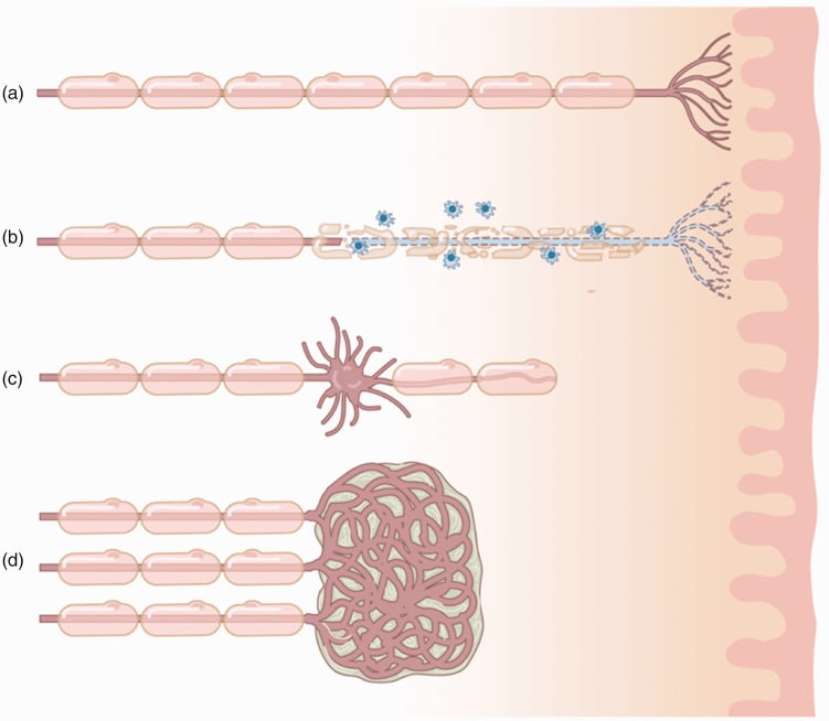
The normal sequence. After axonal disruption the distal segment undergoes Wallerian degeneration: the axon and myelin break down and are cleared by Schwann cells and macrophages over days, leaving the Schwann cells lined up as bands of Büngner to guide regrowth. The proximal stump sends out axonal sprouts (growth cones), and if they enter a distal endoneurial tube they regenerate down it at roughly 1 mm per day (about an inch a month), which is tracked clinically by an advancing Tinel's sign.
Where it goes wrong. A neuroma forms when this regeneration is frustrated. With no distal tube to enter, after a transection or across a gap, the sprouts grow chaotically into scar and produce the disorganised bulb; the connective tissue barrier prevents them advancing, so they turn back on themselves. That is the precise rationale for RPNI and TMR: give the regenerating axons a target so they stop in an organised way rather than forming a neuroma.
Timing. If denervated muscle is not reinnervated within roughly 12 to 18 months the motor end-plates degenerate irreversibly, which is why definitive nerve repair or transfer is not delayed indefinitely.
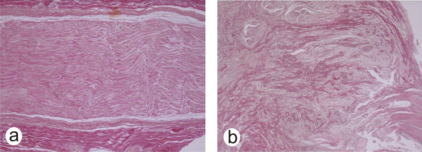
Histology. Three elements make up the bulb: disorganised, entangled axonal sprouts (Zuckerandl's spirals), a stroma of dense fibrosis and scar tissue, and Schwann cells proliferating without guidance.
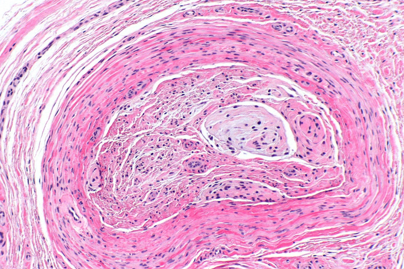
Why it hurts. Three mechanisms, each with a clinical consequence:
- Ectopic pacemakers. Sodium channels (Nav 1.3, 1.7 and 1.8) are upregulated at the nerve tip.
- Mechanosensitivity. The sprout becomes sensitive to pressure, which is what Tinel's sign tests.
- Central sensitisation. Constant bombardment of the dorsal horn leads to chronic pain changes, which is why peripheral blocks sometimes fail in chronic cases (centralisation).
Classification
Terminal neuroma. The end of a cut nerve, the classic "stump" neuroma, produced by unchecked sprout formation. It is the most common type encountered in clinical practice. The amputation neuroma is a terminal neuroma at an amputation stump: often mixed with scar tissue, often involving multiple nerves in the same stump (the sciatic and saphenous, for example), and with a high association with phantom limb pain.
Neuroma-in-continuity. The perineurium and epineurium are intact but the nerve is damaged inside: the axons are disrupted while the tube survives. It often follows a crush injury or a partial laceration, and the challenge is determining whether the axons are conducting through the scar.
- Terminal Neuroma
- Nerve End (Stump)
- Neuroma-in-Continuity
- Along Nerve Course
- Terminal Neuroma
- Zero (Distally)
- Neuroma-in-Continuity
- Variable (May be intact)
- Terminal Neuroma
- Transection
- Neuroma-in-Continuity
- Crush / Stretch / Partial Cut
- Terminal Neuroma
- Excision + Reconstruction
- Neuroma-in-Continuity
- Neurolysis vs Grafting
Seddon. A neuroma is the end result of a particular grade of nerve injury, so examiners expect the Seddon and Sunderland classifications as the substrate. Seddon has three grades. Neurapraxia is a conduction block with the axon intact: full recovery, no Wallerian degeneration and no neuroma. Axonotmesis disrupts the axon and myelin but leaves the connective-tissue tubes intact, so Wallerian degeneration is followed by regrowth along the tubes and good recovery. Neurotmesis is complete disruption including the connective tissue: no useful spontaneous recovery, and the setting in which a terminal neuroma forms.
Sunderland refines this by which layer is breached (Mackinnon added a mixed grade VI):
- I - neurapraxia
- II - axon only, endoneurium intact; recovers
- III - axon and endoneurium, perineurium intact; variable recovery, and can misroute into a neuroma-in-continuity
- IV - axon, endoneurium and perineurium, epineurium intact; a neuroma-in-continuity with no useful recovery that needs surgery
- V - complete transection (neurotmesis)
Why the grade matters at operation. A recovering Sunderland III conducts across the lesion; a Sunderland IV does not. That is exactly why the intra-operative nerve action potential decides what is done to a continuity lesion, as set out under Surgical Technique.
Clinical Assessment
History. The pain is neuropathic in character, electric shock, burning or shooting, and it is triggered by tapping a specific spot. In an amputee separate stump pain from phantom pain, which is felt in the missing limb and is a distinct problem.
Examination. Tinel's sign is pathognomonic: tapping the localised spot reproduces the electric shock in the distribution of the nerve. Check whether the skin over the spot is tethered.
Investigations
The diagnostic block is the gold standard. Inject lidocaine proximal to the neuroma. The block must relieve the pain, at least temporarily: complete relief confirms a peripheral generator, whereas pain that persists despite a perfect block suggests central pain (phantom), an alternative pathology or the wrong diagnosis. If a psychogenic cause is suspected, always use a saline control as a placebo test.
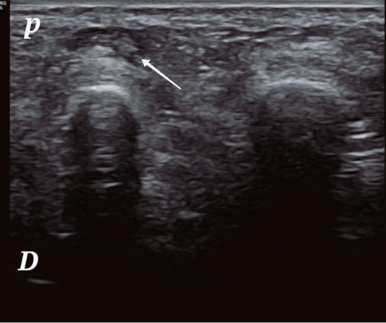
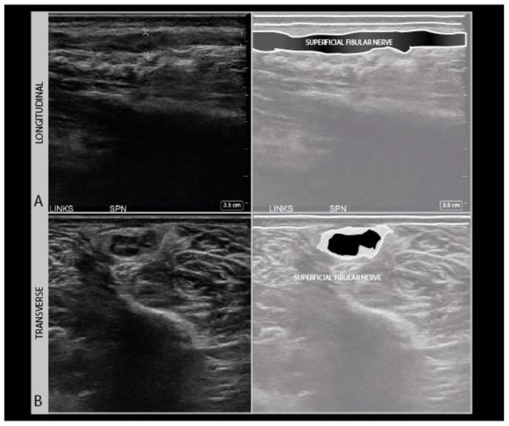
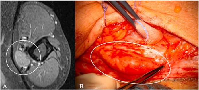
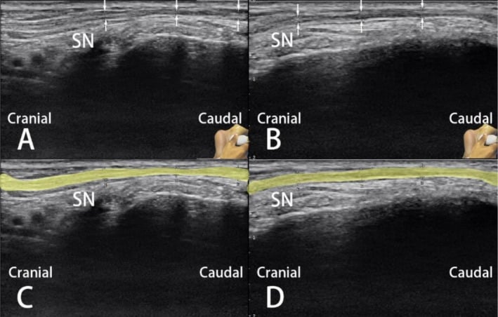
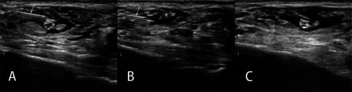
Imaging. Ultrasound can visualise the bulbous nerve end and its relationship to the scar. MRI is useful for deep neuromas (the sciatic, for example) or to rule out other pathology, and MR neurography sequences are increasingly sensitive.
Differential Diagnosis
The examiner's point. The single most important distinction is between a focal, block-responsive neuroma and diffuse or central pain that surgery will worsen, and the response to a diagnostic block is the discriminator. CRPS is a regional pain syndrome with autonomic features, whereas a neuroma causes localised pain with a specific trigger point.
Operating on CRPS makes it worse. Operating on a neuroma can cure it. The block tells you which one you are looking at.
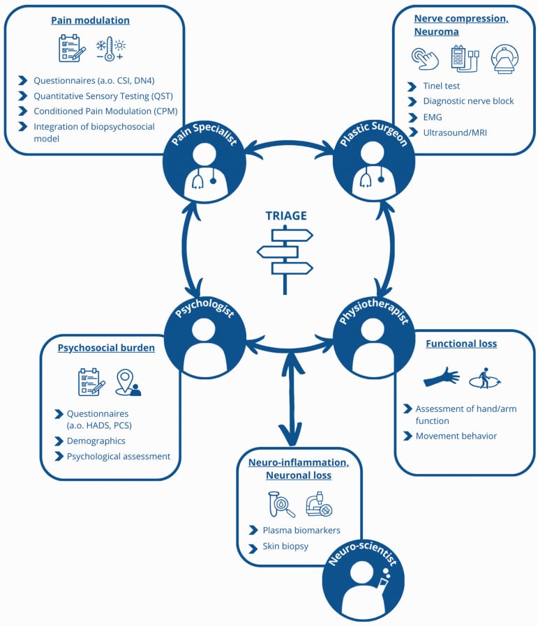
- Pain pattern
- Focal, Tinel-positive trigger point
- Block response
- Complete relief
- Key distinguishing feature
- Reproducible electric pain at one spot
- Pain pattern
- Regional, diffuse
- Block response
- Minimal / no relief
- Key distinguishing feature
- Autonomic and trophic changes, allodynia beyond one nerve
- Pain pattern
- Felt in the missing part
- Block response
- No relief of phantom component
- Key distinguishing feature
- Pain in absent limb, cortical reorganisation
- Pain pattern
- Positional, distribution of one nerve
- Block response
- Relief with proximal block
- Key distinguishing feature
- Provocative compression test, often reversible
- Pain pattern
- Mechanical, load-related
- Block response
- Variable
- Key distinguishing feature
- Linked to prosthetic fit, not a fixed trigger point
- Pain pattern
- Deep, bony, load-related
- Block response
- No relief
- Key distinguishing feature
- Visible on radiograph, palpable mass
Treatment
The ladder. Confirm the peripheral generator first, then climb from conservative care through simple excision to physiological reconstruction. The algorithm below is the whole page in one figure.
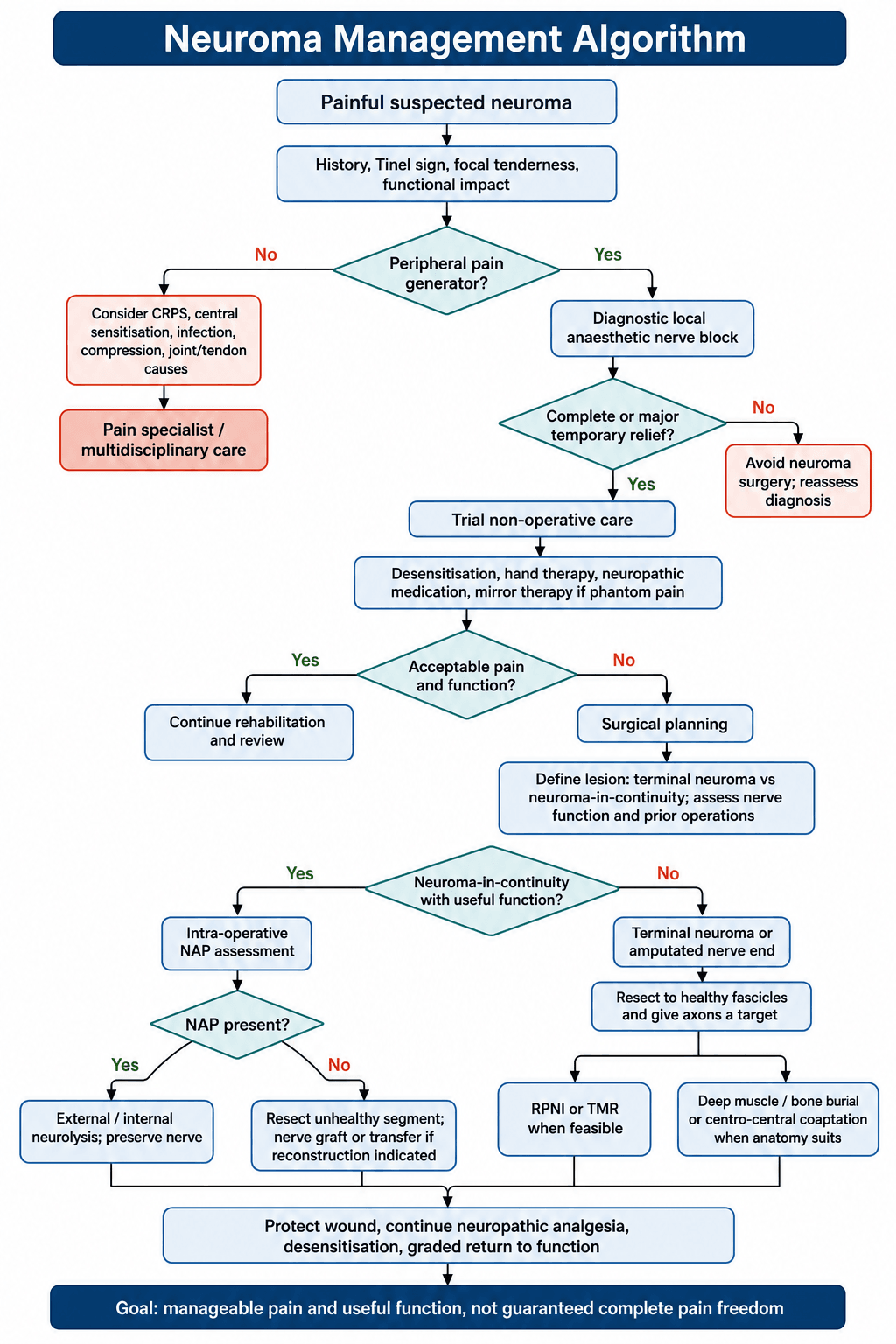
Level 1: non-surgical. Pharmacological management is the first line for neuropathic pain: gabapentin, pregabalin or amitriptyline. Alongside it run desensitisation by massage and texture rubbing, and mirror therapy for phantom pain.
Level 2: simple surgery. Resect and bury: cut the nerve back to healthy tissue and bury it deep in muscle or bone. The failure rate is high, with 20-30% recurrence, because the nerve just grows another neuroma at the new cut end.
Level 3: physiological reconstruction. These techniques give the regenerating axons a "stop signal", and they bring recurrence down to under 10%:
- RPNI gives the nerve a "dummy" muscle target
- TMR transfers the nerve to a "real" muscle target
- Centro-central union connects two nerve ends together in a loop
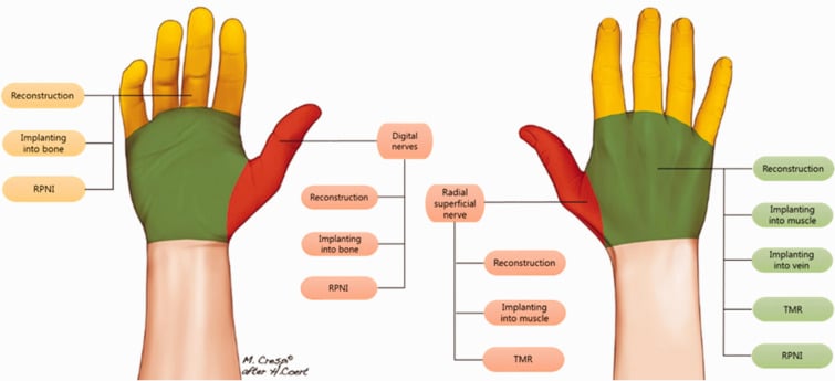
Choosing the operation. The choice is made by level and by whether a usable distal nerve end exists. Sensibility worth restoring favours reconstruction; a non-reconstructable terminal stump needs a protected physiological target rather than a free end left in the pressure zone.
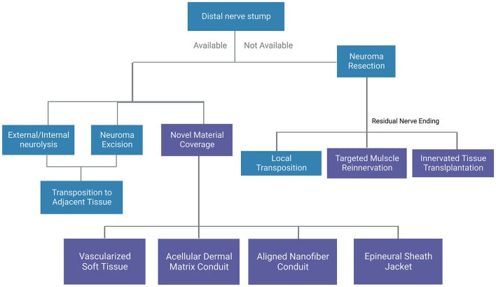
Prevention at amputation. Transect the nerve sharply, bury the end deep in muscle, and give it a target with RPNI or TMR. Prophylactic RPNI at amputation prevented symptomatic neuroma (0% vs 13.3% in controls) and reduced phantom limb pain (51% vs 91%).
DSTNeuroma Prevention
Hook:Don't Suffer Tension.
Surgical Technique
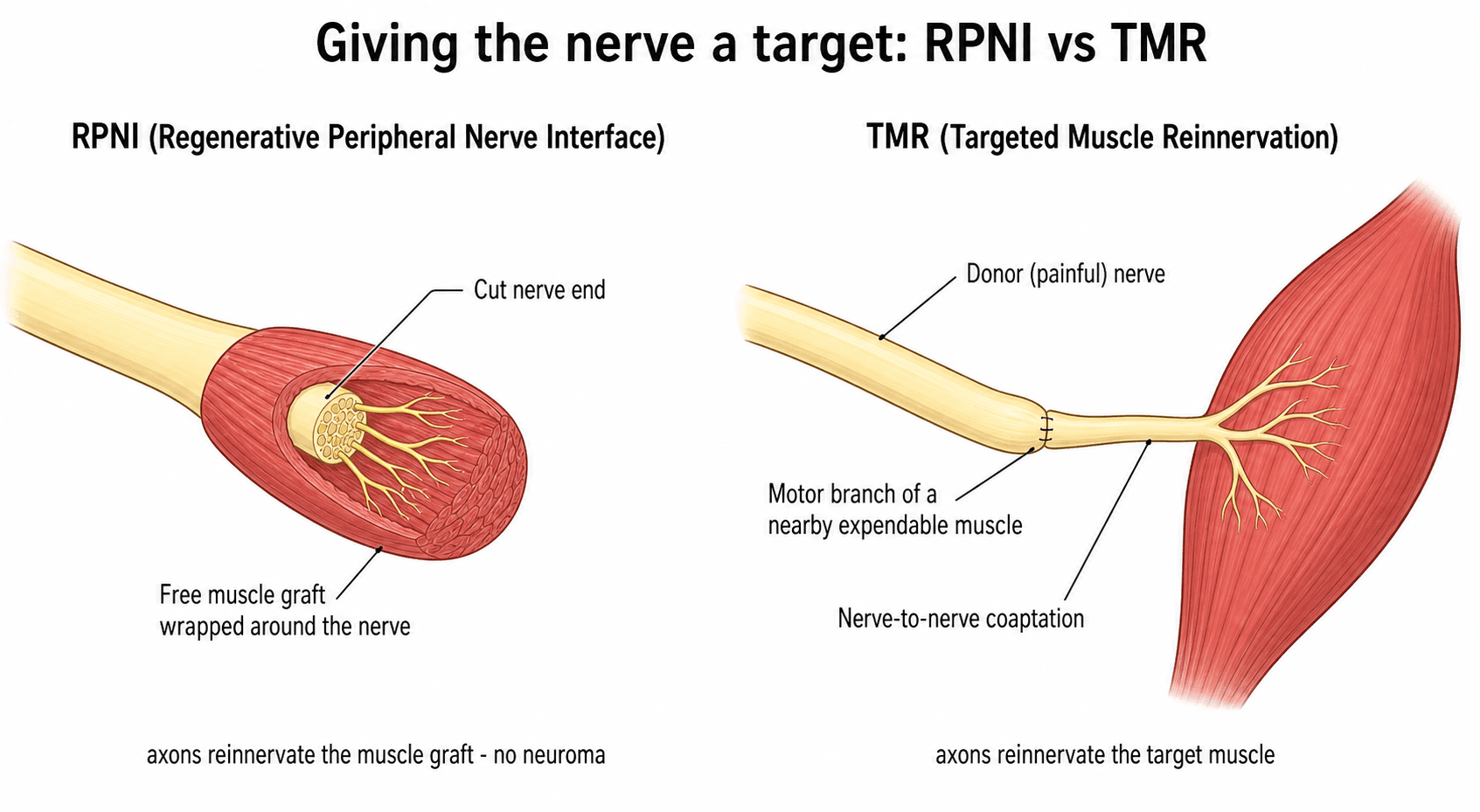
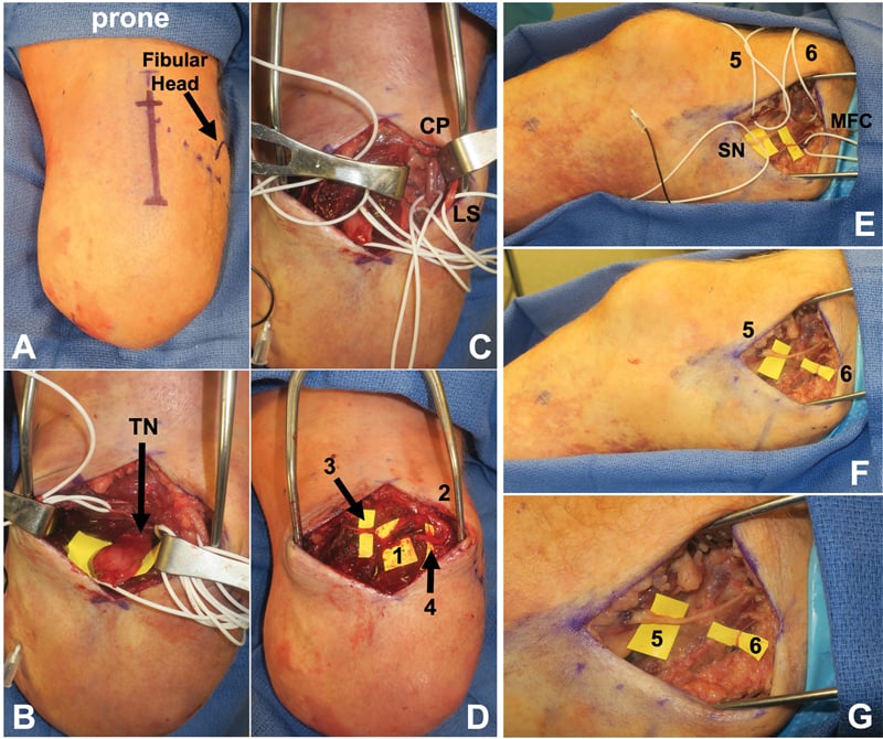
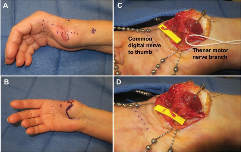
Concept. Provide a physiological target for the nerve end. The muscle acts as a "sponge" for regenerating axons: they grow into the graft and stop (reinnervation), which prevents the formation of a chaotic neuroma bulb.
Technique.
- Harvest a free muscle graft (vastus lateralis or a local muscle), about 3 x 1.5 cm
- Neurolyse the nerve end
- Wrap the muscle graft around the nerve end like a "hot dog in a bun"
- Secure with sutures
- Ensure the graft is well vascularised by the recipient bed, and do not wrap it too tightly (risk of ischaemia)
Specific Scenarios
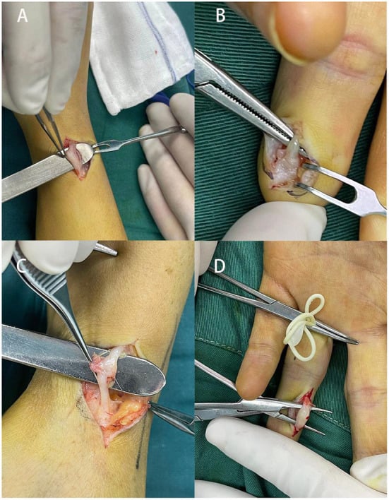
Digital nerve. Very common after finger amputation or crush injury. Resect and bury in the proximal phalanx medullary canal or into an interosseous muscle, taking care to avoid the digital artery; centro-central union is an option if both digital nerves are injured. Dorsal branch neuromas are particularly bothersome because the skin is thin.
Radial sensory nerve. Notorious for painful neuromas (Wartenberg's area); "cheiralgia paraesthetica" is the eponym for RSN compression or neuroma pain. Because the subcutaneous tissue is thin, superficial burial always fails: it often requires resection and burial deep in brachioradialis, or RPNI, and consider transferring it into the deep compartment of the forearm.
Sciatic nerve. Debilitating "sitting pain": the patient is often unable to sit for more than a few minutes, and it is often mistaken for hamstring tendonitis or piriformis syndrome. It needs a transgluteal approach with high exposure, then TMR to gluteal motor branches or burial deep in the pelvis.
Sural nerve. Common after graft harvest or ankle surgery. Resect and bury into the deep posterior compartment (flexor hallucis longus), avoiding the superficial fat where shoes will rub.
Complications
Recurrence. The nerve grows out of the burial site or forms a new neuroma at the cut end; the rates by technique are given under Treatment. Recurrence is frustrating for both surgeon and patient.
Chronic pain. Surgery may trigger CRPS. Excision of a stump neuroma may improve phantom pain by reducing the afferent barrage, but it essentially treats a different pathology: phantom pain is a cortical phenomenon (homunculus reorganisation), and peripheral surgery removes the trigger, not the memory. Do not promise a cure for phantom pain with simple neuroma excision.
Postoperative Care
- Protect the surgical repair with appropriate splinting
- Elevate to reduce swelling and optimise wound healing
- Continue neuropathic pain medications; do not cease them perioperatively
- Start gentle percussion and texture stimulation once the wound heals
- Graded motor imagery, especially important for phantom pain management
- Hand therapy: weekly supervised sessions with a home programme
- Gradual load bearing on the stump
- Prosthetic fitting: socket modifications may be needed after surgery
- Liaise with occupational therapy for graded return-to-work planning
Patient education is essential. Recovery from chronic neuroma pain takes months, not weeks.
Prognosis
Success by technique. The figures to quote, and where each comes from:
- Simple excision succeeds in approximately 60-70%; recurrence at the new transection site is common
- Burial in muscle or bone succeeds in approximately 70-80%; results depend heavily on the location and quality of the burial site
- RPNI gave about a 70% reduction in neuroma pain in the pilot series (Woo 2016), with high satisfaction
- TMR in the only randomised trial (Dumanian 2019) gave significantly better phantom-limb-pain relief than excision-and-burial (about a 3.5-point NRS advantage), with residual-limb pain trending the same way
What to promise. TMR and RPNI outperform simple excision or burial and are now first-line, but the honest endpoint is meaningful pain reduction in most patients, not a 90% pain-free rate. Complete pain freedom is rare; the realistic goal is "manageable pain" and useful function, allowing return to work.
Patient selection is critical. Operating on the wrong patient makes things worse. The features that predict a good result:
- Isolated peripheral neuroma without central sensitisation
- Complete pain relief with the diagnostic nerve block
- Short duration of symptoms (under 12 months)
- Absence of psychological comorbidities
- Single neuroma rather than multiple
The features that predict a poor one:
- Incomplete relief with the diagnostic block (suggests centralisation)
- Duration over 2 years with established chronic pain behaviour
- Associated CRPS features
- Concurrent phantom limb pain (requires separate treatment)
- Multiple previous failed surgeries
- Significant litigation or compensation involvement
Guidelines, Registries & Global Practice
Global Epidemiology:
- Symptomatic neuromas affect an estimated 10 to 30 percent of patients after major limb amputation, and a smaller but significant proportion after digital and peripheral nerve injury.
- The dominant cause of major amputation worldwide is dysvascular disease (diabetes and peripheral arterial disease); trauma-related and combat amputations carry the highest rates of painful neuroma and phantom limb pain.
- Upper-limb and digital amputations tend to produce more functionally disabling neuromas than lower-limb levels, because of superficial nerve position and high sensory demand of the hand.
Side-by-side guideline and consensus positions:
- Position on neuroma / amputation nerve management
- Endorse active nerve handling at amputation; TMR and RPNI increasingly recommended as first-line for prevention and treatment of neuroma-related pain.
- Position on neuroma / amputation nerve management
- Emphasise multidisciplinary limb-loss pathways, diagnostic block before surgery, and targeted reconstruction over repeated simple neurectomy.
- Position on neuroma / amputation nerve management
- Teach sharp transection, tension-free handling and burial principles, with RPNI/TMR as modern target-based reconstruction.
- Position on neuroma / amputation nerve management
- Support TMR and RPNI as evidence-based options; stress patient selection and exclusion of central/CRPS pain.
There is broad agreement across societies that: (1) a diagnostic local anaesthetic block confirming a peripheral generator is mandatory, (2) simple repeated excision has unacceptable recurrence, and (3) target-based reconstruction (RPNI or TMR) is the preferred modern strategy where expertise allows.
- No dedicated international neuroma registry exists; evidence is built from single-centre and multi-institutional cohorts plus one small RCT (Dumanian 2019).
- Amputation and prosthetic registries (e.g. limb-loss and rehabilitation databases) capture amputation volume and prosthetic use but rarely code neuroma pain explicitly, which is a recognised data gap.
- Well-resourced centres: microsurgical expertise, intra-operative nerve action potential testing, RPNI/TMR, MR neurography, and multidisciplinary pain and prosthetic teams.
- Limited-resource settings: reliance on clinical Tinel and diagnostic block, sharp transection with deep muscle/bone burial, and gabapentinoid/tricyclic pharmacotherapy; advanced reconstruction may be unavailable, making meticulous primary nerve handling at amputation especially important.
- Early pain-medicine involvement, structured desensitisation and hand/limb therapy, and psychological support for chronic pain improve outcomes regardless of setting.
- Liaison with prosthetists ensures socket design accommodates surgical reconstruction and any myoelectric interface created by TMR.
Controversies & Areas of Uncertainty
Both give regenerating axons a target, but head-to-head randomised data are lacking. RPNI is technically simpler and uses a free muscle graft; TMR provides a larger motor target and intuitive myoelectric signals. Choice is currently driven by surgeon expertise, nerve calibre, and prosthetic goals rather than high-level comparative evidence.
Evidence (Valerio, Kubiak) supports treating nerves at the index amputation rather than waiting. The uncertainty is cost, added operative time, and whether every amputee benefits or only higher-risk levels.
The field rests on one small RCT and many single-centre cohorts. Heterogeneous outcome measures (NRS, PROMIS, neuroma size) limit pooling, and long-term durability beyond a few years is still being defined.
Peripheral surgery reduces afferent input and can ease phantom pain, but phantom pain is partly a central, cortical phenomenon. Surgery should never be promised as a cure for the phantom component.
MCQ Practice Points
Q: What is the defining histological feature of a neuroma? A: Disorganized proliferation of Schwann cells and axonal sprouts in a dense fibrous stroma.
Q: What clinical test best differentiates a neuroma from CRPS? A: A diagnostic nerve block provides complete relief for a neuroma but minimal/no relief for CRPS.
Q: Which muscle is commonly used for RPNI grafts? A: Vastus Lateralis (free graft) or local potentially expendable muscle.
Q: What surgical technique during amputation has been proven to reduce both phantom and residual limb pain? A: Targeted Muscle Reinnervation (TMR) - transfers nerves to motor branches, providing targets for regenerating axons.
Q: Why does simple neuroma excision often fail? A: The nerve simply grows another neuroma at the new cut end (~20-30% recurrence). Providing a physiological target (RPNI/TMR) reduces recurrence to less than 10%.
Viva Scenarios
Practise clinical reasoning and management decisions out loud
“A 45-year-old carpenter had a amputation of the index finger at the PIPJ level 6 months ago. He complains of excruciating pain when he touches the tip. He cannot work.”
“You are performing a below-knee amputation for trauma. How do you manage the nerves to prevent pain?”
“A 35-year-old woman had a wrist fracture treated with plate fixation 8 months ago. She now has severe burning pain over the radial aspect of the wrist and thumb base. She cannot tolerate her watchband or any pressure. Tinel's is strongly positive over the radial styloid area.”
Principles
- Nerves need a target (or they form neuromas)
- Pain = Mechanical + Ectopic firing
- Diagnostic Block is mandatory before surgery
- Tinel's sign over neuroma = Positive diagnosis
- ALL stumps form neuromas, only some are painful
Techniques
- Simple: Resect and Bury (Muscle/Bone)
- RPNI: Wrap with free muscle graft
- TMR: Transfer to motor branch
- Centro-central: Connect two stumps
Outcomes
- Simple excision: High recurrence (30%)
- Reconstruction (RPNI/TMR): Low recurrence (under 10%)
- TMR now gold standard for major amputation
- RPNI: Newer, promising for minor neuromas
- Centro-central: Requires second nerve stump
Evidence Base
Every card below is anchored to a verified PubMed record. Read the DOI before quoting a figure: the literature is dominated by single-centre cohorts, with one small randomised trial (Dumanian) defining the modern standard.
TMR vs Standard Neurectomy (first surgical RCT)
- Prospective single-blind RCT, 28 major-limb amputees with chronic pain, TMR vs neuroma excision and intramuscular burial
- Longitudinal mixed-model change in phantom limb pain favoured TMR (mean difference 3.5 on NRS, P equals 0.03)
- Residual limb pain trended toward TMR (P equals 0.10); 3 crossover patients also improved after TMR
Preemptive TMR at the Time of Amputation
- Multi-institutional cohort, 51 immediate-TMR amputees vs 438 unselected amputee controls
- Median worst pain over 24 hours was 1 of 10 (TMR) vs 5 (phantom) and 4 (residual) of 10 in controls
- TMR carried roughly 3 to 4 times higher odds of reduced pain severity across all PROMIS domains
TMR Prospective Cohort (CORR)
- Prospective study of 33 major-limb amputees undergoing TMR for established pain
- Residual limb pain fell from 6.4 to 3.6 and phantom limb pain from 6.0 to 3.6 on NRS at 1 year (both P less than 0.001)
- OPUS and Neuro-QOL functional scores also improved significantly
RPNI for Postamputation Neuroma (pilot)
- First clinical series: 46 RPNIs implanted in 16 amputees for symptomatic neuromas
- Patients reported a 71 percent reduction in neuroma pain and a 53 percent reduction in phantom pain
- High satisfaction (94 percent would do it again); complications limited to delayed healing and one new-site neuroma
Prophylactic RPNI Prevents Neuroma
- Case-control study, 45 amputees with prophylactic RPNI vs 45 controls
- Symptomatic neuromas: 0 percent (RPNI) vs 13.3 percent (controls), P equals 0.026
- Phantom limb pain: 51.1 percent (RPNI) vs 91.1 percent (controls), P less than 0.0001
RPNI Biological Basis (mechanism)
- Rat model: divided peroneal nerve neurotised a non-vascularised free muscle transfer (extensor digitorum longus)
- Free muscle grafts revascularised and reinnervated, with axonal sprouting and synaptogenesis confirmed histologically
- Construct remained viable and transduced compound muscle action potentials over 7 months
Surgical Algorithm for Neuroma Management (principles)
- Diagnostic local anaesthetic block confirming a peripheral pain generator is mandatory before any neuroma surgery
- Simple traction neurectomy and re-burial have high recurrence; targeted reconstruction (RPNI/TMR) is preferred where feasible
- Centralised/CRPS pain and incomplete block relief predict surgical failure