Protecting the Most Vulnerable
- Non-Mobile Infants: Any fracture in a non-mobile infant warrants investigation.
- Metaphyseal Corner Fractures: Highly specific (bucket-handle/corner fractures), but not pathognomonic.
- Multiple Fractures: Different healing stages = repeated trauma.
- Rib Fractures: Posterior rib fractures are highly suspicious.
- Reporting: A legal (mandatory reporting) or strong professional duty to report suspected NAI.
- “Say highly specific, never pathognomonic
- “Repeat the skeletal survey at 10-14 days if the first is negative
- “A confirmed mimic such as OI does not exclude coexisting abuse
- “Document meticulously
Overview and Epidemiology
Non-accidental injury (NAI) is physical abuse of a child, often by a caregiver. Fractures are its second most common manifestation, after bruising, which is why it matters to the orthopaedic surgeon, and early recognition can save lives.
Who. The peak age is under 2 years, especially under 1 year, and boys are slightly more affected. The risk factors:
- Young, single parents
- Substance abuse and mental health issues
- A previous history of NAI in the family
- A colicky or difficult child
- Low socioeconomic status, although abuse occurs in all groups
Pathophysiology and Mechanism
Mechanisms of injury. Each mechanism and what it produces:
- Shaking - subdural haematoma and retinal haemorrhages. In infants the head is large and the neck muscles are weak
- Gripping or squeezing - posterior rib fractures, with the thumbs on the spine and the fingers on the ribs
- Twisting or pulling - the classic metaphyseal lesion
- Direct blows - long-bone shaft fractures and skull fractures
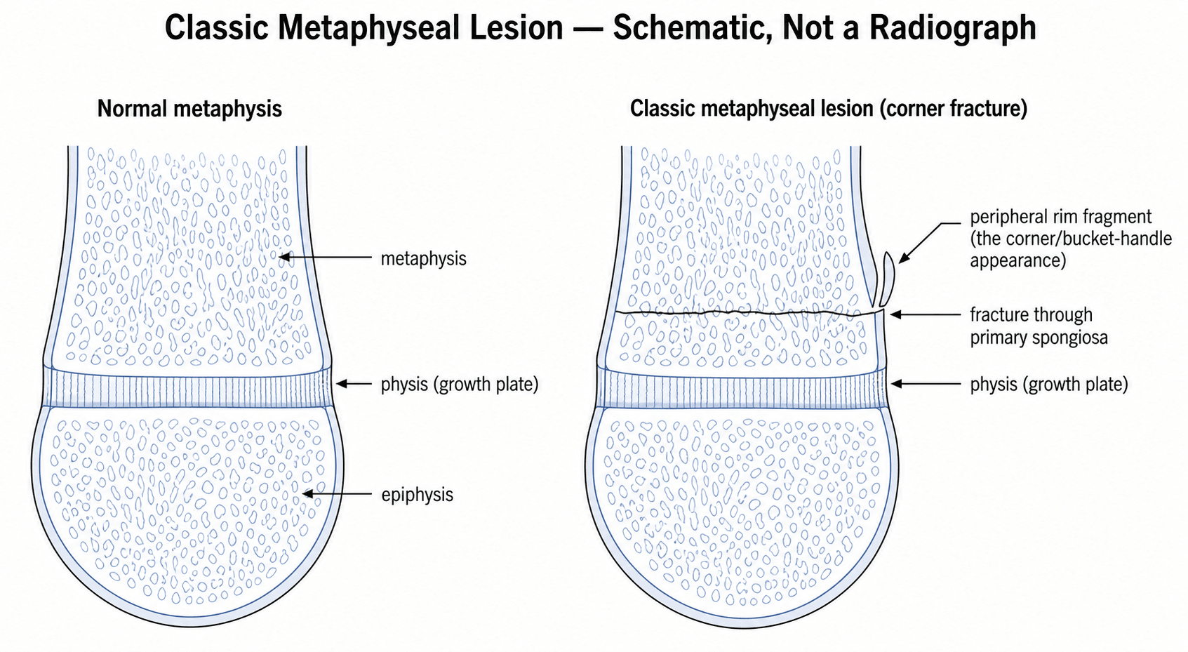
The classic metaphyseal lesion (CML). The primary spongiosa immediately beneath the physis is the mechanically weakest zone of the growing bone. Shear from twisting or pulling propagates a planar fracture across that zone, separating a disc-shaped fragment that is thick at the periphery, where it includes the subperiosteal bone collar, and thin centrally. Micro-CT with histological correlation (Tsai 2014) established that it is a fracture, not an avulsion of tendon or periosteum.
One lesion, two appearances. The disc geometry is why the CML looks like a corner fracture when seen obliquely and a bucket handle when seen en face. The mechanism rarely occurs in accidental falls, and that is the basis of its specificity.
Fracture Patterns and Specificity
Fractures are graded by how strongly they point to abuse. The high-specificity group is rare in accidental trauma, which is what makes it so informative, and when one of these fractures is present NAI is highly likely. Highly specific is not pathognomonic, though, and a differential diagnosis is still required in every case.
High specificity. The metaphyseal corner (bucket-handle) fracture is the classic lesion, and posterior rib fractures come from squeezing. Scapular fractures are very unusual in children and require significant force; spinous process fractures come from a direct blow or hyperflexion, and sternal fractures from a direct blow.
Moderate specificity. Multiple fractures at different healing stages indicate repeated trauma, and bilateral fractures are statistically unlikely from a single accident. Complex skull fractures, with multiple fracture lines or depression, and femoral fractures in a non-ambulatory child also raise strong suspicion and warrant thorough investigation.
Low specificity. Clavicle fractures (common at birth and in falls), linear skull fractures (common in falls), toddler's fractures and subperiosteal injuries are all common in accidental trauma, and a long-bone shaft fracture can be accidental or non-accidental. Here context and mechanism decide.
MRSHigh Specificity Fractures
Hook:MRS - Metaphyseal, Ribs, Scapula/Spinous/Sternum.
Clinical Assessment
History. These features of the history are red flags:
- Delayed presentation - caregivers wait before seeking help
- Inconsistent history - the explanation does not match the severity or pattern of the injury
- Changing story - different versions from different caregivers
- No history - "I don't know how it happened", or a vague explanation
- Inappropriate affect - the caregiver is not appropriately concerned
Examination. The examination covers the whole child:
- Full body inspection for bruises, especially the non-accidental patterns: grip marks, loop marks, bite marks
- Skeletal tenderness - palpate all limbs and the axial skeleton
- Head circumference - increasing head size may indicate a subdural haematoma
- Fontanelle - a bulging fontanelle is concerning
Recognising the Abusive Bruise: Sentinel Injuries and TEN-4-FACESp
A fracture is rarely the first abusive injury; it is often the one that finally reaches an orthopaedic surgeon. Any bruise in a non-mobile infant is suspicious, and two clinical rules turn that instinct into a defensible referral.
The sentinel injury. A minor but unexplained injury in a young, pre-mobile infant, noticed by a caregiver or clinician before a later, more serious abusive event. It is most often a bruise, but it can also be an intra-oral injury or a frenulum tear.
The missed warning. In a case-control study, a prior sentinel injury was reported in a substantial minority of infants with definite abuse but in none of the non-abused controls. The lesion was a bruise in the large majority, most occurred before three months of age, and a medical provider had often been aware of the injury at the time without acting on it. A "trivial" bruise in a very young infant is a chance to intervene before the fracture or the head injury, not a finding to dismiss.
"Those who don't cruise rarely bruise." Accidental bruising tracks motor development. Independently mobile (cruising or walking) toddlers bruise over bony prominences, the shins, knees and forehead, in everyday falls. Bruising in an infant who cannot yet cruise has no innocent mechanical explanation and must be explained or referred, and bruises over soft, protected, non-bony sites (the trunk, ear, neck, cheeks, eyelids, genitalia) are the abusive pattern at any age.
TEN-4-FACESp. The clinical decision rule operationalises this for bruising in children under four years and infants. It flags bruising as high-concern when it involves any of these regions or features:
- Region / feature
- Chest, abdomen, back, flank, buttocks, genitourinary region
- Why it matters
- Soft protected sites do not bruise from ordinary falls
- Region / feature
- Pinna and around the ear
- Why it matters
- Almost never injured accidentally; a 'boxing' or pinch site
- Region / feature
- Anterior or posterior neck
- Why it matters
- Implies grabbing, choking or smothering
- Region / feature
- The rule applies to children under 4 years; ANY bruise in an infant under ~4-5 months
- Why it matters
- The pre-mobile infant who should not have bruises at all
- Region / feature
- Torn frenulum (forced feeding), jaw angle, fleshy cheeks, eyelids, subconjunctival haemorrhage
- Why it matters
- Facial soft tissues that are not struck in everyday play
- Region / feature
- Hand/slap marks, loop or linear ligature marks, bite marks, grip/pinch marks
- Why it matters
- The shape itself encodes the mechanism
What a positive rule means. Applied to bruising in young children the rule is highly sensitive and reasonably specific for abusive injury, which makes it a screening prompt to escalate rather than a diagnosis. A positive TEN-4-FACESp bruise, like a sentinel injury, mandates the full safeguarding pathway and a skeletal survey. It does not on its own prove abuse, and a negative rule does not exclude it.
If asked "what would have prevented this child's femoral fracture?", the answer the examiner wants is recognition of the earlier sentinel injury. Say it explicitly: a pre-mobile infant should not have unexplained bruises, and a TEN-4-FACESp-positive bruise triggers the same admit-protect-investigate-report pathway as a suspicious fracture.
Investigations
Skeletal survey. Full-body radiographs: AP and lateral views of all limbs, with the chest, abdomen, skull and spine. It is mandatory in a child under 2 years with suspected abuse. Repeat it at 10-14 days if the first survey is negative, because healing fractures have become visible by then; the imaging guidance on the evidence cards goes further and makes a follow-up survey at about two weeks standard.
Head imaging. Looks for intracranial bleeding: CT head urgently if there are any neurological signs. MRI brain is more sensitive for subtle injury.
Ophthalmology. Dilated fundoscopy looks for retinal haemorrhages, and multilayer retinal haemorrhages are highly specific for shaken baby syndrome.
Bloods. These rule out the medical mimics:
- Bleeding studies (PT, APTT, platelet count) for a bleeding disorder
- Metabolic bone disease screen (calcium, phosphate, ALP, vitamin D) for rickets and OI
Parent's Guide: Understanding Child Injury Investigations
Why is my child being investigated? When a child has an injury that doctors cannot easily explain, they have a legal and ethical duty to make sure the child is safe. This does not mean they are accusing you of anything - it means they are being thorough.
What investigations will be done?
- X-rays of the whole body (skeletal survey) to check for other injuries.
- A scan of the brain if there is any concern.
- An eye examination to look for signs of bleeding.
- Blood tests to rule out medical conditions.
What happens next? A team of doctors, social workers, and sometimes police will review the findings. Their goal is to ensure your child is safe.
What are your rights? You have the right to legal representation. Cooperating with the investigation is in your child's best interest.
Dating Fractures Radiologically in NAI
Healing fractures can be approximately aged on radiographs. That is the whole reason a skeletal survey is repeated, and the reason multiple fractures at different healing stages is a moderate-specificity flag. The orthopaedic surgeon is repeatedly asked, in clinic, in MDT and in court, "how old is this fracture?", and the honest, defensible answer is a range, never a precise day.
The radiographic features of healing appear in a predictable sequence, but with wide, overlapping windows that depend on the bone, the fracture type and the child's age:
- Typical window
- From the time of injury, resolving over the first 1-2 weeks
- Note
- Non-specific; the earliest and least reliable sign
- Typical window
- Earliest from about 4-7 days, usually well seen by 10-14 days
- Note
- The single most useful early dating sign
- Typical window
- Soft callus over roughly 2-3 weeks, hard callus from a few weeks
- Note
- Becomes denser and better defined as it matures
- Typical window
- From several weeks to many months
- Note
- A remodelled, well-corticated callus indicates an old injury
A fresh fracture has no periosteal reaction. The absence of healing means the injury is recent, broadly within the last week or so. That is why a negative first survey is repeated: by the second, occult rib and metaphyseal fractures declare themselves with periosteal new bone.
CMLs and rib fractures date poorly. Corner and bucket-handle metaphyseal fractures show little or no periosteal reaction and are notoriously hard to age. Rib fractures are frequently invisible acutely and only become apparent as callus forms, which reinforces the value of the repeat survey and of oblique rib views.
Different ages mean different episodes. A non-healing fracture alongside a remodelling one cannot have occurred in a single event. This is the radiological basis of the "different healing stages" red flag.
Bruises cannot be dated by colour. Despite courtroom tradition, the colour of a bruise does not reliably indicate its age; only the presence of yellow has any (weak) discriminating value. Multiple bruises of "different ages" by colour should not be presented as proof of repeated assault.
Do not over-date. State a defensible range and acknowledge the uncertainty. Over-precise dating ("this fracture is exactly 12 days old") is a frequent and successful target for medico-legal challenge.
Never give a single number. The defensible structure is: no periosteal reaction = recent (days); periosteal new bone = roughly 1-3 weeks; mature, dense, remodelling callus = weeks to months. Then add the two caveats that win marks: metaphyseal corner and rib fractures date poorly, and bruise colour does not date a bruise. This honesty about uncertainty is itself the examined competency.
Differential Diagnosis
Highly specific, not pathognomonic. The classic metaphyseal lesion is the most useful radiological finding in this field, and precisely because it is so useful it attracts language stronger than the evidence supports. Highly specific means the finding is rare in the absence of abuse; pathognomonic means its presence establishes the diagnosis. Only the first is true, and the distinction is the difference between evidence and a verdict.
The normal variant. A case report of an 8-month-old who stopped crawling on one leg describes an isolated metaphyseal lesion that prompted transfer for suspected abuse, and the paediatric radiologist specialising in child abuse determined it was a normal variant. The authors' stated conclusion is that an isolated metaphyseal lesion is not pathognomonic for child abuse and a thoughtful differential diagnosis should be considered (DOI).
Syphilis and rickets. A separate report describes a lesion initially diagnosed and treated as a metaphyseal corner fracture that proved to be osteitis of congenital syphilis, a diagnosis whose incidence is rising (DOI). And the existence of a seven-centre study assessing whether radiologists can distinguish rickets from CML tells you the question is live enough to be contested in court (DOI).
What survives, and it is nearly all of it. The CML remains highly specific, its mechanism rarely arises from the falls and handling that parents describe, and a CML in a non-mobile infant demands a full child-protection assessment every single time. Nothing here is an argument for complacency.
Both errors are catastrophic. Under-calling returns a child to a person who is injuring them, and the next injury may be fatal. Over-calling separates a child from parents who have done nothing, destroys a family, and is only partially reversible. Neither error is the graver, and that symmetry is why the finding goes as evidence to a multidisciplinary child-protection process rather than being diagnosed by the orthopaedic surgeon alone.
Word the note accordingly. Write what you see, state that the pattern is highly specific and requires full assessment, and do not write that it is diagnostic of abuse.
- Discriminating Features
- Blue sclerae, dentinogenesis imperfecta, wormian bones, positive family history, low-trauma fractures, normal/osteopenic bone density
- Key Investigation
- Genetic testing (COL1A1/COL1A2)
- Discriminating Features
- Cupped, frayed, splayed metaphyses (not corner fractures), generalised osteopenia, risk factors for vitamin D deficiency
- Key Investigation
- Calcium, phosphate, ALP, vitamin D, PTH; wrist/knee radiographs
- Discriminating Features
- Clavicle or humeral fracture in a neonate, consistent perinatal history, healing already present at first presentation
- Key Investigation
- Birth records; timeline correlation
- Discriminating Features
- Explains bruising and intracranial haemorrhage but NOT fractures; may coexist with abuse
- Key Investigation
- FBC, PT, APTT, fibrinogen, von Willebrand screen, factor levels
- Discriminating Features
- Pathological fractures, metaphyseal lucent bands, systemic features, cytopenias
- Key Investigation
- FBC and film, blood film, marrow if indicated
- Discriminating Features
- Metaphyseal changes in malnourished/preterm or malabsorptive infants; rare
- Key Investigation
- Serum copper and caeruloplasmin
A mimic does not close the case. Investigate mimics thoroughly, but a negative medical workup does not exclude abuse, and a confirmed medical condition such as OI does not exclude coexisting inflicted injury. Report if the clinical picture is suspicious.
Management Algorithm
The child's safety is the absolute priority, and fracture treatment is secondary to it. In order:
- Ensure the child's safety - do not discharge to an unsafe environment
- Admit - for protection and for investigation
- Involve the multidisciplinary team - paediatrician, child protection, social services
- Investigate - skeletal survey, head imaging, ophthalmology review and bloods, as above
- Report and document - see Medico-Legal Considerations
- Treat the fractures as appropriate
Surgical Technique
Non-operative by default. Most NAI fractures can be treated non-operatively, and healing is generally rapid in children.
Femur. A Pavlik harness under 6 months. Older infants and toddlers are treated in a spica cast, applied immediately or delayed, which is the standard treatment; K-wire fixation is rarely needed.
Tibia and fibula. An above-knee cast for most, with conservative treatment preferred.
Humerus. Collar and cuff or an above-elbow cast; ORIF is rarely indicated in infants.
Forearm. Cast immobilisation, accepting more angulation in young children because of remodelling.
Ribs. Supportive care only, with analgesia and respiratory monitoring. No surgical treatment is required.
Skull. Observation for most linear fractures, with neurosurgical involvement if the fracture is depressed or there is intracranial injury.
When surgery is required. Surgery is needed for:
- Open fractures, which are rare in NAI
- Unstable fractures requiring fixation
- Neurosurgical intervention for intracranial haemorrhage
Complications
Physical. Malunion and nonunion are rare with appropriate treatment. Physeal injuries may cause growth disturbance and limb length discrepancy. Brain injury from shaking can cause permanent disability, and retinal haemorrhages may lead to visual problems.
Psychological. Abuse also leaves psychological harm:
- Post-traumatic stress disorder (PTSD) - common in abused children
- Attachment disorders - difficulty forming healthy relationships
- Developmental delay - physical and cognitive
- Behavioural problems - aggression, anxiety, depression
Postoperative Care
Fracture care. Standard fracture aftercare applies: cast care and weight-bearing status, follow-up imaging to confirm healing, and physiotherapy if indicated for stiffness or weakness.
Discharge. Do not discharge without child-protection clearance. Social services must approve the discharge plan, and if home is unsafe an alternative placement must be arranged. Other children in the household must also be assessed.
Follow-up. Follow-up is coordinated between orthopaedics, paediatrics and social services. The discharge summary records all findings and a clear follow-up plan, and goes to the GP and community health services.
Outcomes/Prognosis
The fracture. Most fractures heal well with appropriate treatment, and functional outcomes are generally good if the abuse is recognised early.
The child. Mortality is 10-30% in severe cases (shaken baby syndrome). Without intervention, abuse typically escalates, morbidity approaches 100%, and children who remain in abusive environments have high mortality rates; children removed from abusive environments have better long-term outcomes.
Prognostic factors. The factors that bear on prognosis:
- Age at recognition, younger children being more vulnerable
- Severity of injuries, especially neurological
- Quality of intervention and follow-up
- Availability of safe alternative placement
Medico-Legal Considerations and the Surgeon's Role
You do not need to prove abuse: a reasonable suspicion is the threshold for referral. Even if uncertain, report suspected NAI; you are protected from liability when acting in good faith.
The duty to report. In most jurisdictions clinicians have a legal duty (mandatory reporting) or a strong professional duty to report suspected child abuse. Refer to the local child protection or safeguarding service or social work team and the child protection paediatric lead, and involve police where there is risk of immediate harm. Failure to act on a reasonable suspicion can carry professional and, where mandatory reporting applies, legal consequences.
Documentation. Your notes may be subpoenaed and used in court, so write them meticulously and contemporaneously: factual, objective and comprehensive, with diagrams and photographs, and photographs of the findings before any operation. Document what you observe, not your opinion on who caused it. If specimens or photographs are taken, maintain proper chain-of-custody procedures.
In court. You may be called as a factual or expert witness. Be prepared to explain your findings objectively.
If the child comes back. Monitoring continues after discharge, and a child who returns with new injuries is re-escalated.
Guidelines, Registries & Global Practice
Global epidemiology
- Child maltreatment is reported worldwide; physical abuse disproportionately affects infants, with the highest fracture and fatal-head-injury rates in the first year of life.
- Fractures are a common manifestation of physical abuse and are second only to bruising; up to a third of physically abused children have a fracture, and the proportion is highest in non-ambulant infants.
- Risk is increased by young/sole caregivers, parental substance misuse and mental illness, intimate-partner violence, social isolation and prematurity/disability of the child - but abuse occurs across all socioeconomic groups and no profile is diagnostic.
Side-by-side guideline comparison
- Core position on suspected physical abuse
- Skeletal survey mandatory under 2 years; standardised 2-view survey with follow-up survey at ~2 weeks; neuroimaging for occult head injury in young infants
- Core position on suspected physical abuse
- Joint imaging standards: full survey under 2 years (consider 2-5 years selectively); oblique rib views; repeat survey at 11-14 days; CT then MRI for suspected abusive head trauma
- Core position on suspected physical abuse
- "Suspect" vs "consider" framework for physical abuse; act on unexplained injury in a non-mobile child; multi-agency safeguarding referral
- Core position on suspected physical abuse
- Treat the fracture on standard paediatric principles, but recognition and safeguarding referral take precedence over operative planning
- Core position on suspected physical abuse
- Population-level prevention and a multi-sectoral response; clinical recognition feeds into child-protection systems
Practice variation: high- vs limited-resource settings
- High-resource: dedicated child-protection paediatric teams, on-site paediatric radiology, ophthalmology and forensic input, statutory multi-agency pathways.
- Limited-resource: skeletal survey may be substituted by targeted/limited radiography; ophthalmology and CT/MRI may be unavailable; reporting frameworks and social-care infrastructure vary widely. The clinical threshold for suspicion and the duty to protect the child remain the same regardless of available investigations.
- Reporting is mandatory by law in many countries (e.g. across the US, Australia, parts of Europe and Asia) and professionally expected elsewhere; clinicians must know the local pathway, but the duty to act on reasonable suspicion is universal.
Controversies & Areas of Uncertainty
- Vitamin D deficiency as a cause of fractures: A recurring courtroom argument. Subclinical vitamin D insufficiency is common and does not, by itself, produce the fracture patterns of abuse; radiographic rickets is required before metabolic bone disease can plausibly explain fractures. This remains a contested medico-legal area.
- "Temporary brittle bone disease": A proposed entity used in some defences to explain multiple infant fractures. It is not accepted as a validated diagnosis in mainstream paediatric and radiological practice and lacks a reproducible evidence base.
- The biomechanics of abusive head trauma: The relative contribution of shaking versus impact, and the specificity of the "triad" (subdural haemorrhage, retinal haemorrhage, encephalopathy), remain debated. Current consensus is that the triad raises strong suspicion but is interpreted alongside the whole clinical and radiological picture rather than as standalone proof.
- Dating of fractures and bruises: Radiological fracture dating is broad (ranges, not precise days) and bruise colour does not reliably date injury; over-precise dating in reports is a frequent source of challenge.
- Whole-skeleton imaging in older children: The yield of routine skeletal survey falls sharply after 2 years, so imaging in the 2-5 year group is selective and guideline-dependent rather than uniform.
MCQ Practice Points
Q: Which fracture pattern is MOST specific for NAI? A: Metaphyseal corner (bucket-handle) fractures.
Q: Which location of rib fracture is most specific for NAI? A: Posterior rib fractures (from squeezing).
Q: What is the single most important investigation for suspected NAI? A: Skeletal survey (full body X-rays).
Q: What is the legal obligation when NAI is suspected? A: Mandatory reporting to child protection services.
Q: What condition is most commonly confused with NAI? A: Osteogenesis Imperfecta (OI) - but NAI-specific patterns differ.
Q: What is the mechanism of metaphyseal corner fractures? A: Twisting/pulling forces cause avulsion at the chondro-osseous junction.
Self-Assessment Quiz
Additional Self-Assessment Questions
Viva Scenarios
Practise clinical reasoning and management decisions out loud
“6-month-old brought to ED with a swollen thigh. X-ray shows a mid-shaft femoral fracture. Parents say the baby rolled off the couch.”
“Same infant. Skeletal survey shows healing rib fractures (posterior), a healing radius fracture, and a fresh femur fracture.”
“Parents of the infant claim their child has 'brittle bone disease' (OI). How do you approach this?”
HIGH SPECIFICITY
- Metaphyseal Corner (bucket-handle)
- Posterior Ribs
- Scapula
- Spinous Process
- Sternum
RED FLAGS
- Non-Mobile Infant with fracture
- Delayed Presentation
- Inconsistent History
- Multiple Injuries different stages
INVESTIGATIONS
- Skeletal Survey (repeat at 10-14 days)
- Head CT/MRI
- Dilated Fundoscopy
- Bleeding Studies
DIFFERENTIALS
- Osteogenesis Imperfecta
- Rickets
- Birth Trauma
- Bleeding Disorders
MANAGEMENT
- Admit for Safety
- MDT Involvement
- Mandatory Report
- Meticulous Documentation
LEGAL
- Mandatory Reporting required
- Good faith = liability protection
- Notes may be subpoenaed
- Avoid speculation
Evidence Base
Kemp, Maguire et al (Welsh Child Protection Systematic Review Group)
- 32 comparative studies; once major trauma excluded, rib fractures carried the highest probability of abuse (0.71, 95% CI 0.42-0.91)
- Probability of abuse for a humeral fracture 0.48-0.54, femoral fracture 0.28-0.43 (developmental stage a key discriminator)
- Skull fracture probability 0.30; linear fractures were the commonest skull pattern in both abuse and non-abuse
- No single fracture, in isolation, distinguishes abusive from non-abusive injury
Kleinman & Marks
- Histological study of 40 metaphyseal lesions from 10 abused infants at autopsy
- Fracture runs through the primary spongiosa adjacent to the chondro-osseous junction, undercutting a peripheral fragment
- Inclusion of the subperiosteal bone collar explains the radiographic corner and bucket-handle appearances described by Caffey
Kemp, Butler, Maguire et al
- 34 studies: diagnostic yield of occult fractures is significant in children under 2 years
- A repeat skeletal survey at ~2 weeks provides significant additional information on number and age of fractures
- Skeletal survey commonly misses rib fractures unless oblique rib views are added
Sheets et al
- Of 200 definitely abused infants, 27.5% had a prior sentinel injury versus 0% of 101 non-abused controls (P less than 0.001)
- Most sentinel injuries were bruises (80%); 66% occurred before 3 months of age
- A medical provider was aware of the sentinel injury in 42% of cases yet abuse was not recognised
Pierce et al (TEN-4-FACESp)
- Prospective study of 2,123 children under 4 years presenting with bruising across 5 children's hospitals
- Refined bruising clinical decision rule was 95.6% sensitive and 87.1% specific for abusive trauma
- High-risk regions: Torso, Ear, Neck, Frenulum, Angle of jaw, Cheeks, Eyelids, Subconjunctivae; any bruise in an infant under 5 months; or patterned bruising
Bhardwaj et al
- 20 studies: intraocular haemorrhage had 75% sensitivity and 94% specificity for abusive head trauma
- Bilateral, extensive, multilayered retinal haemorrhages are the most specific pattern
- Traumatic retinoschisis and perimacular folds are seen in a minority of cases but are rarely present in other conditions
Maguire et al
- 14 studies, 1,655 children with intracranial injury (779 inflicted)
- Apnoea (PPV 93%, OR 17.1) and retinal haemorrhage (PPV 71%, OR 3.5) were most predictive of inflicted brain injury
- Gender, seizures and long-bone fractures were not discriminatory; isolated skull fracture was more associated with non-inflicted injury
AAP Section on Radiology / RCPCH-RCR (UK)
- Skeletal survey is mandatory in all children under 2 years with suspected physical abuse
- Standardised survey: dedicated coned views, oblique rib views; follow-up survey at ~2 weeks
- CT/MRI head indicated in infants with neurological signs or in those under 6-12 months with suspected abuse