Protecting the Most Vulnerable
Fracture Specificity
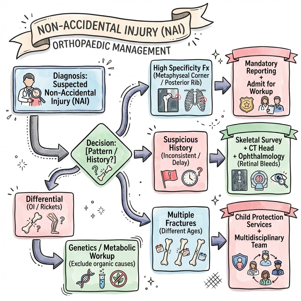
Critical Must-Knows
- Non-Mobile Infants: Any fracture in a non-mobile infant warrants investigation.
- Metaphyseal Corner Fractures: Highly specific (bucket-handle/corner fractures).
- Multiple Fractures: Different healing stages = repeated trauma.
- Rib Fractures: Posterior rib fractures are highly suspicious.
- Mandatory Reporting: Legal obligation to report suspected NAI.
Clinical Pearls
- "Know the high-specificity fracture patterns
- "Any fracture in non-mobile infant = suspect NAI
- "Skeletal survey is essential
- "Document meticulously
Reporting Suspected Abuse
In most jurisdictions worldwide, clinicians have a legal duty (mandatory reporting) or a strong professional duty to report suspected child abuse.
- You do not need to prove abuse - a reasonable suspicion is the threshold for referral.
- Refer to the local child protection / safeguarding service or social work team and the child protection paediatric lead; involve police where there is risk of immediate harm.
- Failure to act on a reasonable suspicion can carry professional and (where mandatory reporting applies) legal consequences.
- Document all findings meticulously and contemporaneously - your notes may be used in court.
Fracture Specificity for NAI
| Specificity | Fracture Patterns | Clinical Context |
|---|---|---|
| Metaphyseal corner (bucket-handle), Posterior rib, Scapula, Spinous process, Sternum | Pathognomonic for abuse | |
| Multiple fractures at different stages, Bilateral fractures, Complex skull | Raise strong suspicion | |
| Clavicle, Long bone shaft, Linear skull, Subperiosteal | Common in accidental trauma too |
MRSHigh Specificity Fractures
| M | Metaphyseal Corner Bucket-handle or corner fractures |
| R | Ribs (Posterior) Squeeze mechanism |
| S | Scapula/Spinous/Sternum Unusual locations |
| M | Metaphyseal Corner Bucket-handle or corner fractures |
| R | Ribs (Posterior) Squeeze mechanism |
| S | Scapula/Spinous/Sternum Unusual locations |
Hook:MRS - Metaphyseal, Ribs, Scapula/Spinous/Sternum.
SHEDInvestigation Checklist
| S | Skeletal Survey Full body X-ray series |
| H | Head CT/MRI Intracranial bleeding |
| E | Eyes (Fundoscopy) Retinal hemorrhages |
| D | Documentation Meticulous notes |
| S | Skeletal Survey Full body X-ray series | E | Eyes (Fundoscopy) Retinal hemorrhages |
| H | Head CT/MRI Intracranial bleeding | D | Documentation Meticulous notes |
Hook:SHED - Survey, Head, Eyes, Document.
NAIRed Flags for NAI
| D | Delay Delayed presentation |
| I | Inconsistent History Story doesn't match injury |
| M | Multiple Injuries Different ages |
| E | Explanation Absent No explanation or vague |
| D | Delay Delayed presentation | M | Multiple Injuries Different ages |
| I | Inconsistent History Story doesn't match injury | E | Explanation Absent No explanation or vague |
Hook:DIME - Delay, Inconsistent, Multiple, Explanation absent.
Overview/Epidemiology
Non-Accidental Injury (NAI) is physical abuse of a child, often by a caregiver.
- Epidemiology:
- Peak age: under 2 years (especially under 1 year).
- Boys slightly more affected.
- Mortality: 10-30% in severe cases (shaken baby syndrome).
- Risk Factors:
- Young, single parents.
- Substance abuse, mental health issues.
- Previous history of NAI in family.
- Colicky or difficult child.
- Low socioeconomic status (although occurs in all groups).
- Importance for Orthopaedic Surgeons:
- Fractures are the second most common manifestation of NAI (after bruising).
- Early recognition can save lives.
Pathophysiology & Pathomechanics
Mechanisms of Injury in NAI
- Shaking: Causes subdural hematoma, retinal hemorrhages. In infants, the head is large and the neck muscles are weak.
- Gripping/Squeezing: Causes posterior rib fractures (thumbs on spine, fingers on ribs).
- Twisting/Pulling: Causes metaphyseal corner fractures (avulsion at the chondro-osseous junction).
- Direct Blows: Long bone shaft fractures, skull fractures.
Why Metaphyseal Corner Fractures are Specific
- The metaphysis is weaker than the shaft.
- Twisting or pulling forces cause avulsion at the periosteal-bone junction.
- This mechanism rarely occurs in accidental falls.
Classification Systems
Fracture Specificity Classification
HIGH SPECIFICITY (Pathognomonic for NAI):
- Metaphyseal corner fractures (bucket-handle)
- Posterior rib fractures
- Scapula fractures
- Spinous process fractures
- Sternum fractures
These fractures are virtually never seen in accidental trauma.
Clinical Assessment
History Red Flags:
- Delayed Presentation: Caregivers wait before seeking help.
- Inconsistent History: Explanation doesn't match injury severity or pattern.
- Changing Story: Different versions from different caregivers.
- No History: "I don't know how it happened."
- Inappropriate Affect: Caregiver not appropriately concerned.
Physical Exam:
- Full Body Inspection: Look for bruises (especially non-accidental patterns like grip marks, loop marks, bite marks).
- Bruises in Non-Mobile Infants: Any bruise in a non-mobile infant is suspicious.
- Skeletal Tenderness: Palpate all limbs and axial skeleton.
- Head Circumference: Increasing head size may indicate subdural hematoma.
- Fontanelle: Bulging fontanelle is concerning.
Fracture Patterns in NAI
HIGH SPECIFICITY (When present, NAI is highly likely):
- Metaphyseal Corner Fractures (Bucket-Handle): Classic. Caused by twisting/pulling.
- Posterior Rib Fractures: Caused by squeezing (thumbs on back).
- Scapula Fractures: Very unusual in children (requires significant force).
- Spinous Process Fractures: Direct blow or hyperflexion.
- Sternum Fractures: Direct blow.
MODERATE SPECIFICITY:
- Multiple Fractures at Different Healing Stages: Indicates repeated trauma.
- Bilateral Fractures: Statistically unlikely from single accident.
- Complex Skull Fractures: Multiple fracture lines, depressed.
LOW SPECIFICITY (Common in accidental trauma):
- Clavicle Fractures: Common in birth and falls.
- Long Bone Shaft Fractures: Can be accidental or non-accidental.
- Linear Skull Fractures: Common in falls.
- Toddler's Fractures: Common accidental injury.
Investigations
Skeletal Survey (MANDATORY):
- Full body X-rays (AP and lateral of all limbs, chest, abdomen, skull, spine).
- Repeat at 10-14 days if initial is negative (allows healing fractures to become visible).
Head Imaging:
- CT Head: Urgent if any neurological signs.
- MRI Brain: More sensitive for subtle injury.
Ophthalmology:
- Dilated Fundoscopy: Retinal hemorrhages (especially multilayer) are highly specific for shaken baby syndrome.
Laboratory:
- Bleeding studies (PT, APTT, platelet count) to rule out bleeding disorders.
- Metabolic bone disease screen (calcium, phosphate, ALP, vitamin D) to rule out rickets, OI.
Documentation:
- Meticulous notes, diagrams, photographs.
- Your documentation may be used in court.
Differential Diagnosis
Conditions that Mimic Non-Accidental Injury
| Condition | Discriminating Features | Key Investigation |
|---|---|---|
| Blue sclerae, dentinogenesis imperfecta, wormian bones, positive family history, low-trauma fractures, normal/osteopenic bone density | Genetic testing (COL1A1/COL1A2); does NOT exclude coexisting abuse | |
| Cupped, frayed, splayed metaphyses (not corner fractures), generalised osteopenia, risk factors for vitamin D deficiency | Calcium, phosphate, ALP, vitamin D, PTH; wrist/knee radiographs | |
| Clavicle or humeral fracture in a neonate, consistent perinatal history, healing already present at first presentation | Birth records; timeline correlation | |
| Explains bruising and intracranial haemorrhage but NOT fractures; may coexist with abuse | FBC, PT, APTT, fibrinogen, von Willebrand screen, factor levels | |
| Pathological fractures, metaphyseal lucent bands, systemic features, cytopenias | FBC and film, blood film, marrow if indicated | |
| Metaphyseal changes in malnourished/preterm or malabsorptive infants; rare | Serum copper and caeruloplasmin |
Clinical tip: Investigate mimics thoroughly, but a negative medical workup does not exclude abuse - and a confirmed medical condition (e.g. OI) does not exclude coexisting inflicted injury. Report if the clinical picture is suspicious.
Controversies & Areas of Uncertainty
- Vitamin D deficiency as a cause of fractures: A recurring courtroom argument. Subclinical vitamin D insufficiency is common and does not, by itself, produce the fracture patterns of abuse; radiographic rickets is required before metabolic bone disease can plausibly explain fractures. This remains a contested medico-legal area.
- "Temporary brittle bone disease": A proposed entity used in some defences to explain multiple infant fractures. It is not accepted as a validated diagnosis in mainstream paediatric and radiological practice and lacks a reproducible evidence base.
- The biomechanics of abusive head trauma: The relative contribution of shaking versus impact, and the specificity of the "triad" (subdural haemorrhage, retinal haemorrhage, encephalopathy), remain debated. Current consensus is that the triad raises strong suspicion but is interpreted alongside the whole clinical and radiological picture rather than as standalone proof.
- Dating of fractures and bruises: Radiological fracture dating is broad (ranges, not precise days) and bruise colour does not reliably date injury; over-precise dating in reports is a frequent source of challenge.
- Whole-skeleton imaging in older children: The yield of routine skeletal survey falls sharply after 2 years, so imaging in the 2-5 year group is selective and guideline-dependent rather than uniform.
Management Algorithm
Immediate Management
- Ensure Child Safety: Do not discharge to unsafe environment.
- Admit to Hospital: For protection and investigation.
- Multidisciplinary Team (MDT): Involve pediatrician, child protection, social services.
The child's safety is the absolute priority.
Surgical Technique
Fracture Management in NAI Context:
- Priority is Child Safety: Fracture treatment is secondary to ensuring the child is protected.
- Conservative Treatment Preferred: Most NAI fractures can be treated non-operatively.
Lower Limb Fractures
Femur Fractures:
- Infants (under 6 months): Pavlik harness
- Older infants/toddlers: Spica cast (immediate or delayed)
- K-wire fixation rarely needed
Tibia/Fibula Fractures:
- Above-knee cast for most
- Conservative treatment preferred
Spica cast application is the standard treatment.
When Surgery is Required:
- Open fractures (rare in NAI)
- Unstable fractures requiring fixation
- Neurosurgical intervention for intracranial hemorrhage
Important Considerations:
- Always document pre-operative findings with photographs.
- Ensure child protection clearance before discharge.
- Post-operative follow-up must be coordinated with social services.
Complications
Physical Complications:
- Fracture Malunion/Nonunion: Rare with appropriate treatment.
- Growth Disturbance: Physeal injuries may cause limb length discrepancy.
- Neurological Sequelae: Brain injury from shaking can cause permanent disability.
- Visual Impairment: Retinal hemorrhages may lead to visual problems.
Psychological Complications:
- Post-Traumatic Stress Disorder (PTSD): Common in abused children.
- Attachment Disorders: Difficulty forming healthy relationships.
- Developmental Delay: Physical and cognitive delays.
- Behavioral Problems: Aggression, anxiety, depression.
Long-Term Outcomes:
- Without intervention, abuse typically escalates.
- Early recognition and intervention can be life-saving.
- Children who remain in abusive environments have high mortality rates.
Postoperative Care
Fracture-Related Care:
- Standard fracture aftercare applies (cast care, weight-bearing status).
- Follow-up imaging to confirm healing.
- Physiotherapy if indicated for stiffness or weakness.
Child Protection Aspects:
- Do Not Discharge Without Clearance: Social services must approve discharge plan.
- Safe Placement: If home is unsafe, alternative placement must be arranged.
- Follow-Up: Coordinated between orthopaedics, pediatrics, and social services.
- Sibling Assessment: Other children in the household must also be assessed.
Documentation:
- Complete discharge summary with all findings.
- Clear follow-up plan documented.
- Communication with GP and community health services.
Outcomes/Prognosis
Fracture Outcomes:
- Most fractures heal well with appropriate treatment.
- Growth disturbance is possible with physeal injuries.
- Functional outcomes are generally good if recognized early.
Child Protection Outcomes:
- With Intervention: Children removed from abusive environments have better long-term outcomes.
- Without Intervention: Mortality rate is 10-30% in severe cases; morbidity approaches 100%.
Prognostic Factors:
- Age at recognition (younger children more vulnerable)
- Severity of injuries (especially neurological)
- Quality of intervention and follow-up
- Availability of safe alternative placement
Key Message: Early recognition by the orthopaedic surgeon can save lives. Even if uncertain, report suspected NAI - you are protected from liability when acting in good faith.
Medico-Legal Considerations
- Mandatory Reporting: Legal obligation. You are protected from liability if acting in good faith.
- Documentation: Your notes may be subpoenaed. Be factual, objective, and comprehensive.
- Avoid Speculation: Document what you observe, not your opinion on who caused it.
- Chain of Custody: If specimens or photographs are taken, maintain proper procedures.
- Expert Witness: You may be called as a witness. Be prepared to explain findings objectively.
Role of the Orthopaedic Surgeon
- Recognition: Identify suspicious fracture patterns.
- Reporting: Notify appropriate authorities.
- Documentation: Meticulous records.
- Treatment: Treat fractures appropriately (usually conservative).
- Court Attendance: May be required as a factual or expert witness.
- Ongoing Monitoring: If child returns with new injuries, re-escalate.
Evidence Base
- 32 comparative studies; once major trauma excluded, rib fractures carried the highest probability of abuse (0.71, 95% CI 0.42-0.91)
- Probability of abuse for a humeral fracture 0.48-0.54, femoral fracture 0.28-0.43 (developmental stage a key discriminator)
- Skull fracture probability 0.30; linear fractures were the commonest skull pattern in both abuse and non-abuse
- No single fracture, in isolation, distinguishes abusive from non-abusive injury
- Histological study of 40 metaphyseal lesions from 10 abused infants at autopsy
- Fracture runs through the primary spongiosa adjacent to the chondro-osseous junction, undercutting a peripheral fragment
- Inclusion of the subperiosteal bone collar explains the radiographic corner and bucket-handle appearances described by Caffey
- 34 studies: diagnostic yield of occult fractures is significant in children under 2 years
- A repeat skeletal survey at ~2 weeks provides significant additional information on number and age of fractures
- Skeletal survey commonly misses rib fractures unless oblique rib views are added
- Of 200 definitely abused infants, 27.5% had a prior sentinel injury versus 0% of 101 non-abused controls (P less than 0.001)
- Most sentinel injuries were bruises (80%); 66% occurred before 3 months of age
- A medical provider was aware of the sentinel injury in 42% of cases yet abuse was not recognised
- Prospective study of 2,123 children under 4 years presenting with bruising across 5 children's hospitals
- Refined bruising clinical decision rule was 95.6% sensitive and 87.1% specific for abusive trauma
- High-risk regions: Torso, Ear, Neck, Frenulum, Angle of jaw, Cheeks, Eyelids, Subconjunctivae; any bruise in an infant under 5 months; or patterned bruising
- 20 studies: intraocular haemorrhage had 75% sensitivity and 94% specificity for abusive head trauma
- Bilateral, extensive, multilayered retinal haemorrhages are the most specific pattern
- Traumatic retinoschisis and perimacular folds are seen in a minority of cases but are rarely present in other conditions
- 14 studies, 1,655 children with intracranial injury (779 inflicted)
- Apnoea (PPV 93%, OR 17.1) and retinal haemorrhage (PPV 71%, OR 3.5) were most predictive of inflicted brain injury
- Gender, seizures and long-bone fractures were not discriminatory; isolated skull fracture was more associated with non-inflicted injury
- Skeletal survey is mandatory in all children under 2 years with suspected physical abuse
- Standardised survey: dedicated coned views, oblique rib views; follow-up survey at ~2 weeks
- CT/MRI head indicated in infants with neurological signs or in those under 6-12 months with suspected abuse
Viva Scenarios
Use these scenarios to practise clinical reasoning and management decisions
The Non-Mobile Infant with a Femur Fracture
"6-month-old brought to ED with a swollen thigh. X-ray shows a mid-shaft femoral fracture. Parents say the baby rolled off the couch."
This is highly concerning for **Non-Accidental Injury (NAI)**. A 6-month-old is non-mobile and cannot generate the force needed for a femur fracture from rolling off a couch. Management: Admit the child for safety. Consult the child protection team immediately. Order a **full skeletal survey**, **head CT**, and **dilated fundoscopy**. Blood tests to rule out OI, metabolic bone disease, and bleeding disorders. Document meticulously. Treat the fracture (Pavlik harness or spica cast), but **do not discharge until the child is safe**. Mandatory reporting to child protection services.
Multiple Fractures at Different Stages
"Same infant. Skeletal survey shows healing rib fractures (posterior), a healing radius fracture, and a fresh femur fracture."
This confirms **repeated Non-Accidental Injury**. The posterior rib fractures are **highly specific** for NAI (squeeze mechanism). Fractures at different healing stages prove multiple episodes of trauma. This is a **child protection emergency**. The child must not be returned to the home. I would escalate to the child protection team, involve police, and ensure the sibling (if any) is also assessed. My documentation must be thorough as this will likely go to court.
Differential Diagnosis: OI
"Parents of the infant claim their child has 'brittle bone disease' (OI). How do you approach this?"
I would take this seriously but not let it prevent a full NAI investigation. I would look for clinical features of OI: **blue sclerae**, **dentinogenesis imperfecta**, **family history**, **wormian bones on skull X-ray**, **short stature**. I would involve a geneticist and order **genetic testing** if clinically indicated. However, even if OI is confirmed, it does not exclude NAI (abused children with OI do exist). The skeletal survey findings (posterior ribs, metaphyseal corners) are not typical of OI and remain highly suspicious.
MCQ Practice Points
High Specificity MCQ
Q: Which fracture pattern is MOST specific for NAI? A: Metaphyseal corner (bucket-handle) fractures.
Rib Fractures MCQ
Q: Which location of rib fracture is most specific for NAI? A: Posterior rib fractures (from squeezing).
Investigation MCQ
Q: What is the single most important investigation for suspected NAI? A: Skeletal survey (full body X-rays).
Legal MCQ
Q: What is the legal obligation when NAI is suspected? A: Mandatory reporting to child protection services.
Differential MCQ
Q: What condition is most commonly confused with NAI? A: Osteogenesis Imperfecta (OI) - but NAI-specific patterns differ.
Mechanism MCQ
Q: What is the mechanism of metaphyseal corner fractures? A: Twisting/pulling forces cause avulsion at the chondro-osseous junction.
Guidelines, Registries & Global Practice
Global epidemiology
- Child maltreatment is reported worldwide; physical abuse disproportionately affects infants, with the highest fracture and fatal-head-injury rates in the first year of life.
- Fractures are a common manifestation of physical abuse and are second only to bruising; up to a third of physically abused children have a fracture, and the proportion is highest in non-ambulant infants.
- Risk is increased by young/sole caregivers, parental substance misuse and mental illness, intimate-partner violence, social isolation and prematurity/disability of the child - but abuse occurs across all socioeconomic groups and no profile is diagnostic.
Side-by-side guideline comparison
| Body / region | Core position on suspected physical abuse |
|---|---|
| AAP (US) | Skeletal survey mandatory under 2 years; standardised 2-view survey with follow-up survey at ~2 weeks; neuroimaging for occult head injury in young infants |
| RCPCH + RCR (UK) | Joint imaging standards: full survey under 2 years (consider 2-5 years selectively); oblique rib views; repeat survey at 11-14 days; CT then MRI for suspected abusive head trauma |
| NICE (UK) | "Suspect" vs "consider" framework for physical abuse; act on unexplained injury in a non-mobile child; multi-agency safeguarding referral |
| AO Foundation / paediatric trauma | Treat the fracture on standard paediatric principles, but recognition and safeguarding referral take precedence over operative planning |
| WHO (INSPIRE) | Population-level prevention and a multi-sectoral response; clinical recognition feeds into child-protection systems |
Practice variation: high- vs limited-resource settings
- High-resource: dedicated child-protection paediatric teams, on-site paediatric radiology, ophthalmology and forensic input, statutory multi-agency pathways.
- Limited-resource: skeletal survey may be substituted by targeted/limited radiography; ophthalmology and CT/MRI may be unavailable; reporting frameworks and social-care infrastructure vary widely. The clinical threshold for suspicion and the duty to protect the child remain the same regardless of available investigations.
- Reporting is mandatory by law in many countries (e.g. across the US, Australia, parts of Europe and Asia) and professionally expected elsewhere; clinicians must know the local pathway, but the duty to act on reasonable suspicion is universal.
NON-ACCIDENTAL INJURY
Clinical summary
HIGH SPECIFICITY
- •Metaphyseal Corner (bucket-handle)
- •Posterior Ribs
- •Scapula
- •Spinous Process
- •Sternum
RED FLAGS
- •Non-Mobile Infant with fracture
- •Delayed Presentation
- •Inconsistent History
- •Multiple Injuries different stages
INVESTIGATIONS
- •Skeletal Survey (repeat at 10-14 days)
- •Head CT/MRI
- •Dilated Fundoscopy
- •Bleeding Studies
DIFFERENTIALS
- •Osteogenesis Imperfecta
- •Rickets
- •Birth Trauma
- •Bleeding Disorders
MANAGEMENT
- •Admit for Safety
- •MDT Involvement
- •Mandatory Report
- •Meticulous Documentation
LEGAL
- •Mandatory Reporting required
- •Good faith = liability protection
- •Notes may be subpoenaed
- •Avoid speculation
Self-Assessment Quiz
Parent's Guide: Understanding Child Injury Investigations
Why is my child being investigated? When a child has an injury that doctors cannot easily explain, they have a legal and ethical duty to make sure the child is safe. This does not mean they are accusing you of anything - it means they are being thorough.
What investigations will be done?
- X-rays of the whole body (skeletal survey) to check for other injuries.
- A scan of the brain if there is any concern.
- An eye examination to look for signs of bleeding.
- Blood tests to rule out medical conditions.
What happens next? A team of doctors, social workers, and sometimes police will review the findings. Their goal is to ensure your child is safe.
What are your rights? You have the right to legal representation. Cooperating with the investigation is in your child's best interest.