Paediatric Spine | CP, DMD, SMA | Pelvic Obliquity | Long Constructs
- Definition: Scoliosis caused by neuropathic or myopathic disorders leading to trunk muscle imbalance.
- Pattern: Typically long, C-shaped thoracolumbar curve with pelvic obliquity.
- Goal: A straight spine over a level pelvis to allow comfortable sitting (and nursing care).
- Pre-Op Optimisation: Nutrition (Pre-albumin), Pulmonary function, Seizure control, Bowel regimen.
- Fixation: Generally T2/T3 to Pelvis (Galveston / Iliac screws).
- “In Duchenne (DMD), fuse EARLY (Cobb greater than 20°) to preserve lung function. Don't wait for 50°.
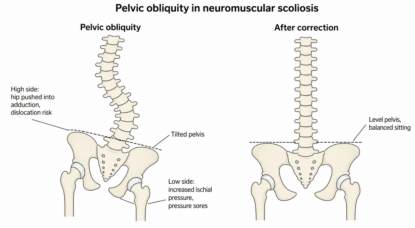
- “Pelvic Obliquity causes ischial pressure sores on the 'down' side.
- “GMFCS Level correlates directly with scoliosis risk (Level I less than 5%, Level V = 90%).
- “Beware of 'Malignant Hyperthermia' in myopathic conditions.
Overview and Epidemiology
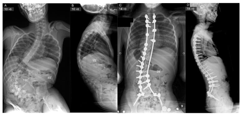
Neuromuscular scoliosis (NMS) is a spinal deformity associated with a heterogeneous group of disorders, including cerebral palsy (CP), Duchenne muscular dystrophy (DMD), spinal muscular atrophy (SMA) and spina bifida. Onset is often early, under 10 years, and most patients are wheelchair-bound (non-ambulatory).
Progression. Unlike idiopathic scoliosis, NMS curves continue to progress after skeletal maturity, because of the loss of trunk control and gravity.
Impact. A severe curve costs the patient three things. Sitting balance goes, so the hands are needed for support; the pelvis tilts, and pressure sores follow; and pulmonary function is restricted, so pneumonia becomes a risk.
- Adolescent Idiopathic
- Structural S-shape (Right thoracic)
- Neuromuscular
- Long C-shape (Thoracolumbar)
- Adolescent Idiopathic
- Rare
- Neuromuscular
- Common (Needs fixation)
- Adolescent Idiopathic
- Stops at maturity
- Neuromuscular
- Continues after maturity
- Adolescent Idiopathic
- Cosmesis + Prevent progression
- Neuromuscular
- Sitting balance + Nursing care
- Adolescent Idiopathic
- Selective (save motion)
- Neuromuscular
- Long (T2-Pelvis)
Pathophysiology and Biomechanics
The driver. The primary driver is muscle weakness in DMD and SMA, or spasticity in CP. Adolescent idiopathic scoliosis is driven by rotation; NMS is often a collapsing deformity under gravity.
Pelvic obliquity. The pelvis is the foundation of the spine, and in NMS it behaves like a tilted table. The tilt may be suprapelvic, produced by the spinal curve itself, or infrapelvic, produced by hip contracture. Either way the consequences are the same: the ischium on the low side bears the weight and ulcerates, and the hip on the high side is driven into adduction and towards subluxation or dislocation.
Classification
Gross Motor Function Classification System (CP)
The risk of scoliosis directly correlates with GMFCS level.
- Description
- Walks without limits
- Scoliosis Risk
- Low (less than 5%)
- Description
- Walks with limitations
- Scoliosis Risk
- Low-Mod
- Description
- Walks with handheld mobility
- Scoliosis Risk
- Moderate
- Description
- Self-mobility with limitations (Wheelchair)
- Scoliosis Risk
- High (~50%)
- Description
- Transported in manual wheelchair (No head control)
- Scoliosis Risk
- Very High (greater than 90%)
Clinical Assessment
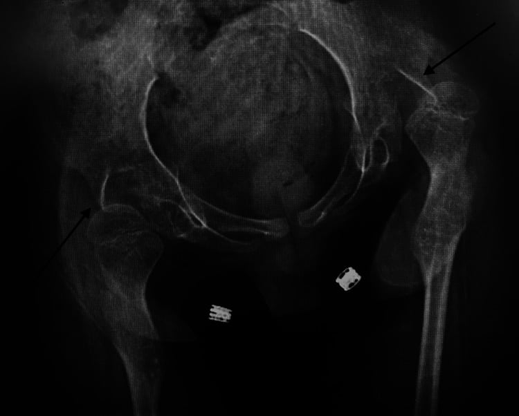
History. Pain is rare in NMS, so a painful child should make you look for a dislocated hip.
- Function: can they sit, and do they use their hands for support?
- Pain: is the curve painful?
- Pulmonary: pneumonia, ICU admissions, CPAP?
- Seizures: frequency and medications
- Nutrition: feeding tube (PEG), weight loss?
Examination. Watch the patient sit: assess sitting balance and head control, and whether they list to one side. Palpate the iliac crests with the patient sitting to judge pelvic obliquity, and look at the ischia for sores. Decide whether the curve is flexible or rigid. Then the hips: a hip flexion contracture greater than 20 degrees exacerbates lumbar lordosis or kyphosis, and look for windswept hips, one abducted and one adducted. Inspect the skin of the back and buttocks for breakdown.
Always check the hips. A dislocated hip can be the primary source of pain, not the spine.
Investigations
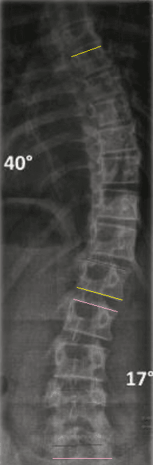
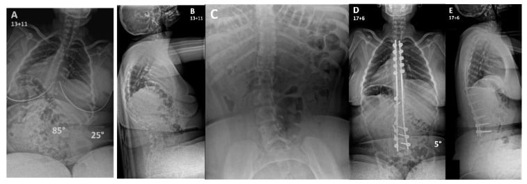
Radiographs. The spine films are taken sitting; the parameters that matter on each view are set out below.
- Parameter
- Cobb Angle
- Significance
- Magnitude of deformity.
- Parameter
- Pelvic Obliquity
- Significance
- Angle of pelvis relative to horizontal. Drives extension to pelvis.
- Parameter
- Kyphosis/Lordosis
- Significance
- NMS often kyphotic (collapsing).
- Parameter
- Flexibility
- Significance
- Determines if anterior release is needed (rare nowadays with pedicle screws).
Flexibility. Traction or bending imaging changes the operative plan by defining how much correction is achievable without excessive force.
Pre-operative Optimisation
Optimisation is a team exercise: ICU, paediatrics, respiratory medicine and a dietician.
Nutrition. The most commonly missed optimisation factor. Malnutrition is rampant in this population and G-tubes are often needed; poor nutrition means infection and wound breakdown. Albumin and pre-albumin (or transferrin) are the markers, with an albumin greater than 3.5 g/dL desired and a total lymphocyte count greater than 1500. Optimise the haematocrit, and check a coagulation profile, because valproate can affect platelets and clotting factors.
Respiratory. Pulmonary function tests and a sleep study. An FVC under 30% predicted means post-operative ventilation must be planned for, and carries a high risk of pneumonia and death; when FVC is low, plan extubation to non-invasive ventilation with mechanical cough assist.
Cardiac. DMD patients have a progressive dilated cardiomyopathy, and it is a fatal risk. A detailed echocardiogram and cardiology review are required, and the ejection fraction determines eligibility for surgery.
Seizures. Ensure valproate levels are therapeutic. Post-operative seizures can break rods.
The rest of the checklist.
- Urine: rule out UTI
- Tone: baclofen pump? spasticity?
- Infection clearance: skin, teeth, ears
- Imaging: traction films, pelvic incidence
- Osteopenia: DEXA? bisphosphonates?
- Bowels: a regimen in place
Anaesthetic Risk in Muscular Dystrophy
Not classical malignant hyperthermia. Boys with Duchenne or Becker dystrophy are not classically malignant-hyperthermia (RYR1) susceptible. What they are at high risk of is an anaesthesia-induced rhabdomyolysis with hyperkalaemic cardiac arrest, triggered by suxamethonium (succinylcholine) and the volatile anaesthetic agents. Clinically it can look MH-like: hyperkalaemia, rising end-tidal CO2, arrhythmia, dark myoglobinuric urine.
The anaesthetic. Avoid suxamethonium absolutely, because of the risk of hyperkalaemic arrest, and avoid or minimise volatile agents in favour of a total intravenous anaesthetic (TIVA) technique. Conditions with a genuine RYR1 link, central core disease for example, are truly MH-susceptible; any crisis is treated with dantrolene, cooling and management of the hyperkalaemia.
Either way the practical message in theatre is the same: declare the dystrophy, plan a trigger-free anaesthetic, and be ready for hyperkalaemia.
Differential Diagnosis
Not every curved spine in a child with disability is "simple" neuromuscular scoliosis. Distinguish the collapsing C-curve from mimics that change management.
- Curve / Clue
- Structural right thoracic S-curve, normal neurology
- Key Differentiator
- No underlying neuromuscular disease; stops at maturity; rarely needs pelvis
- Curve / Clue
- Short, sharp curve; hemivertebra/bar on X-ray
- Key Differentiator
- Vertebral anomaly from birth; screen for VACTERL, cardiac and renal anomalies
- Curve / Clue
- Variable curve plus systemic features
- Key Differentiator
- Dystrophic ribs/scalloping (NF1), arachnodactyly/lens (Marfan); dural ectasia
- Curve / Clue
- Atypical (left thoracic), painful, or rapidly progressive
- Key Differentiator
- Abnormal abdominal reflexes; MRI mandatory for atypical curves
- Curve / Clue
- Postural lean, not a fixed structural curve
- Key Differentiator
- Resolves when underlying pain treated; hip dislocation is the classic NMS trap
An atypical curve (left thoracic), a painful curve, rapid progression, abnormal abdominal reflexes, or a new neurological sign mandates whole-spine MRI to exclude a syrinx, tethered cord, or intraspinal tumour before attributing the deformity to the underlying neuromuscular condition.
Management Algorithm
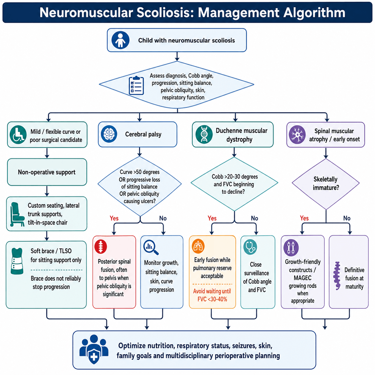
The goal. A straight spine over a level pelvis, so that the patient can sit comfortably and be nursed.
SITSurgical Goals
Hook:The goal is to help them SIT comfortably.
Role of Bracing
Non-operative seating support is the pathway for mild or flexible curves and for poor surgical candidates. Bracing itself is controversial and of limited use, because it does not halt progression in neuromuscular curves. Its purpose is sitting support, a "soft brace" or TLSO, to delay surgery until the child has reached a larger size. The price is pressure sores and a restrictive lung defect from compression of the ribs. Bracing is palliative at best.
Wheelchair Modifications
- Custom moulded seat backs
- Lateral trunk supports
- Tilt-in-space mechanisms
Surgical Technique
The "Unit Rod" vs Pedicle Screws
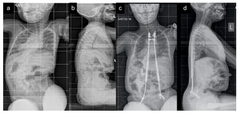
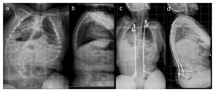
The Unit Rod (Luque-Galveston). The historic gold standard: a pre-bent U-shaped rod fixed with sublaminar wires, its legs driven into the ilium by the Galveston technique. It is cheap, and the segmental wires distribute the corrective force so pullout is less of a problem. Against it are the risk of passing wires in the canal and its limited control of lordosis.
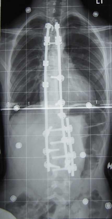
All-pedicle-screw constructs. The modern standard, with iliac screws or S2-alar-iliac (S2AI) screws for the pelvis. They give better correction and three-column fixation without entering the canal; the weakness is pullout in osteoporotic bone.
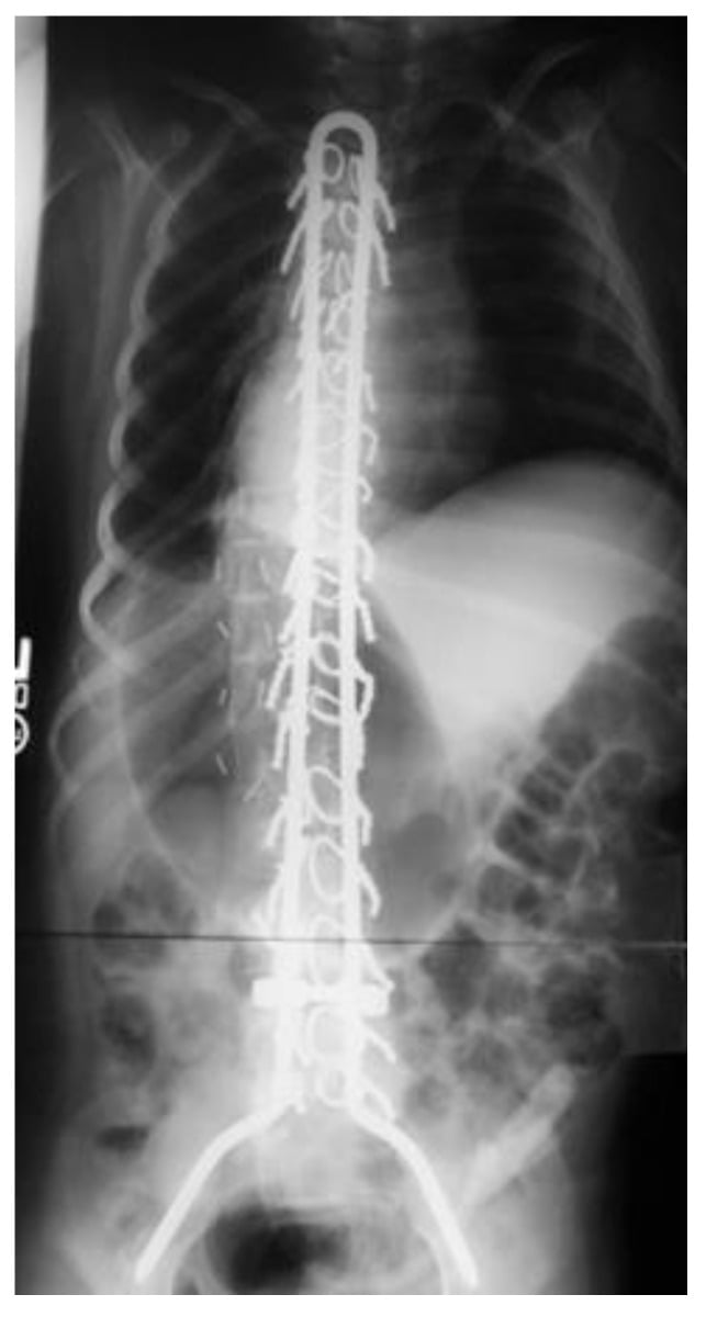
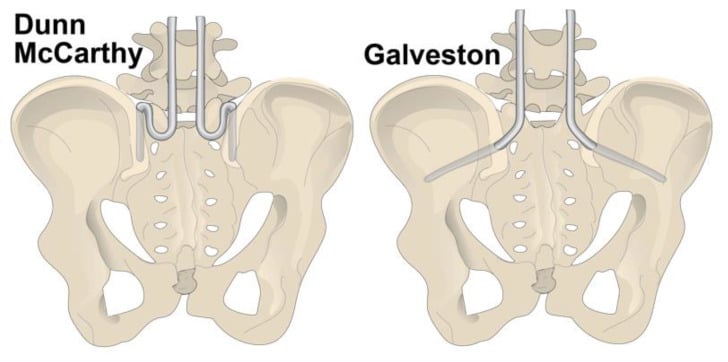
Levels. The upper instrumented vertebra is T2 or T3, to prevent proximal kyphosis. Distally the construct goes to the pelvis when obliquity is greater than 15 degrees (strictly consider it at that figure) or the patient is non-ambulatory, and stops at L5 only if the patient is ambulatory and the pelvis is level. Pelvic fixation is mandatory for obliquity.
NMS has a 5-10x higher infection rate than AIS. Vancomycin powder in the wound, betadine irrigation, optimised nutrition, and minimal theatre traffic.
Intra-operative Neuromonitoring in Neuromuscular Scoliosis
Neuromonitoring is routine in idiopathic scoliosis. The examiner wants you to know why it is different here.
Baseline signals are often unobtainable or unreliable. Many NMS patients (severe cerebral palsy, advanced DMD or SMA, myelomeningocele) have pre-existing motor and sensory deficits, so motor evoked potentials (MEPs) and somatosensory evoked potentials (SSEPs) may be absent or non-reproducible at baseline; in a substantial minority no usable trace can be obtained at all. The wake-up test is rarely feasible either, because of cognitive impairment and the inability to follow commands.
What that means. When signals are obtainable they remain valuable and should be used. When they are not, safety rests on meticulous technique: avoiding over-distraction across a rigid deformity, controlled correction, and accepting less-than-anatomic correction rather than risking the cord. Document the patient's monitorability pre-operatively and counsel the family.
The contrast with AIS. There, reliable MEP and SSEP monitoring is the expected standard, and a true alert mandates the rescue checklist (reduce correction, raise blood pressure, warm, recheck) or a wake-up test.
Complications
- Rate
- 5-15% (High)
- Management / Prevention
- Debridement, Antibiotics, Remove hardware if chronically infected. Prevention is key.
- Rate
- Variable
- Management / Prevention
- Prolonged intubation common. Pre-op PFTs mandatory.
- Rate
- 5-10%
- Management / Prevention
- Poor bone stock. Use aggressive grafting.
- Rate
- Common
- Management / Prevention
- Thin patients. Use low profile screws. Cut rod ends flush.
- Rate
- Rare
- Management / Prevention
- Loss of mesenteric fat pad after straightening. Watch for vomiting.
Postoperative Care and Rehabilitation
Recovery Pathway
- Ventilator wean (crucial in DMD)
- Pain control (epidural or PCA)
- Fluid balance
- Mobilise to wheelchair
- Resume bowel regimen
- Check the fit of the wheelchair (the back support might need modification)
- Home when feeding is tolerated and pain controlled
Outcomes
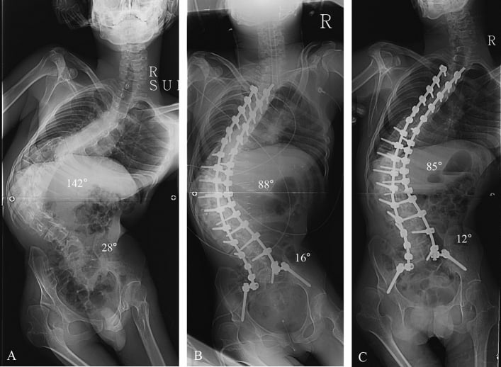
Quality of life. Parents report high satisfaction, primarily because of the ease of nursing care (transfers, bathing) and improved sitting tolerance. Correction of pelvic obliquity is the most impactful factor for sitting.
Guidelines, Registries & Global Practice
Global Epidemiology:
- Scoliosis affects roughly a quarter of all children with cerebral palsy and the large majority of GMFCS IV-V (non-ambulant) children, with prevalence and severity rising with GMFCS level and age (Persson-Bunke, total-population data).
- Essentially all boys with Duchenne muscular dystrophy who lose ambulation develop scoliosis; historically up to 90% before routine glucocorticoid use. Long-term daily corticosteroids have markedly reduced both the incidence and severity of DMD scoliosis, so fewer patients now reach surgical thresholds.
- Spinal muscular atrophy (SMA) types I-II carry the highest and earliest scoliosis burden of the flaccid neuromuscular disorders.
Side-by-Side Guidance (where recommendations differ):
- Position relevant to NMS
- Annual spine surveillance once non-ambulant; daily glucocorticoids reduce scoliosis incidence; consider surgery for progressive curves with attention to cardiac and respiratory status
- Position relevant to NMS
- Spine monitoring from diagnosis; growth-friendly instrumentation for early-onset curves; account for nusinersen intrathecal access when planning fusion
- Position relevant to NMS
- Emphasise pre-operative optimisation (nutrition, pulmonary, cardiac) and fusion to the pelvis for non-ambulant patients with pelvic obliquity
- Position relevant to NMS
- Centralised paediatric spinal services; structured cerebral palsy hip and spine surveillance pathways feed early referral
- Position relevant to NMS
- No randomised evidence; surgery decisions individualised with explicit discussion of uncertainty
- Multicentre prospective databases (e.g. the Harms Study Group / CP spine cohorts informing Yaszay 2020) provide the best available complication benchmarks: roughly a one-in-three major complication rate and a 14% spine-related reoperation rate after CP fusion.
- Population-based CP surveillance registries (Sweden's CPUP, and similar UK/Australian/North American programmes) drive GMFCS-based screening intervals.
- High-resource settings: all-pedicle-screw and S2AI constructs, intra-operative neuromonitoring, cell salvage and tranexamic acid, dedicated paediatric ICU, and multidisciplinary pre-optimisation.
- Limited-resource settings: cost-effective segmental constructs (Unit Rod / sublaminar wire systems remain valuable), selective rather than routine neuromonitoring, and greater reliance on careful patient selection where ICU and blood-bank capacity are constrained.
- Referral principle everywhere: structured hip and spine surveillance pathways frequently detect the curve before it becomes symptomatic, enabling timely referral to a spinal service.
Controversies & Areas of Uncertainty
Quality of evidence. There are no randomised trials of spinal surgery in NMS (Cochrane, DMD). Practice rests on case series and physiological reasoning, so the magnitude of benefit on survival and quality of life is genuinely uncertain.
Fuse to the pelvis or stop at L5? Pelvic fixation reliably corrects obliquity but adds operative time, blood loss and the risk of S2AI or iliac complications. In ambulant CP with a level pelvis, stopping short of the pelvis can preserve motion; in non-ambulant patients with obliquity, the pelvis is included.
Unit Rod versus all-screw. All-pedicle-screw and S2AI constructs dominate modern practice, but no high-level trial proves superiority over the Unit Rod, which remains a cost-effective, well-validated option (Tsirikos: 68% curve and 71% obliquity correction).
Timing in DMD. Earlier surgery preserves a respiratory window, but glucocorticoids have reduced curve incidence/severity, so fewer boys now reach surgical thresholds and some are managed without fusion. The classic "fuse early, by 20-30 degrees" rule is being re-examined in the steroid era.
Growing constructs in SMA. Magnetically controlled growing rods reduce repeat lengthening surgeries but carry implant-related and infection risks, and intrathecal nusinersen access must be planned around any spinal construct.
BMP and graft choice. Osteopenic, low-volume local bone drives interest in BMP and allograft, but BMP is off-label in children and its safety profile in this population is not established.
MCQ Practice Points
Q: What is the operative threshold for scoliosis in Duchenne Muscular Dystrophy? A: Cobb angle greater than 20-30 degrees (much lower than the 45-50 degrees for AIS).
Q: In a non-ambulatory CP patient with pelvic obliquity, where should the fusion stop distally? A: The Pelvis (Iliac/S2AI screws). Stopping at L5 or S1 has a very high failure rate.
Q: How does the infection rate of NMS surgery compare to AIS? A: It is significantly higher (5-15% vs less than 1%).
Q: Which neuromuscular condition is associated with Malignant Hyperthermia? A: Duchenne Muscular Dystrophy (and Central Core Disease). Succinylcholine is contraindicated.
Q: Is spondylolisthesis common in NMS? A: No, spondylolisthesis is associated with walking (repetitive stress). NMS patients usually have long kyphoscoliotic C-curves.
Q: In which NMS condition is cord tethering most common? A: Myelomeningocele (Spina Bifida). Almost all have tethered cords.
Clinical Decision Scenarios
Practise clinical reasoning and management decisions out loud
“A 12-year-old boy with GMFCS V Cerebral Palsy presents with a 60 degree thoracolumbar neuromuscular scoliosis and pelvic obliquity. He is finding it hard to sit in his wheelchair.”
“A 14-year-old boy with Duchenne Muscular Dystrophy (DMD) has a 25 degree scoliosis. His FVC is 50% predicted.”
“You performed a T2-Pelvis fusion on a CP patient. 3 weeks post-op, the wound is dehiscencing and draining serous fluid.”
Optimization Checklist
- Nutrition (Albumin greater than 3.5)
- Lungs (FVC greater than 30%)
- Heart (Echo for DMD)
- Seizures (Controlled)
- Bowels (Regimen)
Surgical Principles
- Fuse T2 to Pelvis
- Correct Pelvic Obliquity
- Use TXA (High blood loss)
- Vancomycin powder (Infection)
Condition Specifics
- CP: GMFCS V most at risk
- DMD: Fuse early (greater than 20 deg)
- SMA: Growing rods often needed
- Myelomeningocele: Latex allergy / Tethering
Evidence Base
GMFCS Level and Scoliosis Risk (Total Population)
- Prospective total-population CP registry of 666 children aged 4-18 years
- 28% had clinical scoliosis (17% mild, 11% moderate/severe)
- Risk of scoliosis rose with GMFCS level and age
- GMFCS IV-V: ~50% risk of moderate/severe scoliosis by age 18; GMFCS I-II: almost no risk. Most curves were diagnosed after age 8
- CLINICAL EXAMINATION OVER-CALLS THE DIAGNOSIS: 28% were scoliotic on clinical assessment, but radiographs confirmed a Cobb angle above 10 degrees in only 54 children (8%) and above 20 degrees in 45 (7%)
Posterior Fusion Slows Respiratory Decline in DMD
- 56 DMD patients; %FVC tracked before and after posterior spinal fusion
- Whole-cohort rate of FVC decline fell from 4% per year pre-op to 1.75% per year post-op (p less than 0.0001)
- Paired subgroup: decline fell from 8.0% to 3.9% per year
- Fusion did not reverse decline but significantly slowed it
No RCT Evidence for DMD Scoliosis Surgery (Cochrane)
- Systematic review seeking RCTs of spinal surgery for DMD scoliosis
- 47 relevant studies screened; none met inclusion (all case series, no randomised/quasi-randomised trials)
- No evidence-based recommendation could be made for or against surgery
- Benefits on survival, respiratory function and quality of life remain unproven by high-level evidence
Major Complications After Spinal Fusion in Cerebral Palsy
- Prospective multicentre database of 257 CP patients with minimum 2-year follow-up
- 78 patients (30%) had at least one major complication; overall 36% complication rate
- Deep infection 4.7% perioperatively plus 3.1% delayed; prolonged ventilation 8.2%
- Spine-related reoperation rate 14.0%; unplanned return to theatre 16% overall (8.6% perioperative, 7.8% delayed)
- ELEVEN PATIENTS (4.3%) DIED between 3 months and 5.6 years after surgery, four of them within the first year - and TWO of those deaths were judged DIRECTLY RELATED TO THE SPINAL DEFORMITY SURGERY
Unit Rod Instrumentation for CP Scoliosis
- Retrospective series of 287 CP children treated with Unit Rod instrumentation to the pelvis
- Scoliosis corrected from mean 76 to 25 degrees (68%); pelvic obliquity from 17 to 5 degrees (71%)
- Deep wound infection 4.2% early and 2.5% late; three perioperative deaths
- Caregiver satisfaction 96%
- THE SCALE OF THE UNDERTAKING, which is usually left out: mean blood loss was 2.8 L for posterior-only and 3.4 L for combined anterior-posterior procedures, with mean ICU stays of 4.9 and 6.7 days and mean hospital stays of 19.6 and 24.5 days
Sacral Alar-Iliac (S2AI) Pelvic Fixation
- Describes the S2-alar-iliac technique: screw one-third in sacral ala, two-thirds in ilium, in line with cephalad anchors
- Low-profile, in-line entry avoids the offset connectors and prominence of classic iliac screws
- Referenced pediatric series reported ~70% pelvic obliquity and ~67% coronal Cobb correction
- Screws of 9 mm or greater outer diameter recommended to prevent breakage
Surgery in Severe DMD With FVC Under 30%
- 14 DMD patients with forced vital capacity under 30% predicted
- Pre-operative inspiratory muscle training raised %FVC before surgery
- All-screw constructs corrected coronal curve and pelvic obliquity, maintained long-term
- Mean %FVC decline 3.6% per year after surgery; high patient and parent satisfaction