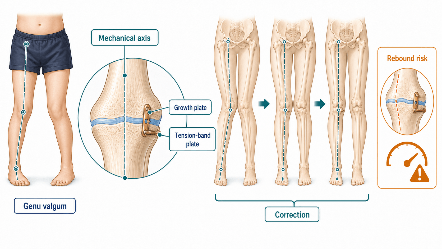
Mechanical axis planning, temporary hemiepiphysiodesis and rebound surveillance
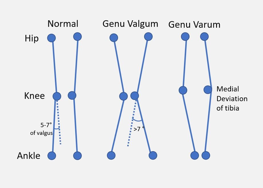
- Separate physiological genu varum or valgum from pathological deformity before planning treatment.
- Plan from standing hip-to-ankle radiographs, not cropped knee images.
- Mechanical axis deviation tells you the effect; LDFA, MPTA and JLCA help identify the source.
- Guided growth requires open physes and enough growth remaining.
- Correction happens over months and follow-up continues after implant removal because rebound can occur.
- “The implant is small; the analysis is the operation.
- “The mechanical axis tells you where load passes, not why it got there.
- “Treat rickets, Blount disease and physeal bars as specific diagnoses, not as simple knock-knee or bow-leg.
- “Guided growth does not correct rotation, severe joint incongruity or a child who is too close to maturity.
A knee that looks valgus or varus is not enough. Confirm mechanical axis, localise the bone and physis causing deformity, assess growth remaining and identify pathological causes before implanting a plate.
Images and Diagrams
- Answer
- Standing hip-to-ankle radiograph.
- Clinical use
- Shows global load axis and both limbs.
- Answer
- How far the mechanical axis is from knee centre.
- Clinical use
- Quantifies the effect on load transfer.
- Answer
- LDFA, MPTA and JLCA.
- Clinical use
- Separates femur, tibia, joint or combined source.
- Answer
- Rebound, undercorrection or overcorrection.
- Clinical use
- Follow-up continues after implant removal.
AXISPlanning Sequence
Hook:AXIS keeps planning grounded in full-length alignment.
PLATEGuided Growth Mechanics
Hook:PLATE links the implant to timing and follow-up.
REBOUNDAfter Removal
Hook:REBOUND makes follow-up after removal explicit.
Overview/Epidemiology
Coronal lower-limb alignment changes normally during childhood. Infants often have genu varum, toddlers pass into genu valgum, and older children gradually settle toward adult alignment. Guided growth is for the child whose alignment is not following that expected pathway or whose deformity is symptomatic, asymmetric, progressive or pathological.
The common clinical problem is genu valgum or genu varum around the knee. The important clinical question is not just whether the knees touch or the ankles separate. The important question is where the mechanical axis passes, what bone creates the deformity, whether the physis is open, how much growth remains, and whether the underlying biology is normal.
Typical indications include persistent genu valgum, pathological genu varum, Blount disease in selected stages, post-traumatic physeal disturbance, skeletal dysplasia and metabolic bone disease after medical optimisation. Guided growth is smaller than osteotomy, but it is not casual. It requires reliable follow-up, timely implant removal and continued surveillance for rebound.
Pathophysiology
Angular deformity develops when growth is asymmetric across a physis or when a bone or joint segment has abnormal orientation. The mechanical axis is the functional consequence: it shows where load passes from the hip to the ankle across the knee. A medial mechanical axis overloads the medial side; a lateral mechanical axis overloads the lateral side.
Guided growth works by temporary hemiepiphysiodesis. A plate, screw construct or other tether slows one side of an open physis. The other side continues to grow, gradually changing the joint orientation and mechanical axis. The correction rate depends on age, remaining growth, which physis is used, the underlying diagnosis and distance from the knee.
Pathological physes behave differently. Blount disease has disordered medial proximal tibial growth and may recur or fail to correct if severe. Rickets and metabolic disease need medical correction before mechanical correction. Post-traumatic physeal bars can create focal angular growth disturbance and may require bar resection, epiphysiodesis or osteotomy rather than a simple plate.
Classification
- Physiological alignment: symmetric, painless, age-appropriate and improving.
- Pathological valgum or varum: persistent, progressive, asymmetric, painful or outside expected developmental range.
- High-risk pattern: lateral thrust, severe obesity, short stature, rickets signs, skeletal dysplasia or prior physeal injury.
- Functional problem: pain, fatigue, patellar symptoms, gait difficulty or sport limitation.
Blount Disease: Langenskiold Staging and the Drennan Angle
Blount disease (tibia vara) is the key pathological cause of genu varum to separate from physiological bowing, and it has its own staging and a measurement that the topic's vivas demand.
the angle between a line across the proximal tibial metaphyseal beaks and a line perpendicular to the tibial diaphyseal axis.
- Under about 10 degrees - usually physiological bowing.
- Over about 16 degrees - likely pathological (Blount disease).
- About 10 to 16 degrees - indeterminate, warranting close follow-up.
a radiographic progression of the medial proximal tibial physis and epiphysis in infantile (early-onset) Blount disease - from medial metaphyseal beaking and irregularity (stages I-II), through progressive medial physeal depression and fragmentation (III-IV), to a bony bar across the medial physis with a sloped epiphysis (V-VI).
- Lower stages in a young child may respond to bracing and then guided growth.
- Higher stages (especially V-VI, with a medial physeal bar) and late-onset / high-BMI disease tend to need osteotomy (often with bar resection), because growth modulation alone undercorrects and the implants break (see the Blount-specific breakage data in the Evidence Base).
Early-onset (infantile, under about 3 to 4 years) versus late-onset (juvenile/adolescent): early-onset is bilateral in up to half, linked to early walking and obesity; late-onset tracks adolescent obesity and is more often unilateral with less growth remaining.
Separate physiological bowing from Blount with the Drennan metaphyseal-diaphyseal angle (over about 16 degrees pathological, under about 10 degrees physiological), then grade infantile disease by the Langenskiold stages I to VI. A medial physeal bar (high Langenskiold stage), late-onset disease and high BMI shift treatment from bracing/guided growth toward osteotomy.
Clinical Presentation
History
Ask about age at onset, progression, symmetry and function. Pain, lateral thrust, fatigue, patellofemoral symptoms and unilateral progression are more concerning than cosmetic concern alone. Ask about previous physeal injury, infection, fracture, metabolic disease, vitamin D risk, renal disease, skeletal dysplasia features, family history and obesity.
Ask the family whether follow-up is practical. Guided growth is a commitment to serial imaging and planned hardware removal.
Examination
Observe the child standing and walking. Look for varus thrust, patellar tracking, foot progression, limb length difference, pelvic level and rotational profile. Measure intercondylar or intermalleolar distance as a clinical baseline, but do not use it as a substitute for full-length alignment.
Assess:
- Hip, knee and ankle range of motion.
- Coronal alignment and patella direction.
- Ligament laxity and joint line opening.
- Lateral thrust in varus knees.
- Rotational profile so torsion is not mistaken for coronal deformity.
- Signs of rickets, skeletal dysplasia or systemic disease.
- Skin, BMI and soft-tissue envelope for implant planning.
A guided-growth plan starts with a standing alignment film and source analysis, not with choosing a plate size.
Investigations
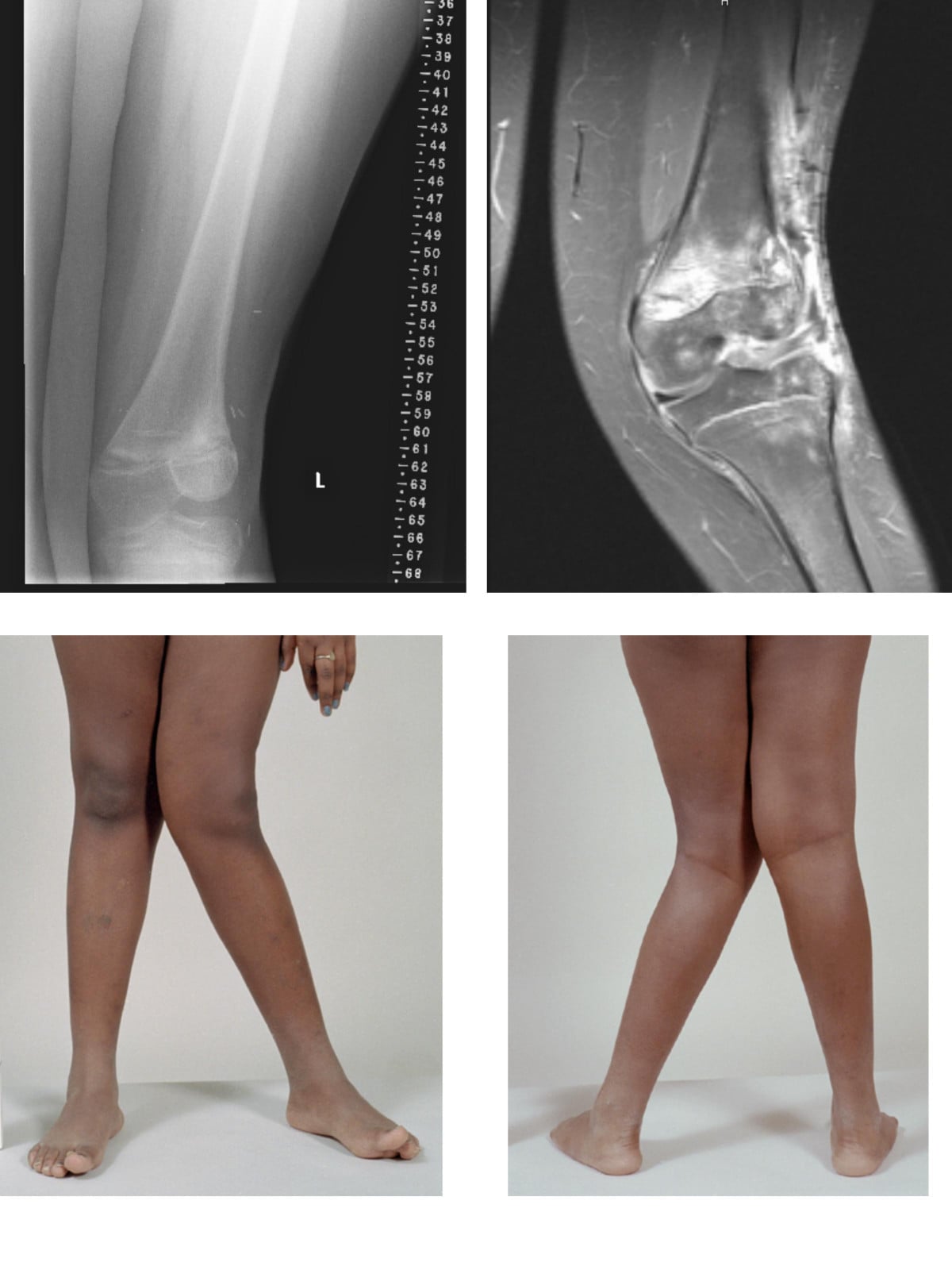
The key investigation is a standing hip-to-ankle radiograph with patellae facing forward where possible. This allows mechanical axis deviation, LDFA, MPTA and joint line convergence to be assessed. A cropped knee film can miss femoral, tibial, limb-length or global alignment contribution.
- Investigation
- Standing hip-to-ankle radiograph
- Decision it informs
- Measures mechanical axis deviation and bilateral alignment.
- Investigation
- LDFA, MPTA and JLCA
- Decision it informs
- Localises femur, tibia, joint or combined source.
- Investigation
- Bone age and maturity assessment
- Decision it informs
- Determines guided-growth feasibility and timing.
- Investigation
- Vitamin D, calcium, phosphate, ALP, renal/endocrine tests when indicated
- Decision it informs
- Optimises metabolic disease before mechanical correction.
- Investigation
- Sagittal and rotational assessment, CT only when needed
- Decision it informs
- Prevents using coronal guided growth for the wrong problem.
Normal Joint-Orientation Angles and the Malalignment Test
The angles named above are only useful against their normal values, and the malalignment test (Paley) is the systematic way to turn a standing film into a deformity diagnosis.
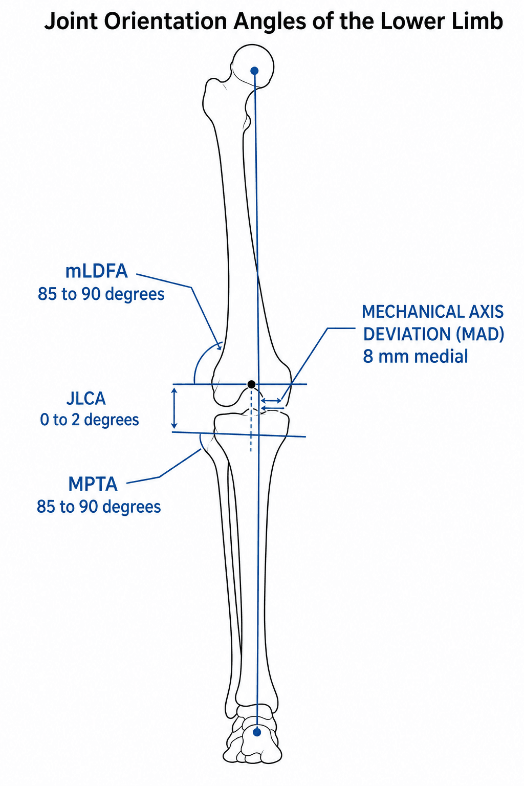
- Approximate normal
- About 8 mm (plus or minus 7) medial to the knee centre
- What it localises
- Overall coronal malalignment / load path
- Approximate normal
- About 85-90 degrees (mean ~88)
- What it localises
- Distal femoral source (raised = femoral varus; lowered = femoral valgus)
- Approximate normal
- About 85-90 degrees (mean ~87)
- What it localises
- Proximal tibial source (lowered = tibial varus; raised = tibial valgus)
- Approximate normal
- About 0-2 degrees
- What it localises
- Intra-articular / ligamentous / cartilage-wear contribution
The malalignment test - a stepwise read:
- Measure MAD on the standing hip-to-ankle film. The mechanical axis normally passes just medial to the knee centre; the tibial plateau can be divided into zones, and a line falling into the medial or lateral outer zones confirms significant deviation.
- Find the femoral contribution with the mLDFA - if abnormal, the distal femur is a (or the) source.
- Find the tibial contribution with the MPTA.
- Account for the joint with the JLCA - a raised value means part of the "deformity" is intra-articular or ligamentous (joint-line opening or cartilage wear), which guided growth will not correct.
mLDFA and MPTA are each about 85-90 degrees; normal MAD is roughly 8 plus or minus 7 mm medial to the knee centre; JLCA is about 0-2 degrees. Work MAD then mLDFA then MPTA then JLCA: this tells you whether the deformity is femoral, tibial, both, or intra-articular - and an elevated JLCA warns that a tension-band plate will not fix a joint-line problem.
Differential Diagnosis
- Physiological genu varum or genu valgum.
- Infantile or adolescent Blount disease.
- Nutritional rickets or hypophosphataemic rickets.
- Renal osteodystrophy or endocrine bone disease.
- Post-traumatic physeal bar.
- Skeletal dysplasia.
- Ligamentous laxity or joint line convergence.
- Rotational profile creating apparent coronal malalignment.
- Limb-length difference with pelvic compensation.
Management

- Best fit
- Observation and explanation
- Reasoning
- Physiological varus and valgus usually improve without surgery.
- Best fit
- Medical optimisation first
- Reasoning
- A plate cannot correct abnormal growth biology if rickets or renal osteodystrophy remains untreated.
- Best fit
- Temporary hemiepiphysiodesis
- Reasoning
- Gradual correction can avoid osteotomy when follow-up and timing are reliable.
- Best fit
- Osteotomy or complex deformity correction
- Reasoning
- Growth modulation is too slow or too limited for the problem.
- Best fit
- MRI or CT mapping, bar resection or epiphysiodesis/osteotomy depending size and growth remaining
- Reasoning
- The growth arrest is focal and may not be solved by a simple tension-band plate.
Observe physiological symmetric age-appropriate alignment. Explain natural history and review triggers. Treat metabolic disease before mechanical surgery. In obesity or Blount risk, address modifiable load and assess for lateral thrust, progression and joint depression.
- What the surgeon does
- Mark the mechanical axis, identify the abnormal physis and choose medial or lateral tethering from LDFA, MPTA and JLCA.
- Safety point
- Wrong-side or wrong-level tethering is a planning error, not a technical complication.
- What the surgeon does
- Supine positioning with fluoroscopy; confirm true AP and lateral views of the target physis.
- Safety point
- Poor intra-operative imaging risks screw placement across the physis or wrong plate position.
- What the surgeon does
- Small medial or lateral incision centred on the physis, protecting periosteum and soft tissues.
- Safety point
- Avoid damaging the physis; the implant should act as a tether, not a physeal bar.
- What the surgeon does
- Place tension-band plate, staple or transphyseal screw according to surgeon preference and diagnosis.
- Safety point
- Screws should hold epiphysis and metaphysis without crossing into the joint.
- What the surgeon does
- Serial standing alignment radiographs until target correction, then planned removal.
- Safety point
- The operation fails if correction is not tracked or removal is missed.
- What the surgeon does
- Continue alignment surveillance for rebound, especially in young children and pathological physes.
- Safety point
- Removal is not discharge.
Complications
Early
- Wrong diagnosis: physiological alignment treated surgically.
- Wrong level or wrong side tethering.
- Infection, wound irritation or hardware prominence.
- No correction because growth remaining is inadequate.
- Family lost to follow-up before planned removal.
Late
- Overcorrection.
- Undercorrection.
- Rebound deformity after removal.
- Hardware breakage or migration.
- Physeal tethering or bar formation.
- Need for repeat guided growth or osteotomy.
Guided growth is not finished in theatre. The correction happens on serial alignment films, and the endpoint is missed if follow-up is missed.
Decision-Making in Practice
Guided growth is a timed growth-modulation operation. It is not an acute correction. The child must have enough growth remaining, the deformity source must be correctly identified, and the family must understand that overcorrection, undercorrection and rebound are surveillance problems.
- How to decide
- Age, symmetry, progression, pain, height, metabolic disease and family pattern
- Management consequence
- Physiological alignment is observed; pathological deformity is measured
- How to decide
- Standing hip-to-ankle film with MAD, LDFA, MPTA and JLCA
- Management consequence
- Implant the correct side of the correct physis
- How to decide
- Bone age, chronological age, pubertal stage and growth velocity
- Management consequence
- Too little growth means osteotomy may be required
- How to decide
- Diagnosis, size, physis, surgeon preference and recurrence risk
- Management consequence
- Tension-band plates, staples or screws have different behaviour
- How to decide
- Serial axis correction and target overcorrection when appropriate
- Management consequence
- Prevents overshoot and manages rebound risk
For genu valgum or varum, treatment begins with the full-length standing film, not a knee-only radiograph. The question is whether deformity arises from distal femur, proximal tibia, both, joint-line obliquity, ligament laxity or extra-articular deformity. Guided growth works poorly if the wrong physis is tethered or if the deformity is mainly intra-articular.
Osteotomy is preferred when correction is urgent, growth remaining is insufficient, deformity is severe and rigid, there is joint depression, or the child is near maturity. Guided growth is preferred when gradual correction is safe, there is enough growth remaining, and the joint surface is acceptable.
Clinical Reasoning Notes
Structured clinical approach
Use a structured sequence:
- Define physiological versus pathological alignment.
- Describe symptoms, progression and asymmetry.
- Obtain standing hip-to-ankle films.
- State mechanical axis deviation.
- Use LDFA, MPTA and JLCA to localise source.
- Check growth remaining and bone age.
- Look for metabolic disease, Blount disease or physeal bar.
- Choose observation, medical treatment, guided growth or osteotomy.
- Plan follow-up, removal timing and rebound surveillance.
Common pitfalls
- Planning from a knee-only X-ray.
- Operating on physiological alignment.
- Ignoring bone age.
- Forgetting rickets or renal bone disease.
- Confusing rotational deformity with coronal deformity.
- Using guided growth when osteotomy is required.
- Not removing implants on time.
- Discharging after removal without rebound surveillance.
Guidelines, Registries and Global Practice
Guided growth is practised worldwide, but the supporting framework is principle-based and society reviews rather than randomised trials or implant registries, because these are paediatric soft-tissue-sparing procedures in growing bone.
Global epidemiology
- Physiological genu varum is usual in infancy and physiological genu valgum peaks around age 3 to 4 years, settling toward adult alignment (mild valgus) by about age 7, across populations.
- Idiopathic genu valgum needing intervention is uncommon; most referrals are physiological and resolve with observation.
- Blount disease prevalence varies by region and body habitus: infantile (early-onset) disease is more frequent where early walking and higher childhood BMI are common, while late-onset (adolescent) disease tracks adolescent obesity.
- Nutritional rickets remains a leading cause of pathological bowing in limited-resource settings, whereas X-linked hypophosphataemic rickets dominates where nutrition is adequate.
Side-by-side guidance
- Emphasis for paediatric angular deformity
- Distinguish physiological from pathological alignment; image with full-length standing radiographs before any intervention.
- Emphasis for paediatric angular deformity
- Reserve surgery for progressive, asymmetric or pathological deformity; observation and natural-history counselling for physiological alignment.
- Emphasis for paediatric angular deformity
- Deformity analysis (mechanical axis, joint orientation angles, CORA) drives the choice between growth modulation and osteotomy.
- Emphasis for paediatric angular deformity
- Endorse temporary hemiepiphysiodesis as first line for suitable coronal deformity with open physes, with planned removal and rebound surveillance.
The common thread across societies is that the analysis (full-length alignment, source localisation, growth remaining and underlying diagnosis) governs the decision, not the appearance of the knee.
Registry and evidence notes
- There is no dedicated international guided-growth implant registry comparable to arthroplasty registries (NJR, AJRR, AOANJRR); evidence comes from multicentre series such as the multinational tension band plating cohort of 967 physes.
- Best available data are cohort-level (Level III to IV), so counselling rests on pooled series rather than registry survivorship.
High- versus limited-resource practice
- Where full-length standing radiography and reliable follow-up are available, tension band plating with planned removal is standard.
- Where imaging or follow-up is limited, percutaneous transphyseal screws (lower cost, low profile) or single-stage osteotomy may be preferred, because guided growth fails without serial alignment review.
- In settings with high nutritional rickets burden, medical optimisation precedes any mechanical correction, and untreated metabolic disease is a frequent reason guided growth fails.
Controversies and Areas of Uncertainty
Guided growth is established, but several questions remain genuinely contested and are favourite viva territory.
- One view
- Aim past neutral to offset expected rebound in young, fast correctors.
- Counter view
- Overcorrection risks creating the opposite deformity and a second operation; aim for neutral and watch.
- One view
- Tension band plates allow faster, growth-arrest-sparing correction than staples.
- Counter view
- Percutaneous transphyseal screws are lower profile and cheaper; staples remain effective in some hands.
- One view
- Remove promptly at neutral to avoid overcorrection.
- Counter view
- Some retain the plate as a guide during continued growth, accepting closer review.
- One view
- Effective in selected early or mild cases and avoids osteotomy.
- Counter view
- High implant breakage and undercorrection in late-onset, high-BMI, severe varus argue for osteotomy.
- One view
- Earlier intervention uses abundant growth and is gentle.
- Counter view
- Very young children rebound more and may simply be physiological; observation may suffice.
Clinical Decision Scenarios
Practise clinical reasoning and management decisions out loud
“A ten-year-old has progressive genu valgum. How do you plan guided growth?”
“A child with genu varum has lateral thrust and obesity. What are you worried about?”
“An eight-year-old corrected to neutral had her tension band plate removed six months ago. The latest standing film shows the valgus is returning. How do you explain and manage this?”
Measure
- Standing long-leg film
- MAD
- LDFA
- MPTA
- JLCA
- Bone age
Indications
- Progressive
- Asymmetric
- Symptomatic
- Outside age range
- Growth remaining
Treatment
- Observe physiological
- Correct metabolic disease
- Guided growth
- Osteotomy
- Monitor rebound
Pitfalls
- Wrong source
- No follow-up
- Overcorrection
- Rebound
- Rotation ignored
Evidence Signals
Guided growth principles and indications
- Temporary hemiepiphysiodesis tethers one side of an open physis to allow differential growth and correct coronal deformity around the knee.
- Staples, percutaneous transphyseal screws and tension band plates are all described constructs.
- Undercorrection and overcorrection are common; careful preoperative planning and follow-up minimise complications.
Multinational tension band plating series defines correction rate
- 967 physes in 537 patients across multiple centres; mean age at implantation 11.4 years.
- Calculated correction rate was 0.77 degrees per month for the femur and 0.79 degrees per month for the tibia.
- Infection rate 1.48 percent and screw breakage 0.55 percent; age at implantation and direction of deformity influenced correction.
Tension band plate corrects faster than stapling
- 34 patients with 65 deformities treated by a non-locking tension band plate; 32 of 34 patients (63 of 65 deformity levels, 97%) corrected to neutral at a mean of 11 months.
- Correction was approximately 30 percent faster than reported with stapling, with no permanent growth arrest.
- Four idiopathic genu valgum patients rebounded and underwent repeat guided growth; two late-onset Blount cases undercorrected.
Evidence Base
Rebound predictors after stapling for genu valgum
- 37 limbs with idiopathic genu valgum followed to maturity or beyond two years after staple removal.
- Rate of correction, BMI, age and initial valgus angle were independent predictors of rebound on multivariate analysis.
- Rapid correctors had a 79 percent rebound incidence (mean 4 degrees); slow correctors with low BMI had less rebound, and none when BMI was at or above 21.
Rebound is common and driven by young age and fast correction
- 100 patients (189 legs) with idiopathic genu valgum analysed on serial long-leg radiographs.
- 59 percent of legs showed rebound growth after plate removal, 30 percent stayed stable and 11 percent continued correcting.
- Sex, age, baseline mechanical axis deviation, mLDFA, mMPTA and the correction rate were significant, with correction rate carrying the highest odds ratio.
Implant breakage is far higher in Blount disease
- 246 tension band implant procedures in 113 patients with Blount disease across 8 centres.
- Overall implant breakage 12 percent (mean 1.6 years after implantation), mostly metaphyseal screws.
- Higher BMI and greater varus deformity raised breakage risk; solid 3.5 mm screws had a 50 percent breakage rate in late-onset Blount disease.
Femoral deformity contributes to malalignment in tibia vara
- 127 limbs with tibia vara reviewed; many had concurrent femoral varus contributing to overall malalignment.
- Femoral lateral tension band plating corrected mLDFA from 98 to 87 degrees, with 80 percent achieving full femoral correction.
- In early-onset tibia vara, 73 percent of femurs corrected with tibial plating alone, supporting observation of the femur in young children.
Physiological alignment and red flags
- Physiological varus and valgus follow a predictable age-related course in young children.
- Pathological features include asymmetry, progression, pain, short stature and systemic disease.
- Treatment depends on diagnosis and growth remaining.