Cuboid Compression | Lisfranc Association | Lateral Column Shortening
- Nutcracker pattern is cuboid compression during forefoot abduction/axial load; real injuries are often more complex than the eponym
- Inspect Chopart, Lisfranc and both columns rather than assuming every cuboid fracture is isolated
- Measure articular congruity and lateral-column length on appropriate radiographs and CT when morphology is uncertain
- ORIF is not required for every displacement label; centring, congruity, shortening, stability, soft tissue and patient factors matter
- Support the actual post-disimpaction void with graft or substitute only when needed
- “Nutcracker describes a compression morphology, not a complete treatment diagnosis
- “Actively assess calcaneocuboid, tarsometatarsal, navicular and medial-column injuries
- “Bone graft is conditional on the defect left after disimpaction
- “Restore articular congruity and lateral-column length without over-lengthening
Overview and Epidemiology
Nutcracker fractures are cuboid compression injuries that may depress articular surfaces and shorten the lateral column. They range from isolated stable fractures to complex Chopart/Lisfranc injuries, and treatment follows actual congruity, length, stability and associated injury rather than the eponym alone. Describe the cuboid impaction, the articular surfaces, lateral-column length and every associated Chopart/Lisfranc component, then justify observation, fixation, distraction and grafting from those findings.
Mechanism. The usual mechanism is loaded forefoot abduction, which can compress the cuboid between the calcaneus and the lateral metatarsal bases. The impaction may involve the calcaneocuboid or fourth/fifth tarsometatarsal surfaces, and the peroneal groove and surrounding midfoot may be involved as well. Lateral-column shortening is possible but not universal, and isolated low-energy and paediatric injuries also occur.
Frequency and evidence. The fracture is rare compared with metatarsal and calcaneal fractures. Published evidence is dominated by case reports and small heterogeneous series, almost entirely Level IV/V, and there are no randomised trials. Associated midfoot injury and intra-articular involvement worsen prognosis more consistently than one displacement number.
Anatomy and Pathophysiology
The cuboid. It lies in the lateral midfoot and is the keystone of the lateral column, maintaining the column's length. Its blood supply comes from branches of the dorsalis pedis and lateral plantar arteries. It articulates with:
- The calcaneus, proximally
- The fourth and fifth metatarsals, distally
- The navicular and the lateral cuneiform, medially
The lateral column. The calcaneus, cuboid and fourth and fifth metatarsals make up the lateral column, which gives the foot its lateral stability and carries weight.
How the fracture does harm. The calcaneocuboid and lateral tarsometatarsal articulations transmit compression across the cuboid. Comminution or depression may create an articular step and shorten the lateral column, and involvement of the peroneus longus groove may add tendon symptoms.
The harm arises from the combination of articular incongruity, instability and length loss, and not every fracture has all three. A shortened lateral column may promote abduction and midfoot imbalance, but over-distraction can also be harmful.
Peroneus Longus and the Peroneal Groove of the Cuboid
Every operative account of a nutcracker fracture says "protect the peroneal tendons", and the os peroneum appears in the differential. The surgical anatomy behind both is what makes the lateral approach and plantar-lateral hardware placement hazardous.
Course. The peroneus (fibularis) longus tendon curves behind the lateral malleolus, passes plantar to the peroneal tubercle of the calcaneus, then turns sharply medially into a distinct groove, the peroneal sulcus, on the plantar-lateral surface of the cuboid. There it runs in a fibro-osseous tunnel roofed by the long plantar ligament, before crossing the sole obliquely to insert on the base of the first metatarsal and the medial cuneiform.
Function. It plantarflexes the first ray and everts the foot. It is an important dynamic stabiliser of the lateral column and transverse arch, the very column the nutcracker fracture shortens.
The os peroneum. This sesamoid within the peroneus longus tendon lies at or just distal to the inferolateral cuboid. It is present (ossified) in a substantial minority of feet and is frequently bilateral. On radiographs it can be mistaken for an avulsion fracture of the cuboid, so a smooth, corticated, often bilateral ossicle at the plantar-lateral cuboid should not be over-called as an acute fragment. Acute rupture of the peroneus longus can fracture or diastase the os peroneum (the "painful os peroneum syndrome"), a separate lateral-foot pathology.
Why it matters in ORIF. The lateral (longitudinal, Ollier-type) approach to the cuboid passes over or between the peroneal tendons, and the peroneus longus, peroneus brevis and sural nerve lie directly in the surgical field, to be identified, protected and retracted. A plantar-lateral impaction fragment can involve the tendon's tunnel, and a prominent plantar-lateral screw or plate edge can impinge on or entrap the peroneus longus, so plantar-lateral hardware is kept low-profile and screws out of the groove. Lateral or plantar-lateral foot pain after cuboid fixation should therefore prompt assessment of the peroneus longus, not just the healing fracture.
Classification Systems
The 2020 critical analysis review by Engelmann, Rammelt and Schepers states explicitly that no cuboid classification has been validated for guiding management or predicting outcome. The Fenton system is descriptive, and was derived in part from paediatric cohorts; treatment is principle-based (restore congruity, length and stability), not classification-driven.
A working grouping. The grouping below is a teaching device for organising an operative plan, not a tool for giving a patient a number. No published series reports outcomes by these groups, and none reports the severe-impaction group separately: the pattern guides the operative approach, it does not give a prognosis.
- What it is
- Impaction with metaphyseal bone loss; lateral column shortened
- Treatment
- ORIF, filling the void if disimpaction leaves one
- What it is
- The commoner situation
- Treatment
- Address both; the midfoot injury governs the construct
- What it is
- Disimpaction leaves a void that will collapse without support
- Treatment
- ORIF with structural graft, bridge plating or distraction
Degree of impaction. Impaction determines the graft type. Severe impaction means significant bone loss and is treated by ORIF with structural graft. Mild impaction means minimal bone loss and is treated by ORIF with cancellous graft.
Clinical Assessment
History. Lateral foot pain, swelling localised to the lateral midfoot and pain on weight bearing follow forced plantarflexion and abduction of the foot. High-energy trauma, a Lisfranc injury mechanism and midfoot instability are the risk factors.
Examination. Inspect for swelling over the lateral midfoot, ecchymosis (which may be delayed) and, if the fracture is displaced, the deformity of lateral column shortening. Then examine for:
- Tenderness over the cuboid, and over the Lisfranc joint if there is an associated injury
- Lateral column instability, and pain on lateral column stress
- Limited, painful midfoot motion, with painful inversion, eversion, plantarflexion and dorsiflexion
- Midfoot alignment, checking for collapse
The associated injury. Isolated cuboid fractures exist, but associated Chopart, Lisfranc, navicular and other column injuries must be actively sought and treated on their own merits. Perform a Lisfranc stress test for midfoot instability, assess midfoot stability and obtain CT to evaluate the whole midfoot. Routinely assess for compartment syndrome in higher-energy injuries.
Do not present fabricated percentages for "Lisfranc association". The honest, defensible statement is that isolated cuboid fractures are rare and most occur within a Chopart or Lisfranc complex, so associated injury must be actively excluded.
Investigations
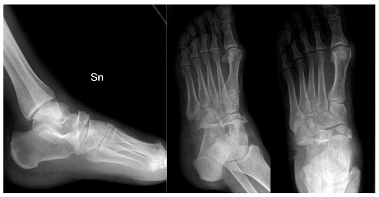
Radiographs. Measure articular congruity and lateral-column length on appropriate radiographs, and on CT when the morphology is uncertain.
- AP - may show the cuboid compression; check for a Lisfranc injury
- Lateral - may show the fracture; assess lateral column alignment
- Oblique - may show the compression better, and views the Lisfranc joint
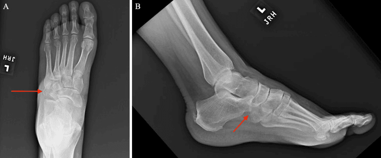
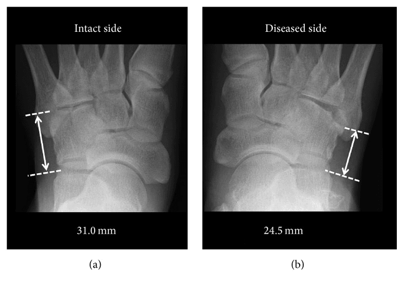
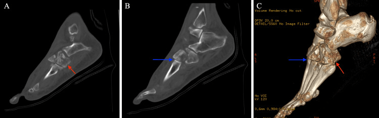
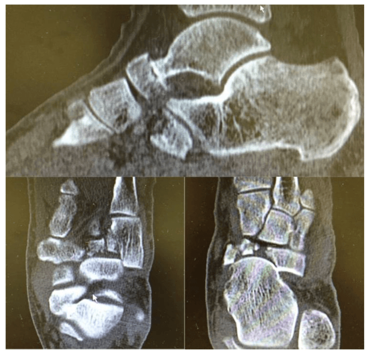
CT. CT is usually required for diagnosis and planning, because nutcracker fractures show impaction and bone loss. It is recommended for any suspected nutcracker fracture, for an associated Lisfranc injury, and when planning surgery. It shows:
- The fracture pattern
- The impaction (bone loss)
- A Lisfranc injury
- Lateral column alignment
CUBOIDWhat to Exclude With Every Cuboid Fracture
Hook:CUBOID = the six things a cuboid fracture obliges you to look for, because it is almost never an isolated injury
Differential Diagnosis
Lateral midfoot pain after a twisting or abduction injury has several causes. The nutcracker fracture is distinguished by cuboid impaction with lateral column shortening on imaging.
- Key Mechanism / Feature
- Forced forefoot abduction, axial load
- Discriminating Finding
- Cuboid compression with lateral column shortening on AP X-ray / CT
- Implication
- ORIF, restore length, graft impaction
- Key Mechanism / Feature
- Low-energy inversion (calcaneocuboid ligament)
- Discriminating Finding
- Small lateral fleck, no shortening, no articular step
- Implication
- Usually non-operative if under 1 mm
- Key Mechanism / Feature
- Axial load on plantarflexed foot
- Discriminating Finding
- Tarsometatarsal malalignment, fleck sign, C1-M2 diastasis
- Implication
- ORIF / fusion; commonly coexists with nutcracker
- Key Mechanism / Feature
- High-energy midtarsal force
- Discriminating Finding
- Talonavicular and/or calcaneocuboid disruption on CT
- Implication
- Urgent ORIF, restore both columns
- Key Mechanism / Feature
- Acute dorsiflexion-eversion
- Discriminating Finding
- Tenderness/clicking behind lateral malleolus, normal bone
- Implication
- Tendon-directed treatment
- Key Mechanism / Feature
- Chronic overload, painful os peroneum
- Discriminating Finding
- Sesamoid within peroneus longus, no acute trauma
- Implication
- Conservative, consider excision if refractory
Management Algorithm
The decision. Nutcracker describes a morphology, not a treatment. Assess both columns and the adjacent joints, compare alignment and length, and restore articular congruity and only the length deficit demonstrated. Fixation is chosen selectively by fragment pattern (screws, plate or bridge construct), and the soft tissue and compartment status affect its timing.
- What it is
- Congruity and column length preserved
- Treatment
- Protection with serial clinical/radiographic review
- Prognostic note
- No validated universal displacement cut-off
- What it is
- Depressed joint surface or shortened column
- Treatment
- Disimpaction and selected fixation; support residual void
- Prognostic note
- Procedure follows actual morphology
- What it is
- Multiple joints or columns disrupted
- Treatment
- Stage and stabilise the complete midfoot injury
- Prognostic note
- Associated injury drives prognosis
Non-operative treatment. It is reserved for minimal impaction without displacement, the patient who prefers it, and medical contraindications to surgery, and it is rarely indicated because of the impaction. The foot goes into a short leg cast, non-weight-bearing, for 6-8 weeks, with serial X-rays or CT to monitor healing.
Surgical indications. Surgery is indicated in most cases. The operative threshold itself is contested (see Controversies).
- Absolute: impaction with bone loss, lateral column shortening, an associated Lisfranc injury
- Relative: patient preference, a high-demand patient
Timing. An isolated fracture is fixed within 2 weeks. With a Lisfranc injury, the cuboid is fixed as part of the Lisfranc ORIF.
The isolated fracture. ORIF restores lateral column length. Disimpact the depressed articular fragment, then fill the resulting void, if there is one, with autograft from the calcaneus or iliac crest, allograft or a substitute; Yu's series used allograft, and only where needed. Where impaction is minimal there may be no defect to fill.
The combined injury. With an associated Lisfranc injury, address every unstable component. Restore cuboid congruity and any measured lateral-column shortening, and support a residual void only when necessary.
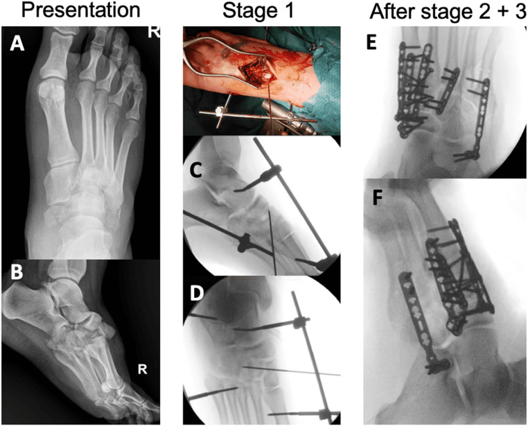
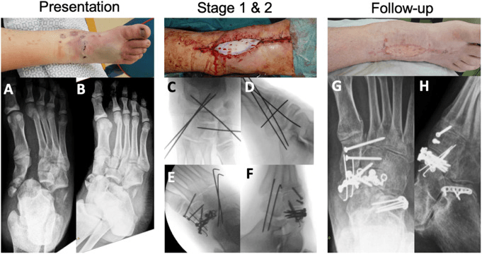
The residual void. After controlled disimpaction, assess the residual void and the stability of the fixation. Graft or substitute is used when structural support is required, and the donor site and material follow the defect, the host and the fixation. Children and minimally impacted fractures may require no graft.
Surgical Technique
ORIF with bone graft. For the impacted, shortened fracture, ORIF with bone graft restores lateral column length, prevents midfoot collapse and allows early motion. The steps:
- Expose the fracture through the lateral approach to the cuboid, protecting the peroneal tendons
- Debride the impacted bone and assess the bone loss
- Restore lateral column length and reduce the fracture anatomically
- Fill the impaction defect with bone graft, autograft from the calcaneus or iliac crest or allograft, using structural graft if the impaction is severe
- Fix with 2.7-3.5mm screws or a 2.0-2.7mm mini-fragment plate
- Confirm the reduction and hardware position fluoroscopically, and verify that lateral column length is restored by comparing with the contralateral foot
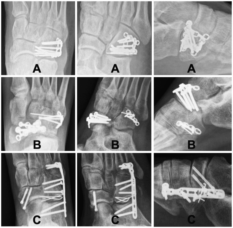
As part of a Lisfranc ORIF. This is the most common scenario. The Lisfranc injury is exposed through a dorsal approach, the primary one, and the cuboid through a lateral approach:
- Fix the Lisfranc injury first, with screws or a plate
- Expose the cuboid through the lateral approach
- Restore lateral column length with bone graft
- Fix the cuboid with screws or a plate
- Confirm that both injuries are fixed and that lateral column length is restored
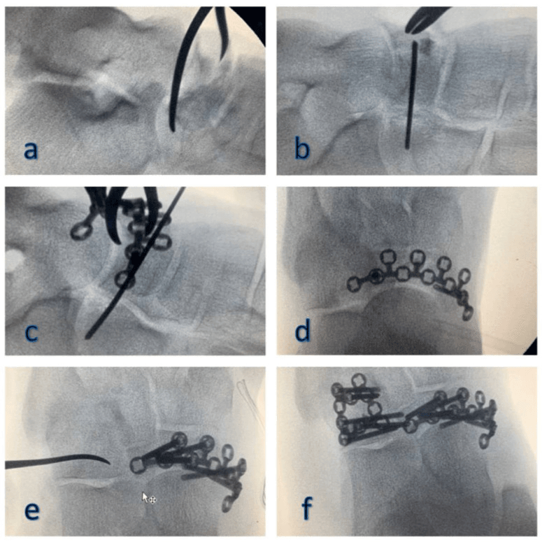
How Lateral Column Length Is Actually Restored: Disimpaction and Distraction
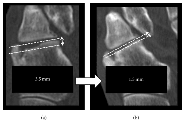
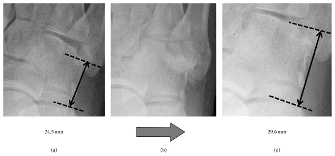
The whole topic turns on the instruction to restore lateral column length, and it is the step trainees most often get wrong. Impaction is a compression injury: the central articular fragment is driven inward and the column collapses. Simply lagging the fragments together reproduces the short, incongruent cuboid, so length cannot be regained with screws alone; it has to be actively regained and then held.
Distraction. A laminar spreader, a small femoral distractor, or a temporary external fixator or K-wire distractor spanning the calcaneocuboid joint (calcaneus to the fourth and fifth metatarsal bases) re-tensions the peripheral fragments and pulls them back out to length by ligamentotaxis. This restores the peripheral rim and column length, but the depressed central articular fragment usually does not follow, just as a depressed tibial plateau fragment will not reduce by traction alone.
Disimpaction and elevation. The central impacted articular fragment is elevated en masse from below, typically through a cortical window in the lateral or dorsal cuboid, under direct or arthroscopic/fluoroscopic control. This restores the congruity of the calcaneocuboid and cuboid-metatarsal surfaces, and leaves a subarticular metaphyseal void.
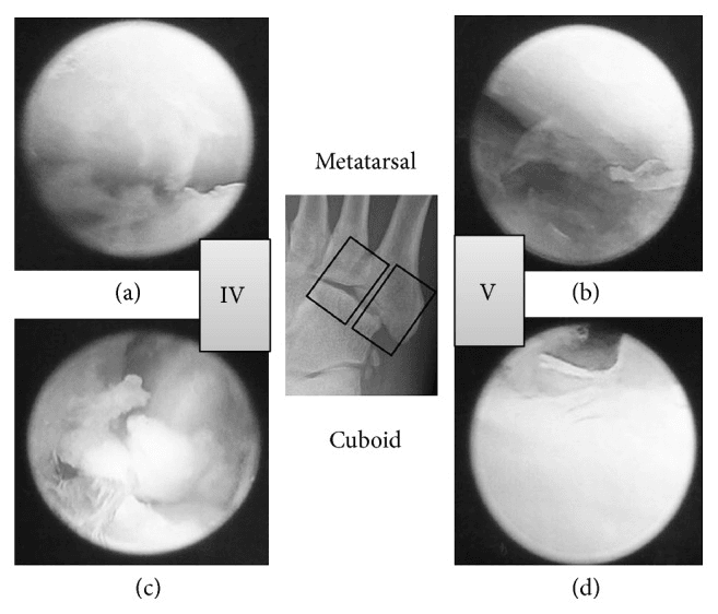
Buttressing the void. When the void needs support, it is filled with structural graft or a structural bone substitute (calcium phosphate or tricortical autograft) to support the elevated surface physically and maintain length. The graft then acts as a strut, not merely a biological filler.
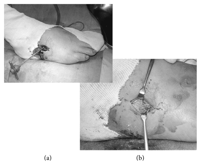
Definitive fixation. A mini-fragment plate used as a buttress, interfragmentary lag screws or, for comminution too severe for stable internal fixation, a bridge plate spanning the calcaneocuboid joint (or retention of the spanning external fixator) holds the reconstruction. Distraction is maintained during fixation, and the restored joint surface and column are measured against intact anatomy: the final result is checked fluoroscopically against the contralateral foot, the practical reference standard for column length. Bridging constructs are typically removed after union.
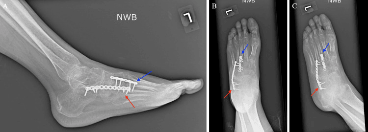
Complications
No cited series reports a rate for lateral column shortening or midfoot collapse, and nonunion, though uncommon, has no verified rate either.
- Incidence
- No verified rate
- Risk Factors
- Inadequate bone graft, severe impaction, bone loss, delayed treatment
- Prevention/Management
- Structural graft, restore length, adequate fixation; revision ORIF with structural graft if symptomatic
- Incidence
- No verified rate
- Risk Factors
- Lateral column shortening, inadequate fixation
- Prevention/Management
- Restore length, adequate fixation; revision ORIF or midfoot fusion if severe
- Incidence
- Common
- Risk Factors
- Focus on cuboid only; injury often missed at first presentation
- Prevention/Management
- CT whole midfoot, exclude Chopart/Lisfranc
- Incidence
- Uncommon (no verified rate)
- Risk Factors
- Inadequate fixation, bone loss
- Prevention/Management
- Rigid fixation, bone graft
Postoperative Care
The foot is immobilised in a short leg cast or boot and kept non-weight-bearing for 6-8 weeks, with ankle range of motion after cast removal and physiotherapy for midfoot range of motion and strengthening.
- Weeks 0-6 - short leg cast, non-weight-bearing; elevation to reduce swelling; ankle range-of-motion exercises if stable
- Weeks 6-8 - CT to confirm healing; cast removal if healing, then a walking boot and progressive weight bearing
- Weeks 8-12 - full weight bearing and progressive activity; return to sport at 3-4 months
Outcomes and Prognosis
What the series show. The cited evidence does not support precise success percentages, so quote the verified series, not invented ranges.
- Adults (Yu, n=6, ORIF and graft): results good in 2 and fair in 4. Imperfect function is the honest expectation, and patients should be counselled realistically
- Children (Ruffing, n=16): a mean AOFAS of 100 for extra-articular lesions, 95 for intra-articular and 66 with midtarsal disruption. Outcome tracks joint involvement and associated midtarsal injury, not the cuboid fracture alone, and associated midfoot disruption is the strongest adverse signal on this page
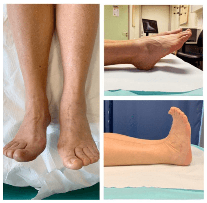
Determinants. Outcome turns on the restoration of lateral column length and articular congruity, and on whether a Chopart/Lisfranc component exists.
Guidelines, Registries & Global Practice
There is no dedicated arthroplasty/implant registry for cuboid fractures, and no randomised trial evidence — guidance is principle-based across societies and synthesised in critical-analysis and systematic reviews.
- Position on Cuboid / Nutcracker Fracture
- Cuboid is the keystone of the lateral column; goals are restoration of length, articular congruity and Chopart/Lisfranc stability. Disimpaction, bone-void filling and buttress fixation (lag screw, mini-fragment plate or temporary CC bridge plate) are described.
- Position on Cuboid / Nutcracker Fracture
- Non-operative care only for non-displaced articular fractures (under 1 mm) or low-energy avulsions; operate to restore congruity, length and stability. No classification validated for clinical use.
- Position on Cuboid / Nutcracker Fracture
- Apply general standards: timely senior assessment, CT for midfoot trauma, soft-tissue-led timing of fixation, compartment-syndrome vigilance in high-energy foot injuries.
- Position on Cuboid / Nutcracker Fracture
- Treat within the Chopart complex: routine CT, exclude compartment syndrome, urgent anatomic reduction maintaining column length gives best long-term outcomes.
- Cuboid fractures: under 1% of foot fractures worldwide; the compression (nutcracker) subtype is rarer still
- Reported across all regions and ages; paediatric cases described with Fenton type 2 predominating
- Evidence base is uniformly low level (case reports, small series) — no geographic registry data
- Well-resourced settings: CT and 3D planning routine; mini-fragment plating, structural graft/substitutes and staged soft-tissue care available
- Limited-resource settings: reliance on plain radiographs (risk of missed injury), K-wire fixation and provisional external fixation; bone substitutes may be unavailable, favouring autograft
- Across all settings the principle is identical: restore lateral column length and articular congruity
Nutcracker fractures are a common viva topic. Know the mechanism (cuboid crushed between calcaneus and metatarsal bases in forced forefoot abduction), that isolated fractures are rare so a Chopart/Lisfranc injury must always be excluded, that ORIF restores articular congruity and lateral column length, that bone graft fills impaction defects to prevent shortening, and that comminuted patterns may need bridge plating or external distraction. Be prepared to discuss the mechanism and why lateral column length restoration is central.
Related pages: Cuboid Fractures for the wider treatment of this bone, of which the nutcracker pattern is the compression subtype, and Chopart Injuries and Lisfranc Injuries for the two complexes the cuboid almost never breaks outside of; Foot Compartment Syndrome for the emergency that must be excluded in every crush pattern; Calcaneal Fractures and Lateral Process Talus Fractures for the hindfoot injuries that share the mechanism; and Calcaneocuboid Arthritis and Midfoot Arthritis for the late consequence of a lateral column left short.
Controversies and Areas of Uncertainty
Threshold for surgery. The widely quoted operative threshold, an articular step or shortening greater than 1-2 mm, derives from small case series and expert opinion, not trials. The displacement at which non-operative care fails is not precisely defined.
Graft type and necessity. Whether autograft, allograft or a synthetic/calcium-phosphate substitute best fills the impaction defect is unsettled: series report all three with acceptable results, and one paediatric series needed no graft at all. The principle, supporting the elevated articular surface and maintaining length, matters more than the material.
Fixation construct. The choice between lag screws, mini-fragment plates, temporary bridge plating across the calcaneocuboid joint and external or internal distraction is individualised to the comminution and the soft tissues. High-quality comparative data are lacking.
MCQ Practice Points
Q: What is the nutcracker mechanism for cuboid fractures? A: Compression between calcaneus and 4th/5th metatarsals - During forced plantarflexion and abduction. Causes impaction and bone loss. Isolated cuboid fractures are rare, so a Chopart or Lisfranc injury must always be excluded.
Q: Why does a nutcracker fracture mandate a search for other midfoot injury? A: Isolated cuboid fractures are rare - The same forefoot-abduction force that crushes the cuboid commonly disrupts the Chopart (midtarsal) or Lisfranc (tarsometatarsal) joints. A nutcracker fracture therefore signals possible midfoot instability — obtain CT, exclude Chopart/Lisfranc injury, and fix all unstable components.
Q: When is graft or substitute used? A: When disimpaction leaves a structural void that threatens articular or column-length maintenance. Small or paediatric defects may need none; material and fixation are chosen from the actual cavity and host.
Q: Why assess lateral-column length? A: Compression may shorten the calcaneocuboid-to-lateral-metatarsal relationship and alter midfoot alignment. Restore a measured deficit while avoiding over-distraction.
Q: When is surgery considered? A: For clinically important articular incongruity, column shortening, instability, open injury or associated midfoot disruption when non-operative care will not maintain alignment. No universal one-millimetre or graft threshold is validated.
Exam Viva Scenarios
Practise clinical reasoning and management decisions out loud
“A 28-year-old patient presents with lateral foot pain after forced plantarflexion and abduction injury. CT shows nutcracker fracture of cuboid with impaction and bone loss. No associated Lisfranc injury.”
“A 32-year-old patient has a nutcracker fracture as part of a Lisfranc injury. The examiner asks you to explain how you manage both injuries and why bone graft is essential.”
“A 40-year-old motorcyclist sustains a high-energy foot injury. CT shows a severely comminuted cuboid with marked articular impaction and lateral column shortening greater than 5 mm, plus a calcaneocuboid joint disruption. The skin over the lateral midfoot is swollen and blistered. The examiner asks how you would assess and manage this.”
Key Concepts
- Nutcracker mechanism = compression between calcaneus and 4th/5th metatarsals
- Isolated cuboid fractures exist but associated Chopart/Lisfranc injury must be excluded
- Support only the residual structural void
- Restore demonstrated lateral-column shortening
Classification
- Type I: Isolated cuboid compression - ORIF with bone graft (80-85% good results)
- Type II: With Lisfranc injury - ORIF both injuries (75-85% good results)
- Type III: Severe impaction - ORIF with structural graft (70-80% good results)
- Key Factor: Impaction severity dictates graft requirement
Treatment
- Stable/minimally displaced: protect and reassess alignment
- Combined injury: restore every unstable joint and column in a soft-tissue-led sequence
- Impacted/displaced: disimpact and fix according to morphology
- Use graft or substitute only when the residual void requires support
Surgical Technique
- Lateral approach: Protect peroneal tendons
- Debride impacted bone, assess bone loss
- Bone graft: Fill impaction defect (autograft or allograft, structural if severe)
- Fixation: Screws (2.7-3.5mm) or mini-fragment plate (2.0-2.7mm)
- Restore lateral column length
- Verify reduction fluoroscopically
Complications
- Lateral column shortening: no verified rate (prevent with bone graft, restore length)
- Midfoot collapse: no verified rate (prevent with adequate fixation)
- Missed associated injury: common, often missed at first presentation (prevent by CT of whole midfoot, exclude Chopart/Lisfranc)
- Nonunion: 5-10% (prevent with rigid fixation, bone graft)
Evidence Base
Named reviews are cited at their stated level; primary case series carry their PubMed identifiers.
Original Description of the Nutcracker Fracture
- Coined the term 'nutcracker fracture' of the cuboid
- Mechanism: cuboid crushed between calcaneus and metatarsal bases
- Caused by indirect violence with forefoot abduction
- Foundational eponym still used in exams and practice
Cuboid Fractures: Critical Analysis Review
- Isolated cuboid fractures are rare — actively exclude Chopart/Lisfranc injury
- Cuboid is the cornerstone of the lateral column
- No validated clinical classification system exists
- Non-operative only for non-displaced (under 1 mm) or avulsion fractures
ORIF and Bone Grafting for Nutcracker Fractures
- All 6 fractures showed lateral column shortening
- Treated with ORIF plus allograft where bone loss present
- Operate if shortening or articular step greater than 1 mm
- Outcomes good in 2, fair in 4 — function often imperfect
Cuboid Nutcracker Fractures in Children: Management and Results
- Fenton classification applied; extra-articular type 2 most common (69%)
- Intra-articular involvement and midfoot disruption worsen AOFAS scores
- Paediatric remodelling means bone grafting often unnecessary
- Articular and associated injuries are the key prognostic drivers
Chopart Dislocations: Systematic Review
- 58 papers synthesised — historically poor outcomes for these injuries
- Routine CT and compartment syndrome assessment advised
- Urgent ORIF restoring column length gives best outcomes
- High rate of missed injury at first presentation
Chopart Joint Injuries — Contemporary Review
- Low incidence but high complication rate if missed or undertreated
- Combined bony and ligamentous injury requires column realignment
- Fixation construct tailored to pattern and soft-tissue envelope
- Prompt recognition and anatomic fixation predict good outcomes