Multinucleated Bone-Resorbing Cells | RANKL-RANK Pathway | Ruffled Border
- Osteoclasts are multinucleated (10-100 nuclei) cells derived from haematopoietic stem cells
- RANKL-RANK pathway is essential for osteoclast differentiation and activation
- Ruffled border creates acidic microenvironment (pH 4.5) to dissolve hydroxyapatite
- Cathepsin K and matrix metalloproteinases degrade organic bone matrix
- Osteoprotegerin (OPG) acts as decoy receptor, inhibiting RANKL-RANK binding
- “Howship lacuna is the resorption pit created by active osteoclasts
- “RANK mutations cause osteopetrosis (marble bone disease)
- “Bisphosphonates induce osteoclast apoptosis by inhibiting farnesyl pyrophosphate synthase
- “Denosumab is monoclonal antibody against RANKL, preventing RANK binding
Overview and Cell Biology
Osteoclasts are the only cells capable of resorbing mineralised bone. They derive from the haematopoietic monocyte-macrophage lineage, not from the mesenchymal stem cells that give rise to the bone-forming osteoblasts, and this dual-origin system is essential for the balance of bone remodelling.
The cell. The osteoclast is a multinucleated giant cell 100-150 μm in diameter carrying 10-100 nuclei, formed by the fusion of mononuclear precursors; multinucleation is required for function. It shares its origin with the macrophage, and its precursors circulate as monocytes.
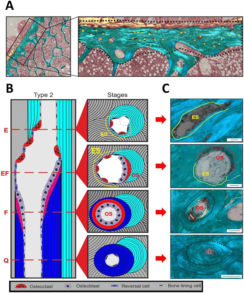
Where it works. An osteoclast lives for approximately 2 weeks, and while active it sits in the resorption pit it has made, the Howship lacuna.
Osteoclast Differentiation and Activation
Osteoclastogenesis Pathway
Haematopoietic stem cells in the bone marrow differentiate into monocyte-macrophage precursors. M-CSF (macrophage colony-stimulating factor) is essential for precursor survival and proliferation.
RANKL (receptor activator of nuclear factor kappa-B ligand), produced by osteoblasts and stromal cells, binds RANK receptors on the precursors. This is the critical commitment step.
Mononuclear precursors fuse to form multinucleated giant cells, a step mediated by dendritic cell-specific transmembrane protein (DC-STAMP).
The cell attaches to bone and develops its ruffled border and sealing zone, creating a sealed resorption compartment.
Bone is dissolved within the sealed compartment, as described under Molecular Mechanisms below. This phase lasts hours to days.
Programmed cell death follows once the resorption cycle is complete, triggered by loss of the RANKL signal or by OPG inhibition.
Regulation of Osteoclast Activity
- Source
- Osteoblasts/stromal cells
- Effect
- Stimulates +++
- Mechanism
- Binds RANK, activates NFκB
- Source
- Stromal cells/osteoblasts
- Effect
- Stimulates ++
- Mechanism
- Precursor survival/proliferation
- Source
- Osteoblasts
- Effect
- Inhibits ---
- Mechanism
- Decoy receptor for RANKL
- Source
- Parathyroid gland
- Effect
- Stimulates (indirect)
- Mechanism
- Increases RANKL expression
- Source
- Kidney (activated)
- Effect
- Stimulates (indirect)
- Mechanism
- Increases RANKL expression
- Source
- Gonads
- Effect
- Inhibits
- Mechanism
- Suppresses RANKL, increases OPG
- Source
- Thyroid C-cells
- Effect
- Inhibits
- Mechanism
- Direct receptor on osteoclast
The master switch. RANKL is the activator; OPG (osteoprotegerin), also produced by osteoblasts, is a decoy receptor that binds RANKL and so prevents RANK activation. The balance between the two determines osteoclast number and activity, and the RANKL:OPG ratio is the key determinant of the rate of bone resorption. It is the target of denosumab therapy.
Oestrogen deficiency. After the menopause, oestrogen deficiency increases RANKL and decreases OPG production, shifting the ratio towards resorption. This explains the accelerated bone loss of postmenopausal women and the efficacy of oestrogen replacement therapy.
Molecular Mechanisms of Bone Resorption
Resorption is a dual process inside one sealed compartment: acidification dissolves the mineral, and enzymes degrade the organic matrix.
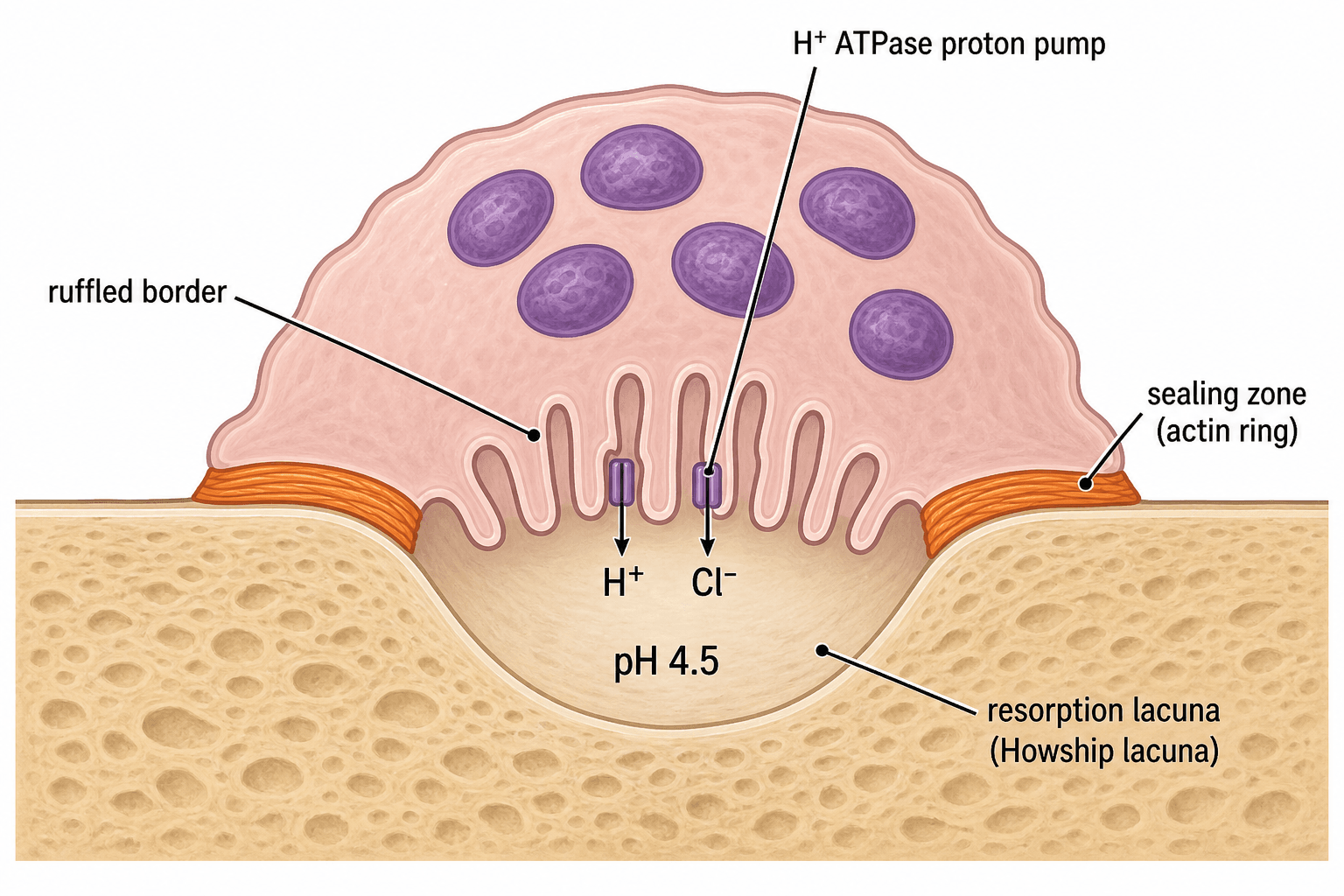
The functional zones. An active osteoclast has distinct functional zones:
- Ruffled border - membrane invaginations facing the bone surface
- Sealing zone - an actin ring that creates a tight seal around the resorption site
- Clear zone - the organelle-free area where the sealing occurs
- Basolateral domain - the opposite surface, where degradation products transcytosed through the cell are released into the circulation
Attachment and sealing. The αvβ3 integrin on the osteoclast surface binds RGD sequences in bone matrix proteins (osteopontin and bone sialoprotein). F-actin filaments then polymerise into a ring within the clear zone, which is densely packed with actin. The resulting peripheral seal isolates the resorption lacuna from the extracellular space; this gasket is what allows acidification and maintains the low pH.
Dissolving the mineral. V-type H+ ATPase pumps in the ruffled border actively drive protons into the resorption lacuna, bringing it to pH 4.5, and ClC-7 chloride channels maintain electroneutrality by carrying Cl- alongside the H+. The protons are generated inside the cell by carbonic anhydrase II (see Sly syndrome below). In this acid the hydroxyapatite crystals dissolve, releasing calcium and phosphate.
Degrading the matrix. Cathepsin K is the dominant collagenase, accounting for most degradation of the organic matrix, and it functions optimally at acidic pH.
- Target
- Type I collagen
- Function
- Major collagenase
- Specificity
- Cleaves telopeptides
- Target
- Gelatin/collagen
- Function
- Matrix metalloproteinase
- Specificity
- Degrades denatured collagen
- Target
- Phosphate esters
- Function
- Tartrate-resistant acid phosphatase
- Specificity
- Serum marker of resorption
Clinical Applications and Pathology
Too much resorption. Osteoclast activity is increased in:
- Osteoporosis (postmenopausal, steroid-induced)
- Paget disease (abnormal osteoclasts)
- Hyperparathyroidism
- Multiple myeloma
- Bone metastases
Too little. Osteoclast activity is decreased in:
- Osteopetrosis (RANK/RANKL/ClC-7 mutations)
- Pycnodysostosis (cathepsin K deficiency)
- Bisphosphonate therapy (excessive)
- Carbonic anhydrase II deficiency
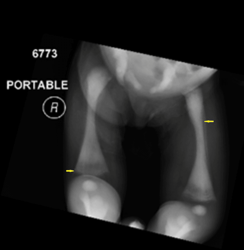
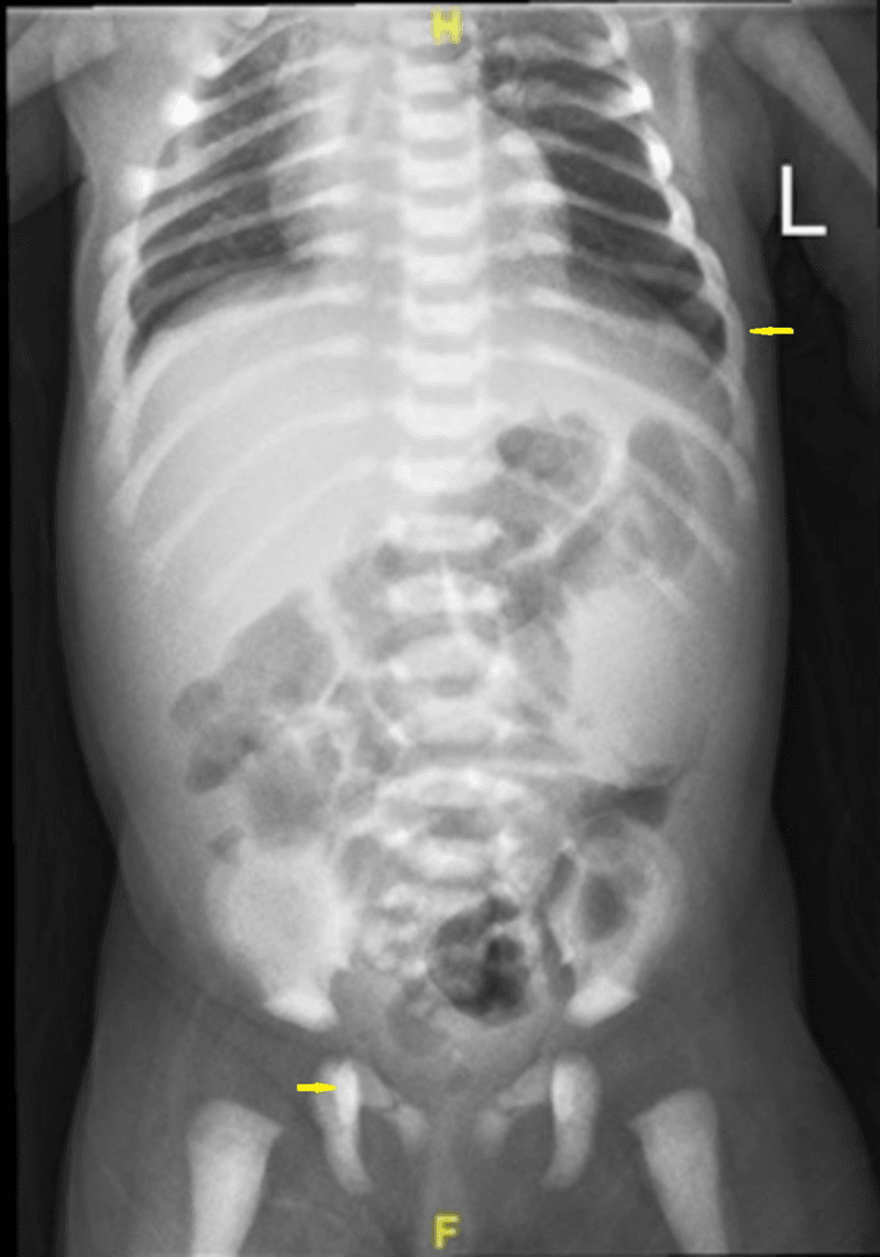
Osteopetrosis results from osteoclast dysfunction. The bone is dense and sclerotic (marble bone) yet paradoxically fragile, and obliteration of the marrow spaces causes cytopenias. Severe forms require haematopoietic stem cell transplantation to provide functional osteoclast precursors, except the RANKL form, in which HSCT is ineffective.
Two kinds of osteopetrosis. In the common osteoclast-rich forms, caused by TCIRG1, CLCN7 or carbonic anhydrase II mutations, osteoclasts are present but unable to resorb. In the osteoclast-poor RANK and RANKL forms, osteoclasts fail to form.
Carbonic Anhydrase II Deficiency (Sly Syndrome)
One enzyme, three tissues. Carbonic anhydrase II (CA-II) catalyses CO2 + H2O into H+ and bicarbonate, generating the very protons the osteoclast's H+-ATPase pumps into the resorption lacuna. The same enzyme is expressed in the renal tubule and the brain, so its autosomal-recessive loss produces a characteristic triad:
- Osteopetrosis - without CA-II-generated protons the osteoclast cannot acidify the lacuna or dissolve hydroxyapatite, so dense bone accumulates; the osteoclasts are present but non-functional
- Renal tubular acidosis - CA-II is required for renal tubular acid-base handling, so its loss impairs both proximal bicarbonate reclamation and distal urinary acidification, a mixed ("type 3") RTA producing a metabolic acidosis
- Cerebral calcification, typically of the basal ganglia, with developmental delay
How it differs. The renal and cerebral phenotype distinguishes CA-II deficiency from the TCIRG1 and CLCN7 acidification defects, which give isolated osteopetrosis. CA-II deficiency is also a milder, often later-presenting form. The detailed osteopetrosis subtypes and the role of HSCT are developed in the osteopetrosis topic.
Differential Diagnosis: Osteoclast-Driven Bone Disorders
When osteoclast number or function is abnormal, several conditions can present with overlapping radiographic or biochemical features. Distinguishing them rests on osteoclast biology.
- Osteoclast Defect
- Excess resorption (high RANKL:OPG)
- Bone Density
- Reduced
- Key Distinguisher
- Low BMD, fragility fractures, high bone turnover markers
- Osteoclast Defect
- Giant, hyperactive, viral-inclusion osteoclasts
- Bone Density
- Mixed lytic/sclerotic
- Key Distinguisher
- Markedly raised ALP, mosaic lamellar bone, bone pain/deformity
- Osteoclast Defect
- Present but cannot resorb (TCIRG1, CLCN7, CA-II)
- Bone Density
- Markedly increased
- Key Distinguisher
- Dense brittle bone, marrow failure, cranial nerve palsies
- Osteoclast Defect
- Osteoclast-poor (fail to form)
- Bone Density
- Markedly increased
- Key Distinguisher
- Few/absent osteoclasts; HSCT ineffective for RANKL form
- Osteoclast Defect
- Cathepsin K deficiency
- Bone Density
- Increased
- Key Distinguisher
- Acro-osteolysis, short stature, retained collagen matrix
- Osteoclast Defect
- PTH-driven focal hyperresorption
- Bone Density
- Focal lytic
- Key Distinguisher
- Raised PTH/calcium, subperiosteal resorption, giant-cell lesion
Bone Resorption Markers (CTX, NTX, TRAP-5b)
The collagen fragments measure how actively osteoclasts are resorbing; TRAP-5b, which the osteoclasts secrete themselves, measures how many there are.
- What it is
- A collagen breakdown fragment released when osteoclasts degrade matrix
- What it reflects
- Osteoclast activity (resorption rate); the preferred serum resorption marker for monitoring antiresorptive response/adherence (drawn fasting; some use it to gauge ONJ risk)
- What it is
- A collagen breakdown fragment (urine or serum)
- What it reflects
- Osteoclast activity; an older resorption marker
- What it is
- An enzyme secreted by osteoclasts themselves
- What it reflects
- Osteoclast number (not just activity), and it is independent of renal function and food intake
On treatment. Resorption markers fall rapidly when an antiresorptive is started, and a drop in CTX confirms response and compliance. They surge above baseline when denosumab is stopped, the biochemical signature of the rebound; its fracture consequence is set out under Denosumab below.
In osteopetrosis. TRAP-5b is high in the osteoclast-rich forms, because there are many osteoclasts, and low in the osteoclast-poor RANK/RANKL forms, because osteoclasts fail to form. It therefore separates the two subtypes and predicts whether haematopoietic stem cell transplantation can work. The bone-formation markers (P1NP, osteocalcin, bone-specific ALP) and the remodelling cycle itself are developed in the osteoblasts-bone-formation and bone-remodeling topics.
Pharmacological Targeting of Osteoclasts
Bisphosphonates, denosumab and calcitonin all target osteoclast activity in osteoporosis.
Bisphosphonates. The nitrogen-containing bisphosphonates (alendronate, risedronate, zoledronic acid) inhibit farnesyl pyrophosphate synthase in the mevalonate pathway, preventing prenylation of the small GTPases essential for osteoclast function. They bind hydroxyapatite with high affinity and become incorporated into the bone matrix, and osteoclasts endocytose the bisphosphonate-containing bone as they resorb it. Inside the cell the drug inhibits the enzyme and disrupts GTPase signalling, and the loss of functional GTPases triggers osteoclast apoptosis, reducing bone resorption.
Adverse effects. Bisphosphonates can cause osteonecrosis of the jaw, which is rare, and atypical femoral fractures with prolonged use (greater than 5 years).
Denosumab. Denosumab is a fully human monoclonal antibody that binds RANKL with high affinity, preventing the RANKL-RANK interaction.
- Denosumab
- Anti-RANKL antibody
- Bisphosphonates
- Osteoclast apoptosis
- Denosumab
- SC every 6 months
- Bisphosphonates
- Oral/IV variable
- Denosumab
- No
- Bisphosphonates
- Yes (years)
- Denosumab
- Yes (discontinuation)
- Bisphosphonates
- No
Stopping denosumab. Bone turnover surges and bone density falls back to baseline within about a year. The vertebral fracture rate returns to that of untreated patients, 7.1 per 100 participant-years after stopping denosumab against 8.5 after stopping placebo, and does not rise above the untreated level. What is specific to denosumab is that patients who do fracture are far more likely to sustain several vertebral fractures at once (60.7% versus 38.7%).
The bridge. Transition to a bisphosphonate is required. Which bisphosphonate, and how and when it is given, is not defined by prospective trials (see Controversies below).
Calcitonin. Calcitonin binds G-protein coupled receptors on the osteoclast surface and inhibits resorption directly, reducing it rapidly (within hours). It also has an analgesic effect, mechanism unclear, but its efficacy is modest compared with bisphosphonates, and tachyphylaxis (a reduced response with repeated dosing) limits long-term use. It is used for acute hypercalcaemia, Paget disease and as an analgesic for vertebral fractures.
Guidelines, Registries & Global Practice
Global Epidemiology
- Osteoporosis affects an estimated 500 million people worldwide; roughly 1 in 3 women and 1 in 5 men over 50 will sustain a fragility fracture.
- Over 8.9 million osteoporotic fractures occur globally each year (a fragility fracture roughly every 3 seconds), with hip fractures projected to rise sharply in Asia as populations age.
- Paget disease shows marked geographic variation — historically common in the UK and populations of British descent, and declining in prevalence in recent decades.
- Infantile (malignant) osteopetrosis has an incidence of approximately 1 in 250,000 births; the autosomal dominant adult form is far more common at around 1 in 20,000.
Major Guidelines, Side by Side
- First-line
- Oral/IV bisphosphonate; denosumab or anabolic for very high risk
- Denosumab Stance
- First-line option in high/very-high risk
- Discontinuation Caveat
- Never stop without follow-on anti-resorptive
- First-line
- Oral bisphosphonate (alendronate/risedronate)
- Denosumab Stance
- Where bisphosphonate unsuitable or higher risk
- Discontinuation Caveat
- Mandatory bisphosphonate bridge on stopping
- First-line
- Bisphosphonate; sequential anabolic-then-antiresorptive in severe disease
- Denosumab Stance
- Recognised potent anti-resorptive
- Discontinuation Caveat
- Structured transition to avoid rebound
- First-line
- Treat above country-specific intervention threshold
- Denosumab Stance
- Reserved per fracture-risk tier
- Discontinuation Caveat
- Uniform rebound warning
- Consensus across societies: nitrogen-containing bisphosphonates remain first-line; denosumab is a potent alternative but must never be stopped abruptly; teriparatide/romosozumab (anabolic) are favoured first for very-high-risk patients, followed by an anti-resorptive to consolidate gains.
- Anti-resorptive "drug holidays" apply to bisphosphonates (which persist in bone for years) but are explicitly contraindicated for denosumab.
Registry and Resource-Setting Notes
- Arthroplasty registries (NJR, AJRR, AOANJRR, SHAR, Norwegian, NZJR) track periprosthetic fractures, which are influenced by underlying bone quality and anti-resorptive status; bisphosphonate use around arthroplasty and its effect on aseptic loosening and revision is an area of ongoing analysis.
- In well-resourced settings, severe infantile osteopetrosis is managed with haematopoietic stem cell transplantation and bone-density-targeted pharmacotherapy is guided by DXA and bone turnover markers.
- In limited-resource settings, DXA access, the cost of denosumab and anabolic agents, and HSCT availability are major constraints; generic oral bisphosphonates and FRAX (which can be calculated without BMD) are the practical mainstays.
Related pages: Osteoblasts and Bone Formation for the cell that makes both RANKL and its decoy OPG, and therefore sets the resorption rate described here, and Bone Remodeling for the coupled cycle the two cells run together; Osteopetrosis for the natural experiments that prove this pathway in humans — TCIRG1, CLCN7 and carbonic anhydrase II mutations produce osteoclasts that cannot acidify, and the RANK/RANKL forms produce no osteoclasts at all, which is why TRAP-5b separates them; Paget's Disease of Bone for pathologically hyperactive osteoclasts; Osteoporosis and Glucocorticoid-Induced Osteoporosis for the diseases these drugs treat; Bisphosphonates for the mevalonate-pathway mechanism only sketched here; and Giant Cell Tumour of Bone for the RANKL-driven tumour in which denosumab is used oncologically rather than metabolically.
Controversies and Areas of Uncertainty
Cathepsin K inhibitors. Odanacatib reduced fractures in the LOFT trial but was withdrawn in 2016 after a signal of increased stroke risk. Whether selective cathepsin K inhibition can be achieved without off-target cardiovascular or cutaneous effects remains unresolved.
The denosumab exit strategy. The rebound phenomenon is established, but the ideal bridging regimen (which bisphosphonate, oral or IV, timing relative to the missed dose, and duration) is not defined by prospective trials and varies between guidelines.
Anti-resorptive drug holidays. Balancing the risks of atypical femoral fracture and osteonecrosis of the jaw, which rise with cumulative exposure, against rebound fracture risk on stopping is contentious. Holidays are reasonable for bisphosphonates but are explicitly unsafe for denosumab.
Osteoclasts as signalling cells. Beyond resorption, osteoclasts secrete "clastokines" that couple resorption to formation. Whether anabolic benefit can be preserved while resorption is suppressed, the basis of interest in coupling-sparing agents, is an active question.
Treatment sequence matters: giving an anti-resorptive (denosumab/bisphosphonate) before the anabolic teriparatide blunts the anabolic response, whereas the reverse sequence (anabolic first, then anti-resorptive to consolidate gains) is preferred. This reflects the dependence of teriparatide on a "remodelling space" created by active osteoclasts.
MCQ Practice Points
Q: Osteoclasts are derived from which cell lineage? A: Hematopoietic monocyte-macrophage lineage - NOT mesenchymal. This is why bone marrow transplantation can cure some forms of osteopetrosis by providing functional osteoclast precursors.
Q: What is the receptor for RANKL on osteoclast precursors? A: RANK (receptor activator of nuclear factor kappa-B). Activation leads to NFκB signaling and osteoclastogenesis. Mutations cause osteopetrosis.
Q: What is the major collagenase enzyme secreted by osteoclasts? A: Cathepsin K - accounts for the majority of type I collagen degradation. Functions optimally at acidic pH. Deficiency causes pycnodysostosis.
Q: How do nitrogen-containing bisphosphonates cause osteoclast apoptosis? A: Inhibit farnesyl pyrophosphate synthase in the mevalonate pathway, preventing prenylation of small GTPases required for osteoclast function and survival.
Exam Viva Scenarios
Practise clinical reasoning and management decisions out loud
“The examiner shows you a histological image of bone tissue with multinucleated cells in Howship lacunae. Describe what you see and explain the cell function.”
“Explain the molecular regulation of osteoclast differentiation and how this relates to osteoporosis treatment.”
“A child presents with dense, sclerotic bones on radiograph, anaemia, recurrent infections and cranial nerve palsies. Bone biopsy shows abundant but dysfunctional osteoclasts. Explain the pathophysiology and how this contrasts with pycnodysostosis, and outline management.”
Key Cell Biology
- Multinucleated (10-100 nuclei) from hematopoietic monocyte lineage
- Lifespan approximately 2 weeks
- Located in Howship lacunae (resorption pits)
- Ruffled border membrane facing bone, basolateral for transcytosis
RANKL-RANK-OPG Axis
- RANKL (osteoblast) + RANK (osteoclast precursor) = activation
- OPG = decoy receptor, blocks RANKL-RANK binding
- RANKL:OPG ratio determines resorption rate
- M-CSF required for precursor survival
Resorption Mechanism
- H+ ATPase pumps create pH 4.5 in sealed lacuna
- Acidic pH dissolves hydroxyapatite mineral
- Cathepsin K degrades type I collagen matrix
- Sealing zone (actin ring) maintains isolation
Pharmacological Targets
- Bisphosphonates: inhibit farnesyl pyrophosphate synthase, induce apoptosis
- Denosumab: anti-RANKL antibody, prevents RANK binding
- Calcitonin: direct osteoclast receptor, rapid inhibition
- Denosumab discontinuation causes rebound resorption
Clinical Conditions
- Osteopetrosis: RANK/RANKL/ClC-7/CA-II mutations, dense fragile bone
- Pycnodysostosis: cathepsin K deficiency
- Postmenopausal osteoporosis: increased RANKL:OPG ratio
- Paget disease: abnormal hyperactive osteoclasts
Key Enzymes and Markers
- Cathepsin K = major collagenase
- TRAP (tartrate-resistant acid phosphatase) = serum marker
- Carbonic anhydrase II = generates H+ from CO2
- ClC-7 = chloride channel for electroneutrality
Evidence Base and Key Studies
Bone Resorption by Osteoclasts
- Landmark synthesis defining the osteoclast as a specialized macrophage polykaryon
- Established M-CSF, RANK ligand and osteoprotegerin as the principal differentiation regulators
- Described integrin-mediated cytoskeletal polarization creating the isolated resorption microenvironment
- Used osteopetrotic mutants to map genes controlling differentiation and resorptive capacity
Osteoclast Differentiation and Activation
- Authoritative review consolidating the RANK signalling pathway in osteoclasts
- Confirmed osteoclasts arise from the monocyte/macrophage haematopoietic lineage
- Positioned OPG as the soluble decoy receptor that neutralises RANK ligand
- Mapped how hormonal signals converge on RANKL to control bone mass
OPG Ligand (RANKL) Is the Osteoclast Differentiation Factor
- Discovery paper identifying OPG ligand (RANKL) as a TNF-family cytokine
- RANKL replaced the requirement for stromal cells, vitamin D3 and glucocorticoids in osteoclastogenesis co-culture
- Directly activated mature osteoclasts and produced hypercalcaemia when given to mice
- OPG blocked all RANKL effects in vitro and in vivo, defining the activator-decoy pair