Pathologic Fracture Management
Visual One-Pager
The pathologic-fracture decision in one view.
- Is it pathologic? A low-energy injury through a pre-existing lesion, with pain preceding the fracture. The commonest causes are metastatic carcinoma (breast, lung, prostate, kidney, thyroid) and multiple myeloma.
- Do you know the diagnosis? Biopsy BEFORE instrumenting any indeterminate or solitary lesion - nailing an unrecognised primary sarcoma is the single most damaging error. Known widely-metastatic disease with a typical lesion is the main exception.
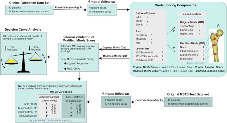
- Impending or complete? For an impending fracture, score fracture risk with Mirels (site, pain, lesion type, size) and stabilise the high-risk lesion before it breaks.
- What construct? Match durability to expected survival (Katagiri/SORG): a load-sharing nail or cement-augmented fixation for a short prognosis, an endoprosthesis for periarticular destruction or a long survivor. Bypass the lesion, span the whole bone, and build a construct that does not rely on biological union.
- Team and adjuvants. Postoperative radiotherapy, bone-modifying agents and systemic therapy - surgery restores function and relieves pain but does not prolong survival.
Definition and Classification
A pathologic fracture occurs through bone weakened by pre-existing disease, typically with minimal trauma that would not fracture normal bone. The underlying pathology compromises the structural integrity and mechanical properties of the bone.
Recognising it. Five features set it apart from an ordinary fracture:
- A low-energy mechanism: normal activities or a minor fall
- A pre-existing bone lesion on imaging
- Pain preceding the fracture, for weeks to months
- An abnormal fracture pattern or location
- Poor healing potential unless the underlying pathology is addressed
The diagnosis rests on correlating the clinical presentation, mechanism, imaging characteristics and patient risk factors, which is what separates it from an insufficiency fracture or an occult traumatic injury.
Causes. The underlying pathology falls into five groups:
- Malignant: metastatic disease (50%), multiple myeloma (20%), primary bone sarcoma (10%)
- Benign: fibrous dysplasia, enchondroma, simple bone cyst, aneurysmal bone cyst
- Metabolic: osteoporosis, osteomalacia, hyperparathyroidism
- Infection: chronic osteomyelitis with bone destruction
- Other: Paget's disease, radiation necrosis
Pathophysiology
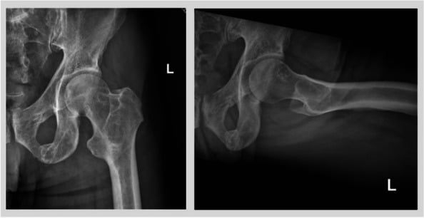
Osteolysis. Tumour cells secrete RANKL (receptor activator of nuclear factor kappa-B ligand), which binds RANK on osteoclast precursors and drives massive osteoclast activation and differentiation. Resorption outpaces formation, the cortex thins, the trabeculae are destroyed and the bone loses its structural integrity.
The osteoblastic response. Some tumours (prostate, breast) stimulate osteoblast activity and produce mixed lesions. The new bone is disorganised, mechanically inferior woven bone without lamellar structure: more of it, but of poorer quality, and still prone to fracture under physiological loads.
The metastatic cascade. Destruction and tumour growth feed each other in sequence:
- Tumour cells preferentially metastasise to red marrow-rich areas
- Adhesion molecules facilitate colonisation of bone
- A tumour microenvironment is established
- A "vicious cycle" of bone destruction and tumour growth sets in
- Cytokines (IL-6, PTHrP) amplify osteoclast activity
- Structural compromise progresses
Primary bone tumours. Tumour replaces normal bone directly, and its expansion thins the cortex, though periosteal reaction may add some strength. Pathologic fracture occurs through the lesion in 5-10% of osteosarcomas and significantly worsens the prognosis through local contamination.
This understanding guides the timing of adjuvant therapy, and explains why radiotherapy or systemic therapy alone rarely prevents fracture once critical bone loss has occurred.
Clinical Presentation
Red flags. Any of these should make you suspect that the bone was abnormal before it broke:
- Progressive bone pain over weeks to months before the fracture
- Night pain unrelieved by rest, characteristic of tumour
- Fracture with minimal trauma or during normal activity
- A history of malignancy, even a remote one
- Constitutional symptoms: weight loss, fatigue, fever, night sweats, anorexia
- Bone pain at multiple sites (polyostotic involvement)
The pain. It is dull, aching and progressive. Establish whether the onset was gradual or sudden (the fracture event), and note that activity modification and NSAIDs are often ineffective.
The mechanism. A detailed account is critical. "Twisted getting out of a chair" or "stepped off a kerb" suggests a pathologic fracture, and pain before the fall indicates pre-existing pathology. High-energy trauma can also occur through pathologic bone, which confounds the picture, and witnesses may clarify what happened.
The medical history. Ask specifically about:
- Current or previous malignancy: type, stage and treatment
- Radiotherapy to bone (radiation-induced fractures)
- Prolonged corticosteroid use (osteoporosis, osteonecrosis)
- Metabolic bone disease
- Previous fractures, for pattern recognition
- A family history of hereditary bone disorders
Function and goals. Record the pre-fracture ambulatory status, weight-bearing ability, how far pain limits function, and the patient's social support and home environment. Establish their goals and expectations, and align them with the prognosis.
Examination priorities. In order:
- Neurovascular status, documented as a baseline
- Skin integrity, because an open fracture can occur with minimal soft-tissue trauma
- Gross deformity and limb alignment
- A palpable mass (soft-tissue extension)
- Regional lymphadenopathy, rare but possible
- A systematic search for additional lesions
Look and feel. Swelling and ecchymosis may be disproportionate. Look for deformity, shortening and rotation, previous scars (biopsy sites), skin changes from radiotherapy, infection or tumour ulceration, and muscle wasting from chronic pain or denervation.
Palpate for point tenderness at the fracture site, a palpable mass or cortical defect, warmth (infection, inflammation) and crepitus with gentle movement, and examine the joints above and below.
Neurovascular examination. Assess the pulses (with an ABI if there is concern) and capillary refill, sensation for nerve compression or injury, and motor function without stressing the fracture. Document the findings precisely, for surgical planning and for medicolegal reasons.
Systematic survey. Palpate and percuss the spine, compress and distract the pelvis, and examine the contralateral limb, the abdomen for organomegaly, and auscultate the chest for lung metastases.
The examination should identify complications of the fracture and gather clues to the underlying diagnosis without adding to the patient's discomfort or displacing the fracture.
Investigations
Views. AP and lateral radiographs of the affected bone, including the joints above and below, with the contralateral limb for comparison and additional obliques if needed to characterise the lesion.
Features of a pathologic fracture. Look for:
- A lytic or sclerotic lesion at the fracture site
- Cortical destruction or erosion
- Pathological periosteal reaction
- A soft-tissue mass
- Bone expansion
- Matrix mineralisation
- Geographic, moth-eaten or permeative destruction
Characterising the lesion. The Lodwick classification grades the pattern of destruction:
- Pattern
- Geographic with a sclerotic border
- Implication
- Benign
- Pattern
- Geographic without a sclerotic border
- Implication
- Benign or low-grade
- Pattern
- Geographic with an ill-defined border
- Implication
- Aggressive
- Pattern
- Moth-eaten
- Implication
- Aggressive or malignant
- Pattern
- Permeative
- Implication
- Malignant
A narrow zone of transition means a slow-growing lesion and a wide one an aggressive lesion, and a cortical breach suggests malignancy. Note the periosteal reaction (onion-skin, sunburst, Codman triangle), any soft-tissue component, and the matrix: rings and arcs for chondroid, cloud-like for osteoid.
Skeletal survey. Indicated when myeloma is suspected, in the metastatic work-up, for assessing metabolic bone disease, and in polyostotic fibrous dysplasia. It identifies synchronous lesions that affect prognosis and may reveal the primary tumour site. The standard views:
- Skull: AP and lateral
- Spine: AP and lateral cervical, thoracic and lumbar
- Pelvis: AP
- Chest: PA and lateral
- Both humeri and femora: AP
- Hands, if myeloma is suspected
Surgical Decision-Making
Biopsy Principles
The golden rules. Five rules govern every biopsy:
- Biopsy before definitive treatment, except in known metastatic disease
- The biopsy tract must be excisable en bloc with the tumour if it is a primary sarcoma
- Avoid contaminating uninvolved compartments
- Coordinate with the treating oncological surgeon before biopsy
- "Whoops" procedures drastically worsen sarcoma outcomes
When biopsy is mandatory. Before any treatment:
- Suspected primary bone tumour
- A solitary bone lesion without a known primary
- Indeterminate radiographic features
- A young patient, in whom sarcoma is more likely than metastasis
- A lesion inconsistent with the known primary tumour
Before instrumentation, biopsy is also needed if there is any possibility of primary sarcoma, if infection cannot be excluded clinically, for a benign aggressive lesion that needs intralesional treatment, and for legal and documentation purposes (a tissue diagnosis).
Timing relative to stabilisation. Which comes first depends on the fracture, the patient and how certain the diagnosis is:
- Stabilise first, biopsy later
- Completed fracture causing severe pain
- Stabilise first, biopsy later
- Neurovascular compromise requiring urgent reduction
- Stabilise first, biopsy later
- Known metastatic disease (diagnostic certainty)
- Stabilise first, biopsy later
- Medical instability requiring damage control
- Stabilise first, biopsy later
When stabilisation comes first, the biopsy can be taken through or adjacent to the incision. The decision balances diagnostic necessity against clinical urgency, with multidisciplinary input whenever possible.
Differential Diagnosis
The first task when a fracture looks pathologic is deciding what the bone is made of. Confusing a primary sarcoma for a metastasis - and nailing it - is the single most damaging error in the whole of metastatic bone surgery: an intramedullary nail seeds the entire canal with tumour and converts a limb-salvageable sarcoma into an amputation. The table below frames the differential the way examiners probe it.
- Typical Patient / Clues
- Over 40, known or occult primary (breast, prostate, lung, kidney, thyroid), often multiple lesions
- Radiographic Signature
- Lytic (renal/thyroid/lung), blastic (prostate) or mixed (breast); geographic to moth-eaten
- Management Implication
- Stabilise + radiotherapy + systemic therapy; biopsy can be at surgery if pattern is typical and primary known
- Typical Patient / Clues
- Over 50, bone pain, anaemia, renal impairment, raised ESR
- Radiographic Signature
- Punched-out lytic lesions, diffuse osteopenia, vertebral collapse; cold on bone scan
- Management Implication
- SPEP/UPEP, marrow biopsy; highly radiosensitive; fixation plus systemic anti-myeloma therapy
- Typical Patient / Clues
- Younger patient OR atypical solitary lesion at any age; no known primary
- Radiographic Signature
- Aggressive periosteal reaction (sunburst, Codman triangle), wide zone of transition, soft-tissue mass, matrix mineralisation
- Management Implication
- DO NOT instrument; refer to sarcoma unit for biopsy through the resection corridor and neoadjuvant chemotherapy
- Typical Patient / Clues
- Young adult, epiphyseal/metaphyseal, eccentric expansile
- Radiographic Signature
- Expansile lytic, fluid-fluid levels (ABC) on MRI, no aggressive periosteal reaction
- Management Implication
- Biopsy first; curettage and grafting, denosumab for GCT - fixation alone inadequate
- Typical Patient / Clues
- Elderly, low BMD, no focal lesion, low-energy mechanism
- Radiographic Signature
- No destructive lesion; diffuse osteopenia; sclerosis along healing trabeculae
- Management Implication
- Standard fracture care plus bone-health workup; treat osteoporosis
- Typical Patient / Clues
- Fever, raised inflammatory markers, immunosuppression, prior open injury
- Radiographic Signature
- Lytic lesion with sclerotic rim, sequestrum, involucrum; can mimic tumour
- Management Implication
- Always send biopsy tissue for culture as well as histology; debride and treat infection before definitive fixation
Treatment Strategies
Non-Operative Management
Observing an impending fracture. Non-operative treatment is reasonable when:
- The Mirels score is 7 or less (low risk)
- The patient is non-ambulatory or non-weight-bearing
- Life expectancy is very limited (weeks)
- Medical comorbidities prohibit surgery
- The patient prefers it after an informed discussion
- The lesion is in the upper limb, where biomechanical demand is lower
Monitor with serial radiographs at 4-6-week intervals, assessing pain, functional status and signs of progression, with a low threshold for surgery if anything changes.
A completed fracture without surgery. This is rarely appropriate. It is considered for a minimally displaced upper limb fracture in acceptable alignment for the patient's functional demands, a prognosis measured in weeks (hospice), medical contraindications to anaesthesia, or a patient who declines surgery.
Care then consists of multimodal analgesia, often requiring opioids; immobilisation with a splint, brace or protected weight-bearing; nursing care (skin, DVT prophylaxis, bowel and bladder); palliative care involvement; counselling the family about expectations; and a focus on quality of life.
What it costs. Non-operative treatment risks persistent, often severe, pain and the complications of immobility (pneumonia, DVT, deconditioning). It also risks nonunion, because healing through tumour is unlikely, progressive deformity, contractures and pressure sores, reduced independence and quality of life, and psychological harm (helplessness, depression).
Shared decision-making is essential, with a realistic discussion of both the surgical and the non-surgical trajectory.
Surgical Management
Core principles. Six concepts shape the construct:
- Stabilise the entire bone (nail versus plate length)
- Bypass the lesion by at least 2 cortical diameters
- Augment with cement when possible
- Make the fixation durable enough to outlive the patient's prognosis
- Restore weight-bearing early, for quality of life
- A single operation if possible, to reduce morbidity
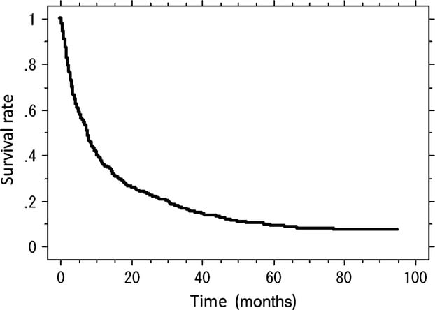
Prognosis first. The Katagiri score predicts survival in skeletal metastases from the primary tumour site, visceral or cerebral metastases, performance status, previous chemotherapy, multiple skeletal metastases and abnormal laboratory data. Expected survival then sets the construct:
- Approach
- Simple stabilisation, maximise function
- Implant selection
- Minimise surgical time and morbidity; a simple locked nail or plate is sufficient, with cement augmentation for immediate stability; a higher later failure rate is accepted because the patient is unlikely to outlive the construct
- Approach
- Durable fixation, possibly with cement augmentation
- Implant selection
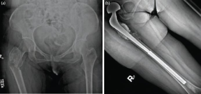
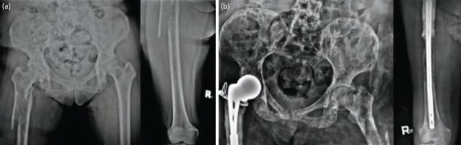
- A durable construct is essential: long-segment fixation, cement augmentation, and an endoprosthesis considered if the lesion is periarticular
- Approach
- Reconstruction as for a primary tumour, considering an endoprosthesis
- Implant selection
- Reconstruct for long-term function: endoprosthetic replacement if bone loss is extensive, allograft-prosthetic composites, and preparation for potential revision
The goal is to match surgical invasiveness and construct durability to a realistic prognosis, avoiding both under- and over-treatment.
Preoperative Embolisation of Hypervascular Metastases
Which tumours. Renal cell carcinoma and follicular thyroid carcinoma are the classically hypervascular skeletal metastases; hepatocellular carcinoma, some neuroendocrine tumours and melanoma can also bleed heavily. Instrumenting these lesions unprepared risks catastrophic, sometimes uncontrollable, intraoperative haemorrhage.
What it is. Transarterial (angiographic) embolisation devascularises the lesion before open surgery. Interventional radiology selectively catheterises the tumour's feeding vessels and occludes them with particles (PVA), gelfoam, coils or a liquid embolic (Onyx).
Timing. It is best performed within about 24-48 hours before surgery. If the delay is longer, collateral revascularisation returns and the benefit is lost.
Benefit. Embolisation substantially reduces intraoperative blood loss and transfusion requirement in hypervascular lesions, making curettage and instrumentation safer, cleaner and quicker. For fixation of a renal cell metastasis it is the single preoperative step that most reduces catastrophic bleeding.
Caveats. Post-embolisation syndrome (pain, fever, nausea) and non-target embolisation can occur. Embolisation reduces but does not abolish bleeding, so crossmatched blood, careful cell-saver use (tumour) and meticulous haemostasis are still needed. Where embolisation is unavailable (limited-resource settings), the threshold to operate on a renal or thyroid metastasis rises.
Intraoperative Embolism and Cardiopulmonary Collapse
The mechanism. Instrumenting a tumour-filled medullary canal - reaming, nail insertion and especially pressurised cementation - can force tumour thrombus, marrow fat and PMMA monomer or cement into the venous circulation. The emboli lodge in the lungs, causing acute pulmonary hypertension, hypoxia, hypotension and, at worst, cardiac arrest. This is a tumour, fat and cement variant of bone-cement implantation syndrome (BCIS).
Who is at risk. Patients with a high tumour burden, hypervascular lesions, long-segment or bilateral instrumentation, and pre-existing cardiopulmonary disease.
Mitigation. Reduce the risk at every step:
- Warn the anaesthetist before reaming and cementing
- Ream gently and vent the canal (a distal drill hole) to lower intramedullary pressure
- Use a cement gun with retrograde filling and avoid over-pressurisation
- Lavage the canal before cementing
- Maintain euvolaemia
- In very high tumour burden, consider a construct that minimises canal pressurisation
Recognition and management. Watch for the tell-tale sudden fall in end-tidal carbon dioxide and blood pressure during instrumentation; the scenario to recognise is sudden desaturation and hypotension during cephalomedullary nailing of a femoral metastasis. Manage the event with resuscitation, high-flow oxygen and haemodynamic support. Cement embolism, though rare, is potentially fatal, and management is supportive; it rarely requires embolectomy.
Complications
Haemorrhage. The risk comes from hypervascular tumours (renal cell, thyroid), large lesions with soft-tissue extension, coagulopathy (liver metastases, chemotherapy) and vascular injury during the approach. Prevent it with preoperative embolisation of hypervascular lesions, crossmatched blood, a cell saver if the tumour burden is low, meticulous haemostasis, and a tourniquet considered for extremity lesions.
Control bleeding with direct pressure and packing, bipolar or monopolar cautery, bone wax for cortical bleeding, and gelfoam or other haemostatic agents. Massive haemorrhage calls for damage control: pack, stabilise temporarily and resuscitate in ICU.
Cement extravasation. Cement can penetrate the joint (arthrotomy, synovitis), compress neurovascular structures (nerve palsy, vascular injury) or extrude into the soft tissues (wound complications). Prevent it with careful attention to cement viscosity and fluoroscopic monitoring.
Thermal injury. Excessive necrosis of normal bone raises the risk of fracture, extruded cement can burn the skin, and heat conduction can injure nerves. Irrigate during polymerisation and minimise contact with the soft tissues.
Fracture propagation. Iatrogenic fracture can occur during reduction manoeuvres, reaming for an intramedullary nail, or implant insertion in difficult anatomy. Prevent it with gentle technique and image guidance, and consider prophylactic cerclage wires.
Neurovascular injury. Direct injury during the approach (anatomy altered by tumour), compression by cement or haematoma, and ischaemia from prolonged tourniquet or retraction. Prevention is careful dissection, frequent neurovascular checks and appropriate retraction.
Anticipation and prevention matter most, because intraoperative complications in oncological surgery often have limited salvage options.
Prognosis and Outcomes
Katagiri Revised Prognostic Scoring System for Skeletal Metastasis
Guidelines, Registries & Global Practice
Global Epidemiology
- Bone is the third most common site of metastasis after lung and liver; skeletal metastases develop in 30-40% of patients with advanced solid-organ cancer.
- The five carcinomas that account for the great majority of bone metastases are breast, prostate, lung, kidney and thyroid (mnemonic "BLT with Kosher Pickle"). Breast and prostate dominate because of their high prevalence and prolonged survival.
- Multiple myeloma is the commonest primary malignancy of bone and a major cause of pathologic fracture, with bone involvement in the majority at diagnosis.
- Roughly one in five patients with symptomatic skeletal metastases will require surgery for a skeletal complication during their disease course.
- An ageing global population and improving systemic cancer survival are increasing the prevalence of metastatic bone disease and the lifetime burden of pathologic fracture.
Side-by-Side Guideline Comparison
- Region
- USA
- Core Recommendation Emphasis
- Tissue diagnosis before instrumenting indeterminate lesions; biopsy via the future resection corridor; load-sharing fixation that spans the whole bone
- Region
- UK
- Core Recommendation Emphasis
- Early specialist (MDT) discussion; 'fix it once, fix it to outlive the patient'; avoid solo fixation of indeterminate solitary lesions; refer suspected primary sarcoma before surgery
- Region
- UK
- Core Recommendation Emphasis
- Whole-spine MRI within 24h of suspected MSCC; treat definitively (surgery or radiotherapy) within 24h of confirmed cord compression; flat bed-rest until stability assessed
- Region
- International consensus
- Core Recommendation Emphasis
- Structured assessment of Neurology, Oncology, Mechanical instability and Systemic disease to drive radiotherapy vs separation surgery vs stabilisation
- Region
- International
- Core Recommendation Emphasis
- Construct must achieve immediate full weight-bearing without reliance on union; cement augmentation and full-length implants for diaphyseal disease
- Region
- Europe
- Core Recommendation Emphasis
- Bone-modifying agents (zoledronate or denosumab) to reduce skeletal-related events; coordinate radiotherapy and systemic therapy around surgery
Where guidance genuinely converges, it is on three principles: obtain a diagnosis before instrumenting any indeterminate lesion, manage through a multidisciplinary team, and build a durable, immediately weight-bearing construct because biological union is not expected. Divergence is mostly about thresholds and pathways (e.g. NICE mandates 24-hour timelines for cord compression that other bodies state only as principle).
Registry and Outcome Evidence
- National arthroplasty registries (NJR for England/Wales, AOANJRR in Australia, the Swedish SHAR, the Norwegian and New Zealand registries) increasingly capture tumour/oncology endoprostheses, informing revision and infection rates for megaprostheses used in periarticular metastatic disease.
- Dedicated bone-tumour databases such as the Scandinavian Sarcoma Group registry generated much of the implant-failure data above (e.g. higher failure of plating versus nailing in the humerus, and of osteosynthesis versus endoprosthesis in good-prognosis patients).
- Registry signals consistently show that long expected survival is the dominant risk factor for construct failure, reinforcing prognosis-matched reconstruction.
High- vs Limited-Resource Practice Variation
- Well-resourced settings: ready access to MRI/PET-CT staging, image-guided core biopsy, modular tumour prostheses, custom 3D-printed implants, preoperative embolisation for hypervascular tumours, and stereotactic body radiotherapy (SBRT) enabling "separation surgery" for the spine.
- Limited-resource settings: diagnosis may rest on plain radiographs and clinical judgement; intramedullary nailing with cement is often the workhorse because megaprostheses and SBRT are unavailable; embolisation and on-table cell salvage may be inaccessible, raising the threshold for operating on hypervascular renal/thyroid lesions; non-operative palliation with conventional radiotherapy carries greater weight.
- Across all settings the exam-relevant principles are identical - the trade-offs change with available technology and realistic prognosis, not the underlying decision logic.
Related pages: Metastatic Bone Disease for the underlying disease this page manages the mechanical consequences of; Describing a Bone Tumour Radiograph and Predicting Bone Tumour by Age for the systematic reading that separates a metastasis from a primary before anyone reaches for a nail; Osteosarcoma for the primary sarcoma that must never be intramedullary-nailed, and the reason biopsy precedes fixation; Primary Bone Tumors of the Spine for the same problem in the vertebral column, where SINS and the Patchell evidence apply; Femoral Shaft Fractures for the nailing technique that is modified here by reaming, cement and the need to protect the whole bone; Fat Embolism Syndrome for the cardiopulmonary collapse that reaming a metastatic canal can precipitate; Radiotherapy for Musculoskeletal Tumours for the adjuvant that both protects the construct and, on this page's own evidence, raises nonunion and stress-fracture risk; and Osteoporosis for the commoner cause of a fragility fracture in the same demographic, which must be excluded before a solitary lucency is called metastatic.
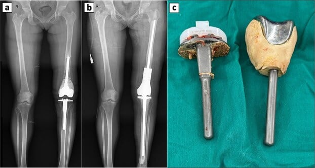
Endoprosthetic Reconstruction More Durable Than Internal Fixation for Pathologic Proximal Femur Fractures
Implant Choice and Failure in Metastatic Humeral Disease (Scandinavian Sarcoma Group)
Failures After Operation for Skeletal Metastases of Long Bones
SORG Machine-Learning Algorithm for Survival in Extremity Metastases (International Validation)
Key References:
-
Mirels H. Metastatic disease in long bones: a proposed scoring system for diagnosing impending pathologic fractures. Clin Orthop Relat Res. 1989;(249):256-264.
-
Wedin R, Bauer HC, Wersäll P. Failures after operation for skeletal metastatic lesions of long bones. Clin Orthop Relat Res. 1999;(358):128-139.
-
Harrington KD. The management of acetabular insufficiency secondary to metastatic malignant disease. J Bone Joint Surg Am. 1981;63(4):653-664.
-
Steensma M, Boland PJ, Morris CD, Athanasian E, Healey JH. Endoprosthetic treatment is more durable for pathologic proximal femur fractures. Clin Orthop Relat Res. 2012;470(3):920-926.
-
Weber KL, Lewis VO, Randall RL, et al. An approach to the management of patients with metastatic bone disease. Instr Course Lect. 2004;53:663-676.
Special Populations
Pelvic and Acetabular Lesions
The Harrington classification of acetabular metastases grades the defect and points to the reconstruction:
- Defect
- Intact acetabulum: metastasis in periacetabular bone, medial wall and dome intact
- Management
- Curettage, cement and screws, with or without radiotherapy
- Defect
- Medial wall destroyed, superior dome intact
- Management
- Medial wall reconstruction (cement and mesh), total hip arthroplasty
- Defect
- Superior dome (the weight-bearing surface) destroyed, requiring structural support
- Management
- Protrusio cup with cement, custom triflange implant, or cup-cage construct
- Defect
- Massive destruction: medial wall and dome both destroyed, possibly extending to the ilium or ischiopubic rami
- Management
- Complex reconstruction, often a saddle prosthesis or custom implant
Fixation or arthroplasty. The decision depends on:
- The class of acetabular involvement
- Whether there is a femoral head or neck lesion
- Expected survival (over 6 months for THA)
- The patient's ambulatory status and goals
- The severity of hip pain
For an isolated periacetabular lesion without articular involvement, percutaneous screw fixation with cement augmentation may suffice.
Spine Metastases with Pathologic Fracture
SINS. The Spinal Instability Neoplastic Score predicts spinal instability in metastatic disease on a scale of 0-18, from six components:
- Location (junctional vs non-junctional)
- Pain (mechanical vs positional vs none)
- Bone lesion (lytic vs mixed vs blastic)
- Spinal alignment (subluxation, kyphosis, scoliosis)
- Vertebral body collapse (over 50%, under 50%, none)
- Posterolateral involvement (bilateral, unilateral, none)
- Interpretation
- Stable: radiotherapy with or without chemotherapy
- Interpretation
- Potentially unstable: an individualised decision
- Interpretation
- Unstable: surgical stabilisation indicated
Spinal Instability Neoplastic Score (SINS): Spine Oncology Study Group Consensus
Surgical indications. Surgery is indicated for:
- Spinal instability (SINS 13 or more)
- Neurological deficit (cord compression, radiculopathy)
- Intractable pain despite radiotherapy
- Radioresistant tumour
- A tissue diagnosis is needed
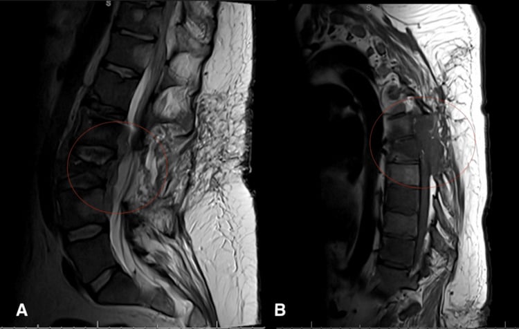
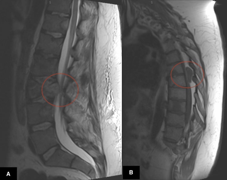
Decompression and instrumented fusion. This relieves neural compression, provides immediate stability and allows early mobilisation, but does not address anterior column destruction, and may fail for that reason.
Separation surgery and stereotactic radiotherapy. A minimal decompression creates space for radiation, followed by short-segment stabilisation and high-dose focused radiotherapy after surgery. It is an emerging paradigm for radioresistant tumours.
Vertebroplasty and kyphoplasty. Cement stabilisation for compression fractures without instability or cord compression. Pain is relieved in 70-90%; the complications are cement leak and, rarely, neural injury.
Corpectomy and reconstruction. Anterior column structural support with an expandable cage or cement, combined with posterior stabilisation. This is major surgery, reserved for patients with longer expected survival.
Spinal pathologic fractures need neurosurgical or spinal surgical expertise, with a multidisciplinary oncology team.
Paediatric Pathologic Fractures
Aetiology. The causes differ from those in adults:
- Benign lesions are more common (unicameral bone cyst, fibrous dysplasia, enchondroma)
- Primary malignant tumours (osteosarcoma, Ewing sarcoma) vs metastatic disease
- Langerhans cell histiocytosis
- Leukaemia and lymphoma
Growth. Avoid physeal injury if possible, and anticipate the potential for limb-length discrepancy and the long-term effects of implants. The remodelling potential is greater than in adults.
Benign lesions. Many heal spontaneously after fracture (unicameral bone cyst 15%). The initial treatment is immobilisation of the fracture, with definitive treatment of the lesion (curettage, grafting) after healing; avoid unnecessary surgery during the acute phase.
Malignant lesions. Treatment is multimodal (chemotherapy and surgery, with or without radiotherapy), with limb salvage preferred when feasible, physeal-sparing techniques when possible, and expandable prostheses in young children to accommodate growth.
The paediatric population requires subspecialty expertise and different treatment paradigms, focused on cure and long-term function.
Controversies & Areas of Uncertainty
- Mirels threshold for prophylactic fixation. A score of 9 or more is the classic cut-off, but the score is sensitive and poorly specific, so it over-predicts fracture and may lead to over-treatment. There is growing interest in CT-based structural metrics (axial cortical involvement greater than 30 mm, circumferential cortical destruction) and CT-based finite-element ("rigidity") analysis as more specific predictors, though none has displaced Mirels in routine practice.
- Biopsy before fixation in the "obvious" metastasis. When widely metastatic disease is established and the lesion is typical, many surgeons proceed to stabilisation with intra-operative sampling. The counter-argument is that solitary or atypical lesions in a cancer survivor occasionally turn out to be a second primary or sarcoma; the safe default in any doubt is biopsy first.
- Endoprosthesis versus intramedullary nailing for the proximal femur. Registry and cohort data show endoprostheses fail far less often than ORIF and somewhat less than nails, but they are bigger operations with higher early dislocation and infection risk. The decision hinges on expected survival, extent of bone loss and joint involvement rather than a single rule.
- Separation surgery plus SBRT versus aggressive decompression for the spine. The Patchell trial established surgery plus conventional radiotherapy for cord compression, but the modern paradigm increasingly favours minimal "separation" surgery followed by high-dose stereotactic radiotherapy, especially for radioresistant tumours - access to SBRT drives this variation.
- Role and duration of bone-modifying agents. Bisphosphonates and denosumab reduce skeletal-related events, but optimal agent, dosing interval and how to manage rebound hypercalcaemia after stopping denosumab remain debated, as does their role around the time of surgery.
- Cemented intramedullary augmentation. Cement improves immediate fixation and adds a tumoricidal thermal effect, but extravasation, embolism and the loss of future MRI surveillance are real trade-offs; routine versus selective use is not standardised.
MCQ Practice Points
Q: What is the Mirels scoring system for impending pathological fractures?
A: Scores 1-3 points for each: Site (upper limb/lower limb/peritrochanteric), Pain (mild/moderate/functional), Lesion type (blastic/mixed/lytic), Size (less than 1/3, 1/3-2/3, greater than 2/3 of cortex). Total ≥9: prophylactic fixation; 8: borderline, weigh clinically; ≤7: irradiate. Sensitivity ~90%, specificity ~35% for predicting fracture. Guides surgical decision-making for impending fractures.
Q: What are the principles of surgical fixation for pathological fractures?
A: Load-sharing constructs preferred - assume tumor will NOT heal. Locked intramedullary nails for long bone diaphyses. Endoprosthetic replacement for periarticular lesions or extensive bone loss. Cement augmentation fills defects and provides immediate stability. Span entire bone - protect against additional lesions. Radiation post-operatively.
Q: When should pathological lesions be biopsied before fixation?
A: Biopsy first if: Unknown primary, solitary lesion in patient without cancer history, atypical features, suspicion of primary bone tumor. Direct to surgery if: Known cancer with typical metastatic pattern, multiple skeletal lesions, characteristic imaging (renal, breast, lung, prostate patterns). Wrong diagnosis changes entire treatment approach.
Q: What is the life expectancy consideration in pathological fracture surgery?
A: Surgery indicated if expected survival greater than 6 weeks. Prognosis scoring (eg, Katagiri, PATHFx) helps estimate survival. Poor prognosis tumors: Lung, melanoma, hepatocellular. Better prognosis: Breast, prostate, renal (may survive years). Match surgical complexity to prognosis - endoprosthesis for long survivors, simple fixation for poor prognosis.
Q: What is the role of radiation in pathological fracture management?
A: Postoperative radiation (typically 8Gy single fraction or 30Gy/10 fractions) for: Tumor control, pain relief, prevention of progression. Not a substitute for adequate fixation. Begin 2-4 weeks post-op to allow wound healing. Preoperative radiation occasionally used for highly vascular tumors (renal) to reduce intraoperative bleeding.
Clinical Decision Scenarios
Practise clinical reasoning and management decisions out loud
“A 68-year-old woman with metastatic breast cancer (ER+/PR+) presents with 3 months of progressive right thigh pain. She is ambulatory with a cane. Radiograph shows a 4cm lytic lesion in the subtrochanteric region with greater than 50% cortical destruction on the lateral cortex. She is currently on an aromatase inhibitor. How do you approach this case?”
“A 55-year-old man presents with acute pain and deformity of his left arm after lifting a bag of groceries. Radiograph shows a mid-diaphyseal humeral fracture through a 3cm lytic lesion. He has no history of malignancy. Radial nerve is intact. How do you manage this patient?”
“A 62-year-old man with known metastatic prostate cancer presents with 2 weeks of progressive thoracic back pain, now with bilateral leg weakness (power 3/5), a sensory level at T6, and difficulty voiding. MRI shows a T6 metastasis with vertebral body collapse, epidural extension and cord compression. How do you manage him?”
Mirels Score - Know Cold
- Site: Upper limb (1), Lower limb (2), Peritrochanteric (3)
- Pain: Mild (1), Moderate (2), Functional (3)
- Lesion: Blastic (1), Mixed (2), Lytic (3)
- Size: Less than 1/3 (1), 1/3-2/3 (2), Greater than 2/3 (3)
- Score greater than or equal to 9 = Prophylactic fixation indicated
- Score 8 = Individualized decision, consider fixation
- Score less than or equal to 7 = Observation acceptable with monitoring
Biopsy Principles (Common Exam Question)
- Biopsy before treatment EXCEPT: known metastatic disease with typical lesion, life/limb emergency
- Coordinate with treating oncologic surgeon BEFORE biopsy
- Biopsy tract must be excisable en bloc with tumor
- Longitudinal incision in line with definitive surgery
- Core needle preferred (80-90% diagnostic, minimal contamination)
- Never violate adjacent compartments or joint
- Mark tract with clip/suture for later excision
- Whoops procedures devastate sarcoma outcomes
Surgical Construct Principles
- Stabilize entire bone (nail) vs lesion only (plate)
- Bypass lesion by minimum 2 cortical diameters each direction
- Load-sharing (nail) preferred over load-bearing (plate)
- Cement augmentation: immediate stability, thermal necrosis, improved screw purchase
- Early weight-bearing priority (quality of life in palliative care)
- Durability must match prognosis: short (less than 6m) simple, intermediate (6-12m) durable, long (greater than 12m) prosthesis
- Nail advantages: minimally invasive, entire bone, allows radiation
- Plate advantages: periarticular, allows curettage, upper extremity
Staging Workup Sequence
- Radiographs: AP/lateral affected bone + joints above/below
- Skeletal survey: multiple myeloma or identify additional lesions
- CT chest/abdomen/pelvis: identify primary tumor, visceral mets
- MRI affected bone: marrow involvement, soft tissue extent, skip lesions
- Bone scan or PET-CT: systemic skeletal survey
- Labs: CBC, CMP, LFTs, calcium, ALP, SPEP/UPEP, PSA (if appropriate)
- Biopsy: tissue diagnosis (coordinate with oncologist)
- MDT discussion: oncology, radiation oncology, radiology
Prognosis by Primary Tumor
- Long survival (greater than 12m): Breast (ER+), Prostate, Thyroid, Myeloma
- Intermediate (6-12m): Renal, Colon, Melanoma, Unknown primary
- Short (less than 6m): Lung (especially small cell), Pancreas, Liver
- Presence of visceral metastases cuts survival in half
- Ambulatory status crucial predictor of functional recovery
- Surgery improves quality of life but NOT survival duration
- Use Katagiri score or similar to guide reconstruction durability
Complications to Discuss
- Intraoperative: hemorrhage (hypervascular tumors - embolize), cement extravasation (joint/nerve), fracture propagation
- Early: infection (5-15%, higher with chemo/radiation), wound dehiscence, VTE
- Late: implant failure (5-15% at 1 year), nonunion (expected, construct must not rely on healing), local progression
- Cement complications: thermal necrosis, embolization (rare, potentially fatal), joint penetration
- Revision: escalate construct durability (plate to nail to prosthesis), higher complication rate, balance with prognosis
Special Situations
- Pelvis/Acetabulum: Harrington classification (I-IV), Class III-IV need THA/custom implant
- Spine: SINS score (greater than or equal to 13 unstable, needs fixation), vertebroplasty for compression fractures without instability
- Pediatric: benign lesions common, fracture may induce healing (UBC), malignant needs multimodal treatment
- Radiation: adjuvant post-op (2-3 weeks delay for wound healing), NOT substitute for fixation in high-risk lesions
- Hypervascular tumors: renal, thyroid - preoperative embolization, crossmatch blood, anticipate hemorrhage